Introduction
Total mesorectal excision (TME) is the gold standard for curative rectal cancer surgery worldwide, which reduces local recurrence rates and improves survival rates [
1,
2]. In TME, surgeons excise the mesorectum including the lymphovascular tissue which is considered as the possible source of pelvic recurrence [
3]. Lymphatics from the rectum drain along rectal arteries to internal iliac nodes. The middle rectal artery (MRA) is regarded as an integral part of the lymphatic drainage to the lateral pelvic nodes, and is the only artery encountered during TME running into the rectum through the proper rectal fascia in the pelvis [
4]. Therefore, understanding the anatomy of the MRA is necessary for surgeons to perform TME with efficacy and safety.
Previous studies have provided anatomical knowledges about the MRA: its frequency, origins, courses, relationships to the lateral ligament, and distributions to the rectum [
5-
7], however it is still not easy to grasp the schema of the MRA. One of the major causes for this is based on the complexity of the pelvic vasculature. In previous reports investigating the origins of the MRA, whole detailed vasculature from the internal iliac artery to the MRA have not been described. Adoption of oversimplified schema may limit comprehensive understanding about pelvic vasculatures including the MRA.
The aim of this study was to categorize MRAs regarding their detailed vasculature patterns with identification of their frequencies, and to provide schematic information that would improve clinical applicability.
Go to :

Materials and Methods
The institutional review board of the Catholic University of Korea ruled that a cadaveric study was beyond its review authority. All cadavers used in this study were donated to the Catholic Institute for Applied and Clinical Anatomy under informed consent. Exclusion criteria included any gross evidences of pelvic surgeries or malignancies, which could affect normal pelvic anatomy.
Forty sides of the pelvis from 20 cadavers (10 male, 10 female) were investigated. Ages at death of the specimens were 85.5±7.4 years (range, 68.8–98.9 years). For purposes of the study, the pelvis was sectioned in the midsagittal plane with preservation of intrapelvic organs and embalmed in 10% formalin solution. The peritoneum was detached and removed from the pelvic brim down to the pelvic floor. All the vascular branches from the internal iliac artery were carefully dissected, focusing on identification of the MRA. When the MRA was present, all the arterial vasculatures related to its origin from the level of the internal iliac artery were investigated.
The MRAs were grouped into major groups depending on their origins. Each major group was subdivided into minor groups as the MRA-originating artery also presented various branching patterns from the internal iliac artery.
Go to :

Results
Frequencies of the MRA according sex and side of pelvis are described in
Table 1. The MRA was identified in most cadavers, and commonly in the pelvic sides. More cases of MRA were identified in males or the right pelvis, but a considerable number of cases were also observed in females or the left pelvis. The MRA was present bilaterally in seven cadavers (35.0%, 7/20 cases), and unilaterally in 11 cadavers (55.0%, 11/20 cases).
Table 1
Frequencies of middle rectal artery regarding sex and side of pelvis
|
Variable |
Cadaver |
Pelvis |
|
Sex |
|
|
|
Male |
10/10 (100) |
15/20 (75.0) |
|
Female |
8/10 (80.0) |
10/20 (50.0) |
|
Side |
|
|
|
Right |
- |
14/20 (70.0) |
|
Left |
- |
11/20 (55.0) |
|
Total |
18/20 (90.0) |
25/40 (62.5) |

A total of seven types of MRA origin were identified, thus, each major group was designated as type I to VII in order from the most frequent to the least. The origin of MRA was mostly related to the gluteal-pudendal arterial system (
Table 2). The most common origin of the MRA was the internal pudendal artery (type I,
Fig. 1), followed by the inferior gluteal artery (type II,
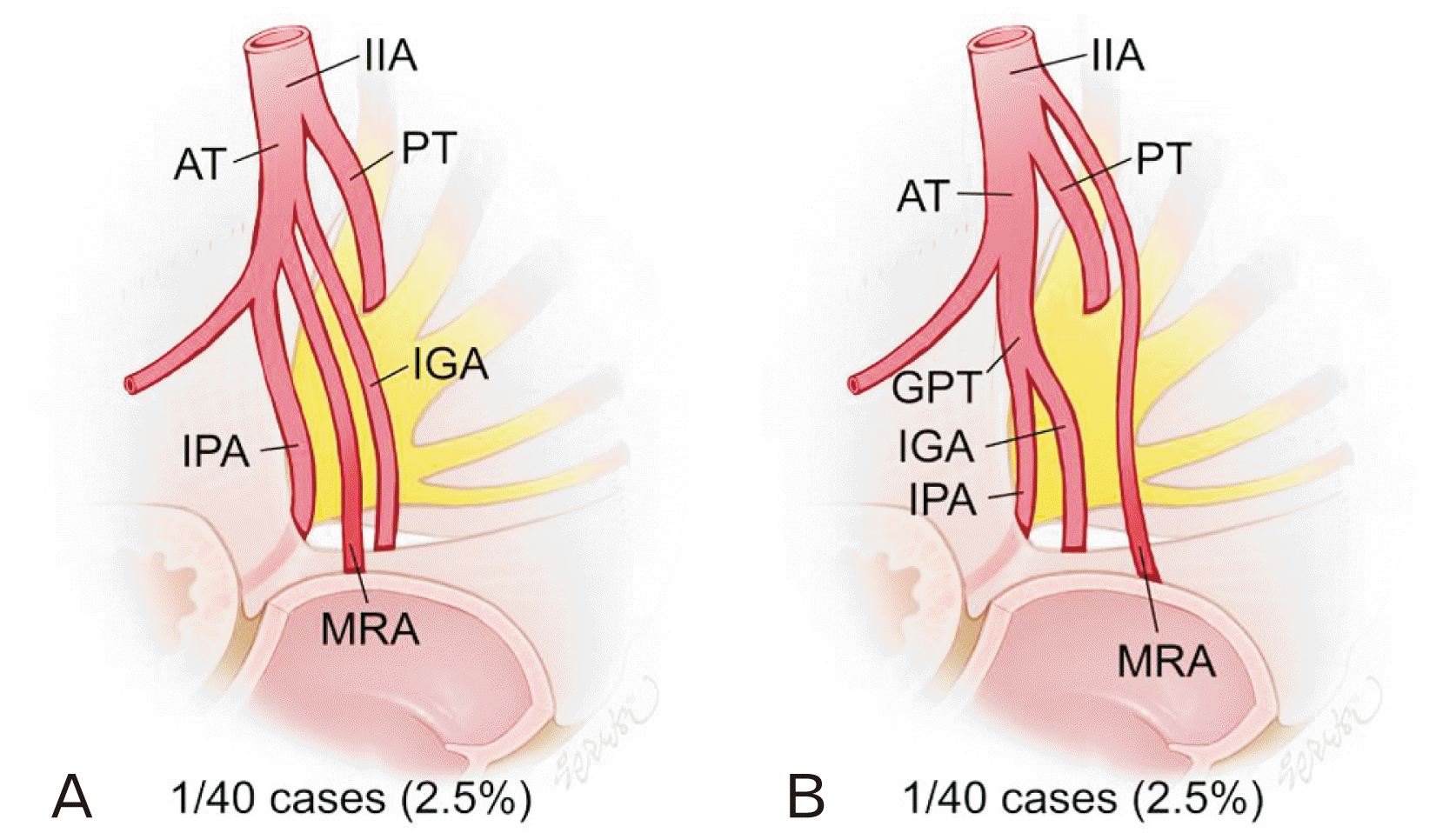
Fig. 2). Most of the internal pudendal artery and inferior gluteal artery, in these two major types, arose from the anterior trunk of the internal iliac artery either directly (type Ia and IIa;
Figs. 1A,
2A) or formed a gluteal-pudendal trunk before bifurcation (type Ib and IIb;
Figs. 1B,
2B). Notably, in two cases, these arteries originated from the posterior trunk of the internal iliac artery, not from the anterior trunk (type Ic and IIc;
Figs. 1C,
2C). The MRA arising directly from the gluteal-pudendal trunk (type III;
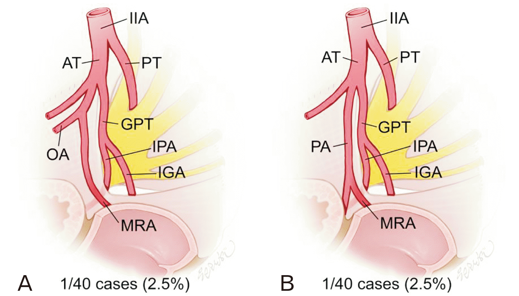
Fig. 3) was also identified in two cases. In type IVa, MRA arose from the inferior vesical artery which had an origin from the gluteal-pudendal trunk (
Fig. 4A).
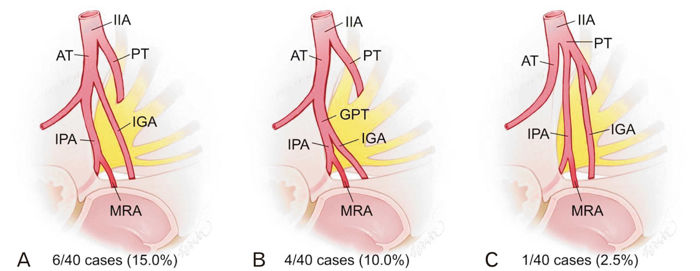 | Fig. 1Middle rectal artery arising from internal pudendal artery (type I). (A) Internal pudendal artery arises directly from anterior trunk of internal iliac artery and gives off middle rectal artery (type Ia). (B) Internal pudendal artery arises from gluteal-pudendal trunk and gives off middle rectal artery (type Ib). (C) Internal pudendal artery arises from posterior trunk of internal iliac artery and gives off middle rectal artery (type Ic). IIA, internal iliac artery; AT, anterior trunk of internal iliac artery; PT, posterior trunk of internal iliac artery; IPA, internal pudendal artery; IGA, inferior gluteal artery; MRA, middle rectal artery; GPT, gluteal-pudendal trunk. 
|
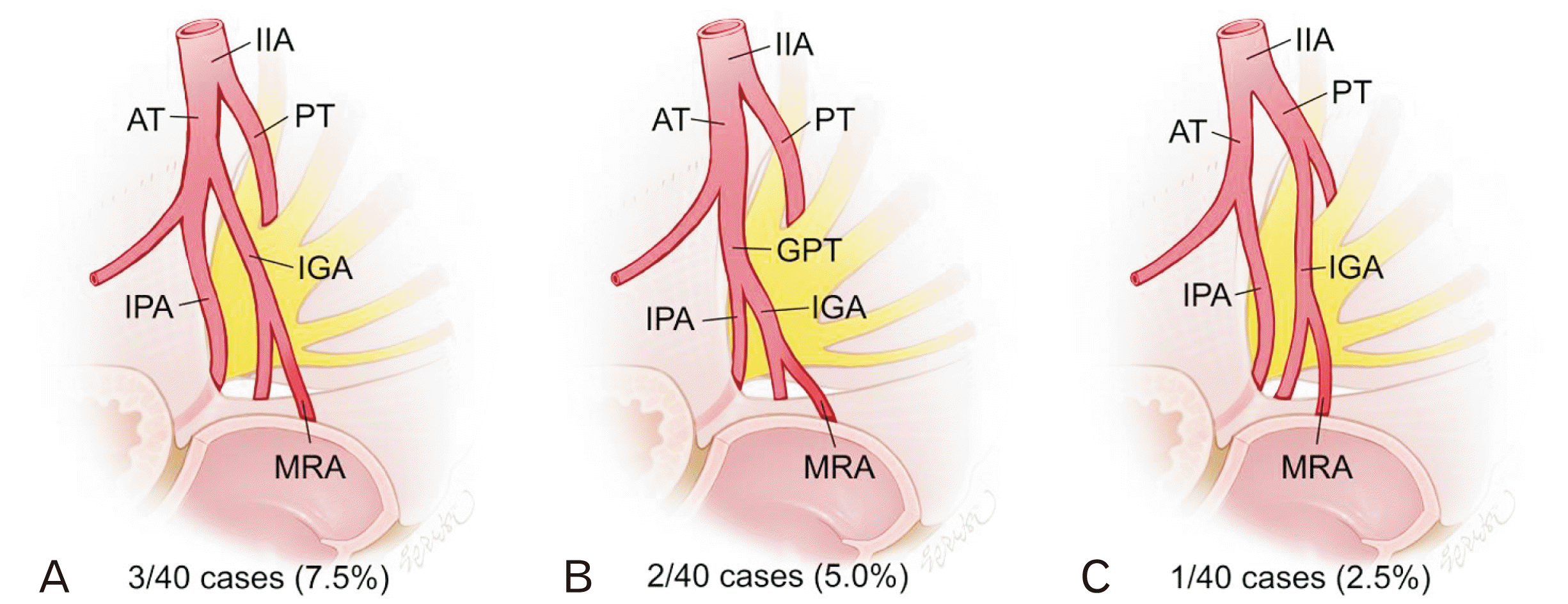 | Fig. 2Middle rectal artery arising from inferior gluteal artery (type II). (A) Inferior gluteal artery arises directly from anterior trunk of internal iliac artery and gives off middle rectal artery (type IIa). (B) Inferior gluteal artery arises from gluteal-pudendal trunk and gives off middle rectal artery (type IIb). (C) Inferior gluteal artery arises from posterior trunk of internal iliac artery and gives off middle rectal artery (type IIc). IIA, internal iliac artery; AT, anterior trunk of internal iliac artery; PT, posterior trunk of internal iliac artery; IPA, internal pudendal artery; IGA, inferior gluteal artery; MRA, middle rectal artery; GPT, gluteal-pudendal trunk. 
|
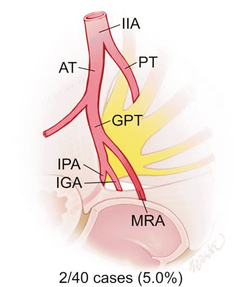 | Fig. 3Middle rectal artery arising directly from gluteal-pudendal trunk (type III). IIA, internal iliac artery; AT, anterior trunk of internal iliac artery; PT, posterior trunk of internal iliac artery; GPT, gluteal-pudendal trunk; IPA, internal pudendal artery; IGA, inferior gluteal artery; MRA, middle rectal artery. 
|
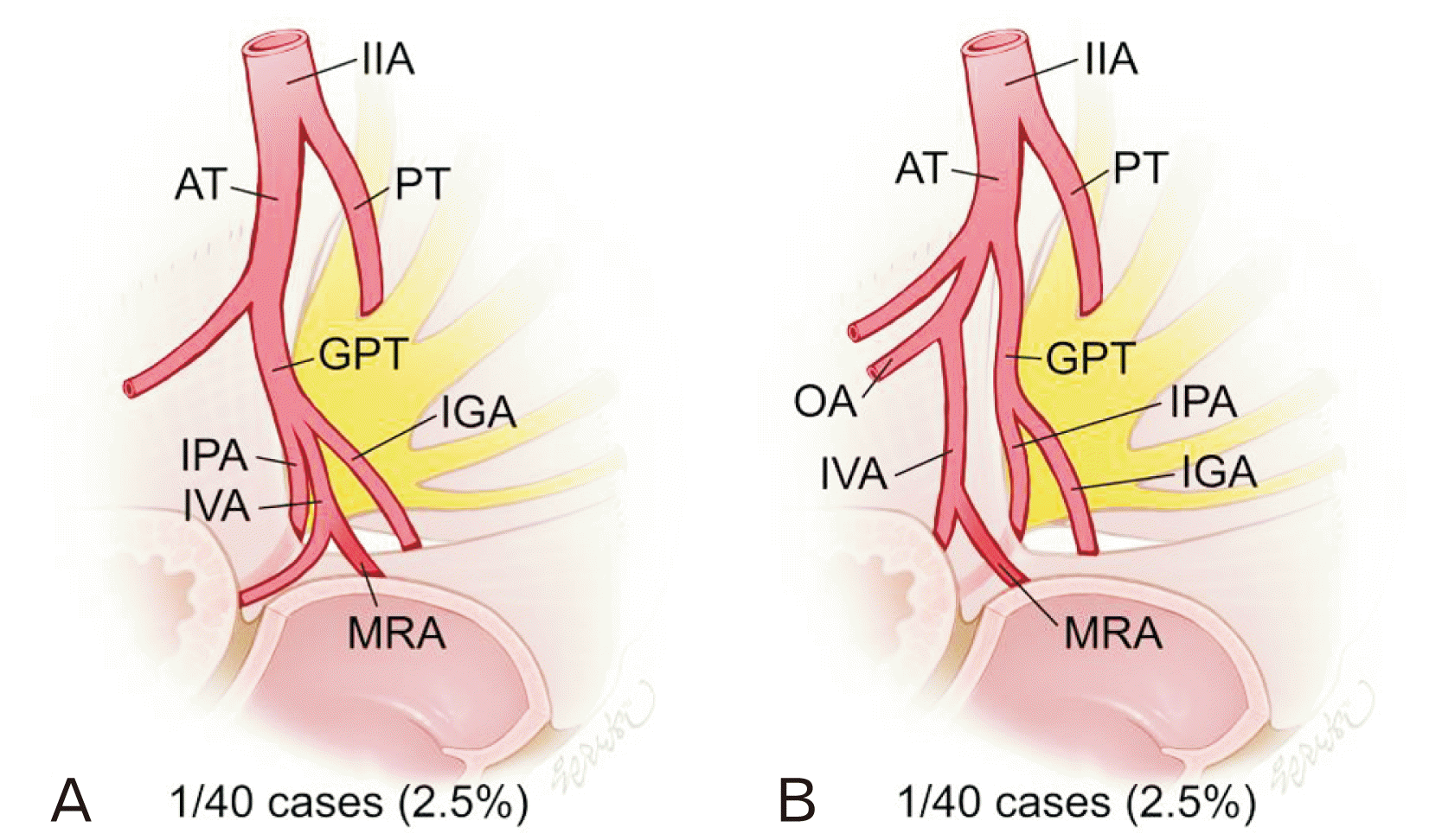 | Fig. 4Middle rectal artery arising from inferior vesical artery (type IV). (A) Inferior vesical artery arises from gluteal-pudendal trunk and gives off middle rectal artery (type IVa). (B) Inferior vesical artery arises from obturator artery and gives off middle rectal artery (type IVb). IIA, internal iliac artery; AT, anterior trunk of internal iliac artery; PT, posterior trunk of internal iliac artery; GPT, gluteal-pudendal trunk; IPA, internal pudendal artery; IGA, inferior gluteal artery; IVA, inferior vesical artery; MRA, middle rectal artery; OA, obturator artery. 
|
Table 2
Origins of the middle rectal artery
|
Sex |
IPA |
IGA |
GPT |
IVA |
IIA |
OA |
PA |
Total |
|
Male |
7 |
2 |
2 |
1 |
1 |
1 |
1 |
15 |
|
Female |
4 |
4 |
0 |
1 |
1 |
0 |
- |
10 |
|
Total |
11 |
6 |
2 |
2 |
2 |
1 |
1 |
25 |

Other origins of the MRA except the gluteal-pudendal arterial system include the inferior vesical artery arising from the obturator artery (type IVb;
Fig. 4B), internal iliac artery (type V;
Fig. 5), obturator artery (type VI;
Fig. 6A), and prostatic artery (type VII;
Fig. 6B). In the cases of type V, the MRA arose either from the anterior trunk (type Va,
Fig. 5A) or from the main trunk of the internal iliac artery (type Vb,
Fig. 5B). From their origin, the MRAs coursed inferiorly and then medially to approach the rectum through the proper rectal fascia.
 | Fig. 5Middle rectal artery arising from internal iliac artery (type V). (A) Middle rectal artery arises from the anterior trunk of internal iliac artery, and presents independent course from internal pudendal artery and inferior gluteal artery (type Va). (B) Internal iliac artery gives off middle rectal artery before it is separated into anterior and posterior trunk (type Vb). IIA, internal iliac artery; AT, anterior trunk of internal iliac artery; PT, posterior trunk of internal iliac artery; IGA, inferior gluteal artery; IPA, internal pudendal artery; MRA, middle rectal artery; GPT, gluteal-pudendal trunk. 
|
 | Fig. 6Additional origins of middle rectal arteries. (A) Middle rectal artery arises from obturator artery (type VI). (B) Middle rectal artery arises from prostatic artery (type VII). IIA, internal iliac artery; AT, anterior trunk of internal iliac artery; PT, posterior trunk of internal iliac artery; GPT, gluteal-pudendal trunk; OA, obturator artery; IPA, internal pudendal artery; IGA, inferior gluteal artery; MRA, middle rectal artery; PA, prostatic artery. 
|
Go to :

Discussion
In this study, the anatomy of the MRA was provided in a manner applicable to the TME. A complete TME is defined as “complete removal of the lymph node bearing mesorectum along with its intact enveloping fascia,” and includes anterolateral division of the lymphatics and middle rectal vessels. For this, the dissection plane is extended from the mesentery root above the aortic bifurcation downward into the pelvis and in a lateral to medial fashion to open the lateral pelvic wall to identify MRA [
3]. Through dissection of 40 hemi-pelvises, vasculature patterns related to MRAs with variability were identified. The schematic figures for these were presented to display detailed courses from the internal iliac artery to the origin of the MRA for application to the step of lateral pelvic dissection and identification of the MRA.
The present study identified MRA in 90% of the cadavers and in 62.5% of the pelvic sides. The frequencies of MRA in previous studies ranged from 12% to 97% in subjects, and from 8% to 95% in pelvic halves [
4]. According to Kiyomatsu et al. [
4] (2017), this variety of frequency may not be based on the detection method, either dissection or imaging technique, considering that the accuracy of detecting MRA has not improved over time. This implies that precise dissection is not inferior to imaging technology when identifying the small vasculature. The MRA, as a result of dissection in this study, was identified as a common structure in both sexes and bilateral sides; thus, surgeons should be reminded in order to safely perform rectal surgery.
In the present study, more cases of MRAs were identified in male pelvic sides (75.0%, 15/20 cases) than in female pelvic sides (50.0%, 10/20 cases), and in right (70.0%, 14/20 cases) than in left side (55.0%, 11/20 cases). In the study of Didio et al. [
5], no significant differences of MRA frequencies in both sexes and bilateral pelvic sides were identified. Sato and Sato [
6] identified the MRA more frequently in male (25.9%, 14/54 pelvic sides) than in female (14.8%, 4/27 pelvic sides), whereas Jones et al. [
8] observed more MRAs in female samples (26.5%, 9/34 pelvic sides) than in male samples (36.4%, 8/22 pelvic sides). These discordances among the abovementioned studies are based on the random selection of small-sized samples from different population groups, which implies needs for follow-up study with a larger sample size.
In 10% of the cases in this study, MRA was not identified in any of bilateral sides, and the superior rectal artery was the only feeding artery above the pelvic diaphragm. Among rectal arteries, the superior rectal artery is considered to be the main artery supplying the rectum, with the MRA as the supplementary artery [
9-
11]. Therefore, the superior rectal artery was considered to compensate for the blood-supply of the absent MRA in 10% of the cases.
Multiple anatomical textbooks describe the MRA as an artery arising directly from the anterior trunk of the internal iliac artery or from the inferior vesical artery [
12,
13]. However, the results of the current anatomical investigation suggest that the most common origin of the MRA is the internal pudendal artery, followed by the inferior gluteal artery. The internal iliac artery and inferior vesical artery constitute the less common among origins of the MRA. These results correspond to the findings of previous studies [
5-
7]. The gluteal-pudendal trunk, which was identified to give off a MRA in two among 40 pelvic halves in the present study, was the third most frequent origin of the MRAs in the study of Bilhim et al. [
7] (2013). The MRAs arising from the obturator artery and prostatic artery have also been previously reported in the literature [
7,
14].
Despite the consistent reports about the origins of the MRA, identification of this vessel in rectal surgery is still a laborious step due to the complex pelvic vasculature. In this study, the internal pudendal artery and inferior gluteal artery, which primarily gave off the MRA, usually arose directly from the anterior trunk of the internal iliac artery (type Ia and IIa) as classical anatomical findings, but they formed gluteal-pudendal trunk before bifurcation with a considerable frequency in other cases (type Ib and IIb). Moreover, there were rare cases of the internal pudendal artery and the inferior gluteal artery originating from the posterior trunk (type Ic and IIc), not from the anterior trunk. In the other types of MRAs (type III to VII), pelvic vasculatures from the internal iliac artery to the MRA also presented complexity. Even the less common of the MRA origins and pelvic vasculature patterns should be considered in planning the surgical approach to prevent unexpected vascular injury. Therefore, the schematic information of this study, which included detailed courses of the MRA and pelvic vasculature, is valuable for rectal surgeons in minimizing the risk of vascular injury during the TME.
The anatomical findings of this study are also applicable to radiologic interventions. To perform arterial embolization procedures in cases of severe rectal hemorrhage [
15], recognition of the MRA anatomy and possible variations of the pelvic vasculature are required. Schematic information about the possible vasculature patterns found in this study would help to minimize time consumption during intervention procedures.
This study had several limitations. First, the distribution of rectal arteries within the submucosal layer of the rectum was not investigated. For the purpose of application in rectal surgeries, this study focused on the vasculature pattern of MRA from the internal iliac artery to the entry site to the rectum. Second, among the MRA origins reported in previous studies, the median sacral artery giving off the middle rectal branch was not identified in this study. DiDio et al. [
5] (1986) reported frequencies of this type as 3.3% (1/30 cases). Follow-up study with a larger sample size would possibly find other minor types and help provide illustrations for them. Despite these limitations, the advanced anatomical findings of this study related to MRA and pelvic vasculature is considered noteworthy for its improved clinical applicability.
Go to :


 PDF
PDF Citation
 Print
Print


 XML Download