We have read the article entitled “Splanchnic nerve neurolysis via the transdiscal approach under fluoroscopic guidance: a retrospective study”, for which we congratulate its authors for their results and recommendations [1].
In 2003, Plancarte-Sánchez et al. [2] described a CT-guided transdiscal technique in 64 abdominal cancer patients, reporting a reduction in complications in the approach compared to the conventional technique (paraplegia, pneumothorax, and liver or renal puncture). Considering the level of radiation used in CT procedures, it is important to have a fluoroscopy-guided technique to access the splanchnic nerves.
A double transdiscal access for neurolysis of the splanchnic nerves using fluoroscopy was recently reported with the intention of covering more splanchnic fibers using a smaller volume [3].
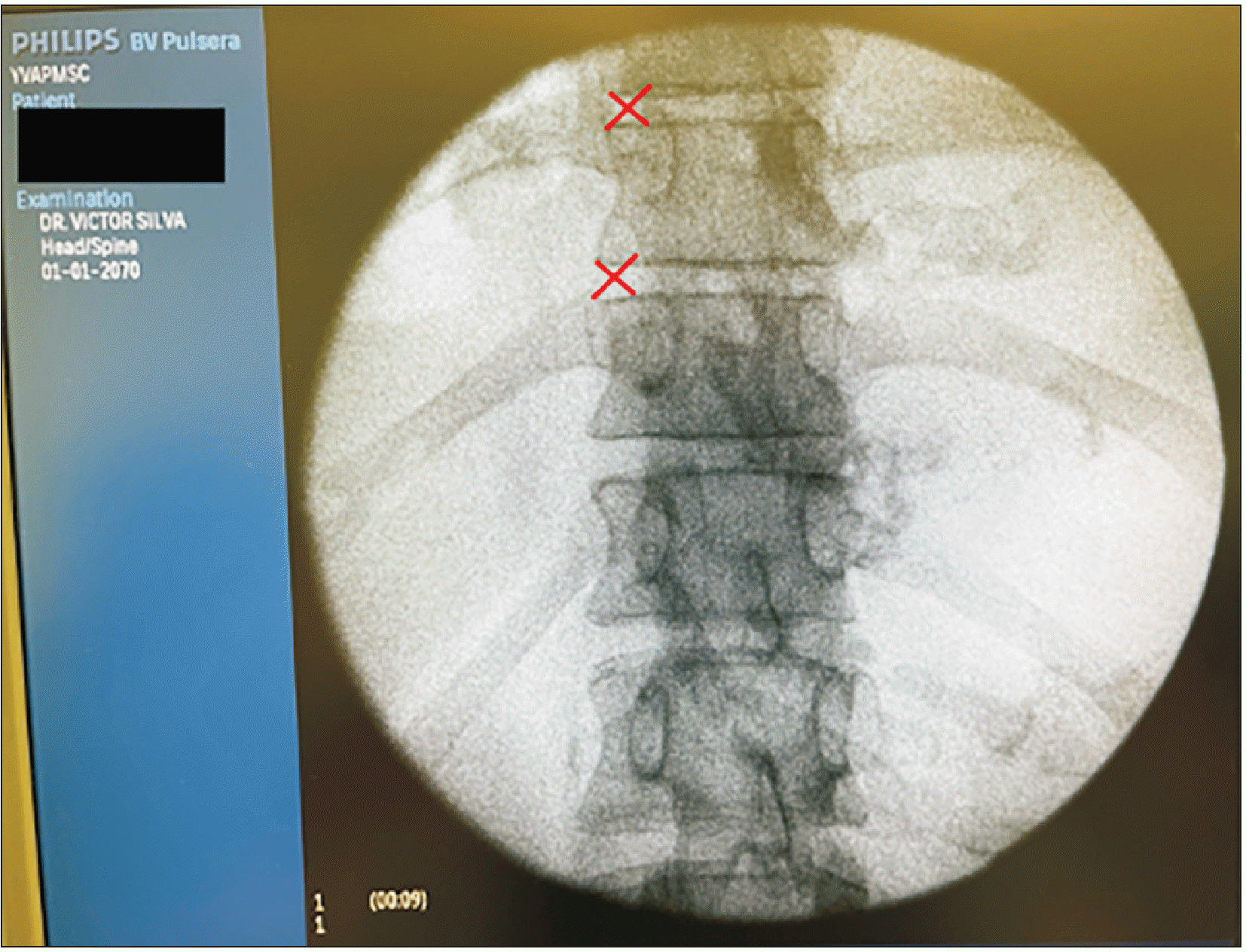
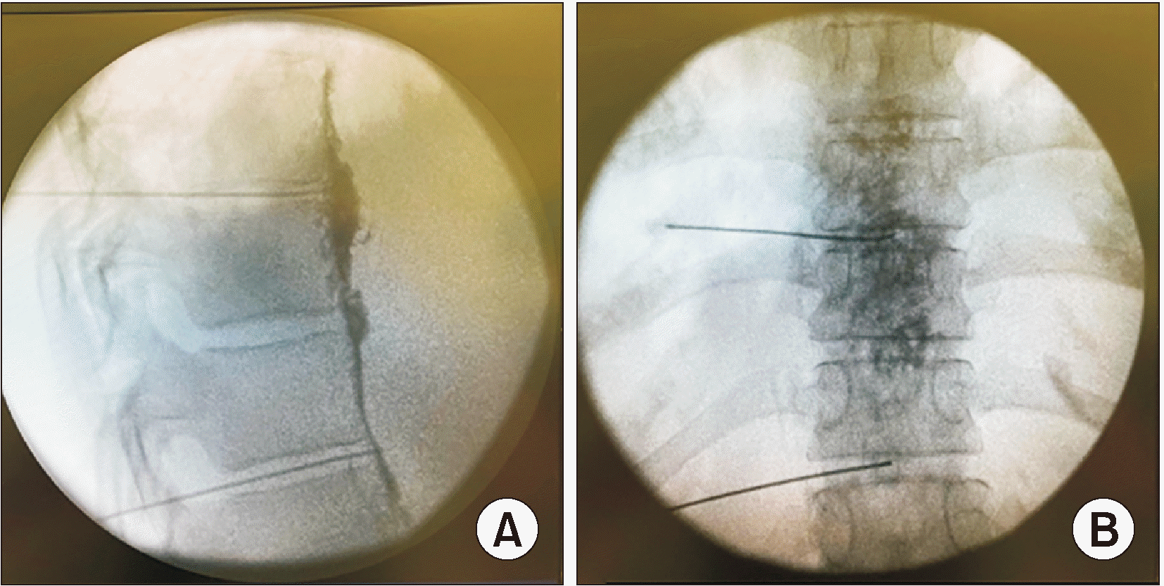
It is true that to date a fluoroscopy-guided transdiscal technique has not been described for access to the splanchnic nerves, with only the paravertebral technique having been described [4]. The images and recommendations regarding the transdiscal technique are amazing. We would like to augment the information in this great paper by adding a way to initially access the disc. With a 15-degree curved-tip needle, in a tunnel access, we visualize the space between the rib and the superior articular process. In this way, the obliquity needed is minimal, approximately 5 degrees ipsilateral (Fig. 1). Usually, the access requires 20 degrees of obliquity, but in this technique description, the 15 degrees of the tip of the needle allows us to reduce the angle from 20 to 5 degrees since, once the disc is contacted, the tip of the needle is advanced medially, allowing it to finish in the central and anterior part of the disc, which is the target of this procedure (Fig. 2). With this technique we diminish the risk of puncturing the pleura, which is a potential complication of this approach (Fig. 2).
Notes
DATA AVAILABILITY
Data sharing is not applicable to this article as no datasets were generated or analyzed for this study.
REFERENCES
1. Cai Z, Zhou X, Wang M, Kang J, Zhang M, Zhou H. 2022; Splanchnic nerve neurolysis via the transdiscal approach under fluoroscopic guidance: a retrospective study. Korean J Pain. 35:202–8. DOI: 10.3344/kjp.2022.35.2.202. PMID: 35354683. PMCID: PMC8977204.

2. Plancarte-Sánchez R, Máyer-Rivera F, del Rocío Guillén Núñez M, Guajardo-Rosas J, Acosta-Quiroz CO. 2003; Transdiscal percutaneous approach of splanchnic nerves. Cir Cir. 71:192–203. Spanish. PMID: 14617407.
3. Silva V, López AG, Martínez L. 2021; Splanchnic nerve neurolysis: double access for abdominal cancer pain. BMJ Support Palliat Care. doi: 10.1136/bmjspcare-2021-003216. DOI: 10.1136/bmjspcare-2021-003216. PMID: 34083320.

4. Ahmed A, Arora D. 2017; Fluoroscopy-guided neurolytic splanchnic nerve block for intractable pain from upper abdominal malignancies in patients with distorted celiac axis anatomy: an effective alternative to celiac plexus neurolysis - a retrospective study. Indian J Palliat Care. 23:274–81. DOI: 10.4103/IJPC.IJPC_28_17. PMID: 28827930. PMCID: PMC5545952.


 PDF
PDF Citation
 Print
Print


 XML Download