Journal List > Korean J Pain > v.35(3) > 1516078755

John, Sohn, and Kim: Relationship between needle depth for lumbar transforaminal epidural injection and patients’ height and weight using magnetic resonance imaging

Abstract

Background

Optimal needle depth in transforaminal epidural injection (TFEI) is determined by body measurements and is influenced by the needle entry angle. Physician can choose the appropriate needle length and perform the procedure more effectively if depth is predicted in advance.

Methods

This retrospective study included patients with lumbosacral pain from a single university hospital. The skin depth from the target point was measured using magnetic resonance imaging transverse images. The depth was measured bilaterally for L4 and L5 TFEIs at 15°, 20°, and 25° oblique angles from the spinous process.

Results

A total of 4,632 measurements of 386 patients were included. The lengths of the left and right TFEI at the same level and oblique angle were assessed, and no statistical differences were identified. Therefore, linear regression analysis was performed for bilateral L4 and L5 TFEIs. The R-squared values of height and weight combined were higher than the height, weight, and body mass index (BMI). The following equation was established Depth (mm) = a – b (height, cm) + c (weight, kg). Based on the equation, maximal BMI capable with a 23G, 3.5-inch, Quincke-type point spinal needle was presented for three different angles (15°, 20°, and 25°) at lumbar levels L4 and L5.

Conclusions

The maximal BMI that derived from the formulated equation is listed on the table, which can help in preparations for morbid obesity. If a patient has bigger BMI than the one in the table, the clinician should prepare longer needle than the usual spinal needle.

INTRODUCTION

Epidural injections such as the interlaminar epidural block and transforaminal epidural injection (TFEI) have been widely used for the treatment of patients with low back pain and lumbosacral radicular pain [14]. As injection agents, corticosteroids and local anesthetics are commonly used to reduce pain and inflammation [57]. Both interlaminar and transforaminal block techniques are effective for lumbar radiating pain and have low complication rates, but the transforaminal technique can be more effective than the interlaminar [8,9]. An interlaminar epidural injection spreads into the dorsal epidural space and diffuses to the ventral target site [6,10]. Consequently, the concentration of the injecting agent is inadequate [11] or an epidural catheter should be inserted and positioned in the ventral epidural space [12]. TFEI can deliver the injectate precisely to the damaged nerve root and anterior epidural space, resulting in better outcomes, such as long-term efficacy and improved effects [1,6,12,13]. The optimal needle depth in TFEI may be determined by the patients’ height, weight, and anatomical differences, and is also influenced by needle entry angle [14,15]. Needle localization close to the desired site requires anatomical knowledge and procedural expertise [16]. Procedure accuracy is important to avoid adverse events [1719], and prior magnetic resonance imaging (MRI) analysis of epidural depth is helpful in this regard [20].
Some studies have directly and indirectly measured the depth of the epidural space and reported their relationship to body measurements [1,14,21]. However, most calculated the depth of the epidural space by converting the needle length used for the injection and did not provide an exact description of the lumbar level. Further, the majority of studies conducted are limited to interlaminar epidural injection [22], and there is a lack of research on depth in TFEI [14]. Commonly used needles for TFEI are too short to approach the target points or target lesions in some patients. However, if the needle depth could be predicted in advance, the physician could prepare a needle of the proper length and inject the drugs more effectively to the target point.
Therefore, the purpose of this study was to investigate the relationship between patients’ height, weight, and body mass index (BMI) and needle depth for TFEI with different angles of approach using lumbar spine MRI images.

MATERIALS AND METHODS

1. Study design and participants

This was a retrospective cohort study, including patients who visited the pain clinic and had lumbar spine MRI images from July 1st, 2005, to March 31st, 2020. This study was conducted in a single university hospital. Ethical approval for this study was waived by the Institutional Review Board of Konkuk University Hospital in Seoul, Republic of Korea (IRB file no. 2020-04-011).

2. Exclusion criteria

Exclusion criteria included a history of lumbar spine surgery, post-state lumbar spine vertebroplasty or kyphoplasty, history of a lumbar spine compression fracture, and absence of height and weight measurement within six months before or after the day the patient underwent MRI.

3. Measurement protocols

Demographic data such as sex, age (years), height (cm), weight (kg), and BMI (kg/m²) were collected. The length (mm) from the skin to the target point on the posterior vertebral body below the pedicle was measured using MRI images. The measurement was performed at the time of bilateral L4 and L5 TFEI implementation with a given angle of 15°, 20°, or 25° oblique from the spinous process. These three angles are the most frequently used oblique fluoroscopic angle for lumbar TFEI in clinical situations.
The depth (mm) from the skin to the target point was measured using T1-weighted transverse images by four anesthesiologists experienced in TFEI and with in-depth knowledge of the anatomical structure of the lumbar spine.
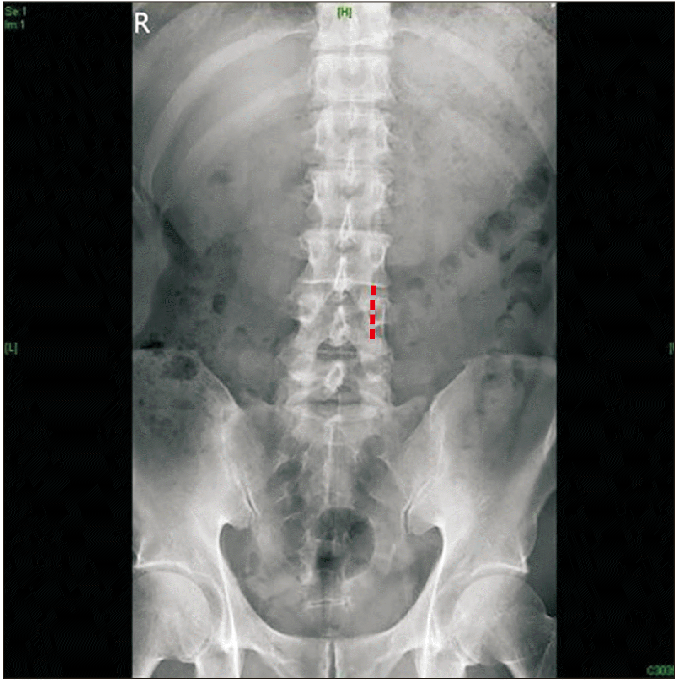
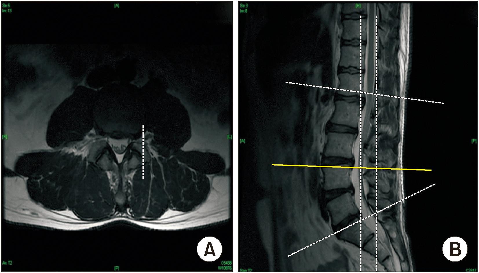
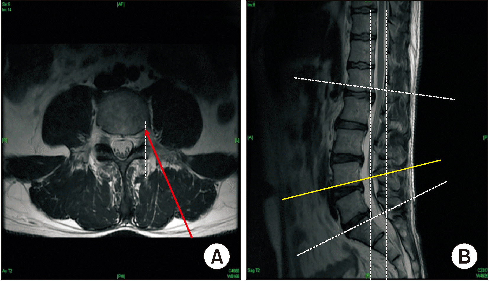
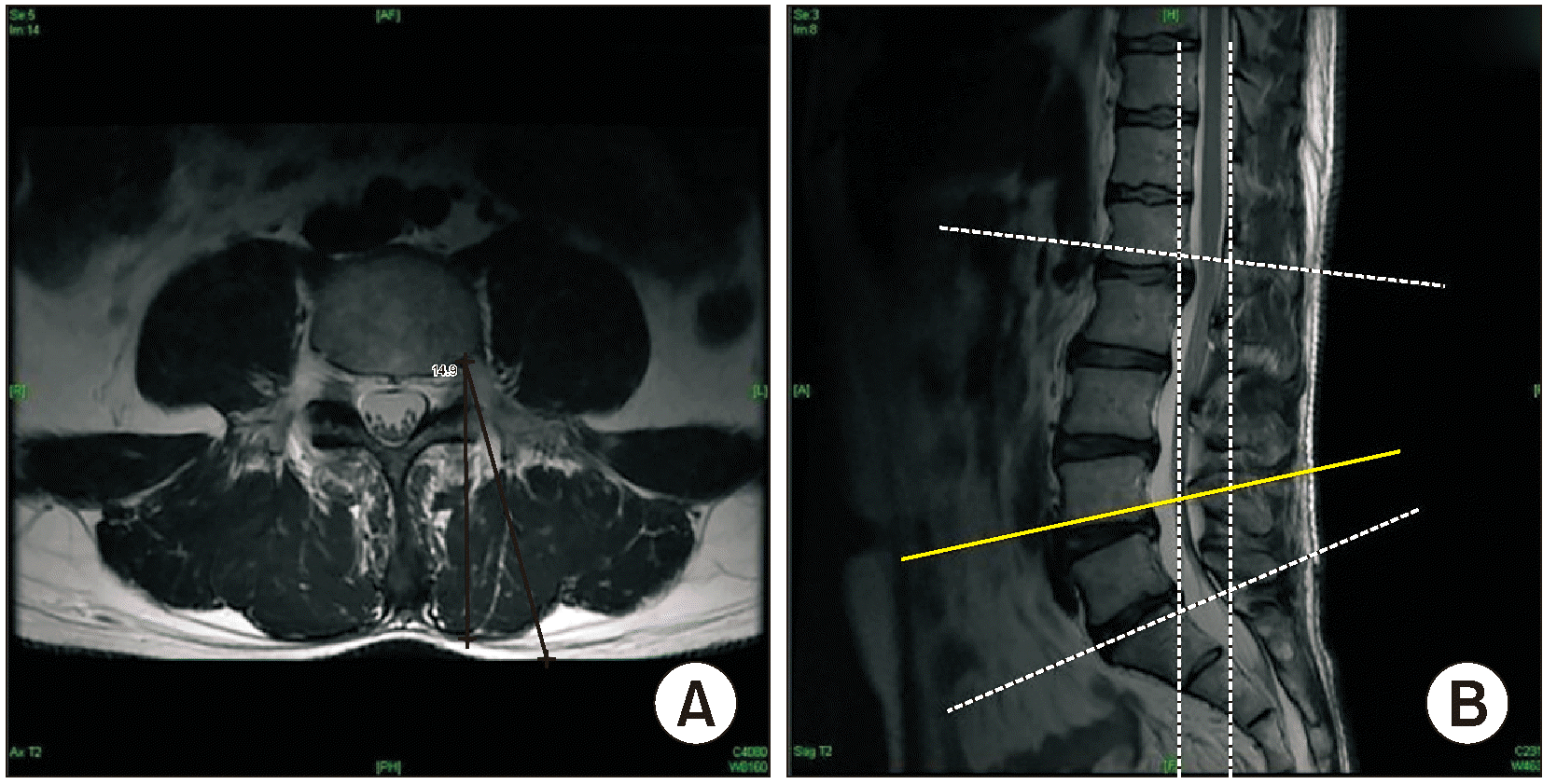
The target point of TFEI is where the line perpendicular to the center of the pedicle encounters the rear part of the vertebral body adjacent to the pedicle, which the nerve root passes underneath the target point (Fig. 1). The target needle point is not exactly found in the MRI image, however it is speculated through the superior articular process of the upper lumbar level (Fig. 2). When measuring the length of the L4 TFEI, the transverse image of the lower L4 vertebral body related to the target point was checked to measure the length from the skin to the target point (Figs. 3, 4). The length was measured bilaterally for three different angles at 15°, 20°, and 25° oblique from the spinous process, respectively, in the picture archiving and communication system (PACS, GE Healthcare, Chicago, IL). The same process was performed for the L4 and L5 TFEI.

4. Statistical analyses

Statistical analyses were performed using SPSS version 17 (SPSS Inc., Chicago, IL). The obtained lengths between left and right TFEI at the same level and oblique angle were assessed with Student t-test to compare significant differences (P < 0.05). The measured data were evaluated through linear regression analysis to correlate patients’ height, weight, and BMI with the distance from skin to target point (P < 0.05). After comparing the coefficient of determination for each parameter, an equation for this dependent variable was obtained after selecting a dependent variable whose coefficient of determination was close to 1.

RESULTS

A total of 550 patients (351 female and 199 male) were initially evaluated, of which 164 (121 female and 43 male) were excluded due to a history of lumbar spine surgery, post-state lumbar spine vertebroplasty or kyphoplasty, a history of compression fracture of the lumbar spine, or absence of height and weight data. The final analysis included 4,632 measures of 386 patients (230 female and 156 male). The mean age of patients was 63.2 (21–95) years. The mean height was 161.3 (138.5–187.0) cm, the mean weight 63.1 (34.1–108.0) kg, and the mean BMI 24.2 (15.8–35.3) kg/m2 (Table 1).
The lengths between left and right TFEI at the same level and oblique angle were assessed, and no statistically significant differences were identified between each side (Table 2). Therefore, a linear regression analysis was performed for bilateral L4 and L5 TFEI at the oblique angles of 15°, 20°, and 25°, respectively.
Linear regression analysis was performed for every level and angle, as well as height, weight, BMI, and height and weight, all correlated with the length from the skin to the target point. The results showed that the R-squared values of height and weight combined were higher than those of height, weight, and BMI (Table 3). Based on the result from linear regression analysis, an equation of 15° oblique L5 epidural needle depth (mm) = 68.11 – 0.18 × height (cm) + 0.53 × weight (kg) was established (Table 4). Similarly, a linear regression analysis was performed at different oblique angles and levels, and the results showed height and weight combined correlated significantly with needle depth, allowing the development of an equation for each particular case (Table 5).
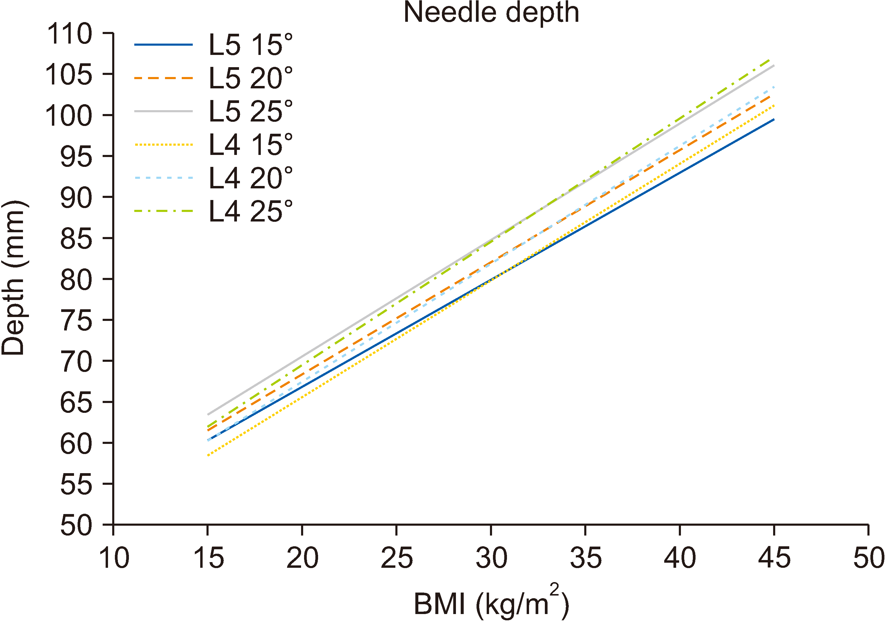
We formulated another predictive equation of needle depth based on linear regression analysis (Table 6). BMI was also correlated with needle depth and the equation of BMI is easy to use. A graph based on the equation from Table 6 shows estimated needle depth based on BMI at three different angles (15°, 20°, and 25°) at lumbar levels L4 and L5 (Fig. 5). Based on the equation from Table 6, the maximal BMI that could possibly be treated with the commonly used 88.9 mm needle were calculated (Table 7).

DISCUSSION

In this study, height, weight, BMI, and combined height and weight all correlated with the distance from the skin to the target point. The R-squared values of height and weight combined were higher than the others. Based on the results from linear regression analysis, an equation was established: needle depth (mm) = a – b [height (cm)] + c [weight (kg)], where “a” is the constant and “b” and “c” are regression coefficients, which differ with lumbar level and the oblique angle from the spinous process.
Many patients suffer from low back pain with radiculopathy resulting from intervertebral disc herniation and spinal stenosis. Spinal stenosis and intervertebral disc herniation are different diseases, but share common causes of pain [16,23]. Pain can be due either to mechanical pressure on the nerve roots from anatomical structures, or secondary problems associated with the release of inflammatory and nociceptive mediators around the nerve sheath [16,24]. TFEI showed beneficial outcomes for both lumbosacral radicular pain secondary to herniated intervertebral disc and degenerative lumbar spinal stenosis [25,26]. The transforaminal approach can deliver local anesthetics and steroids closer to the nerve root or ventral to the epidural space than the interlaminar approach, and it is expected to provide better pain relief [8,27,28].
Accuracy is the main point of injection success, and has improved with the use of fluoroscopy [11,26]. However, even the most precisely executed injection could still fail to provide analgesic benefits, because of failure in accurate targeting [1]. In particular, a short needle in obese patients cannot be positioned at the correct ventral epidural space. In some cases, the needle tip is placed outside the foramen despite complete insertion. Therefore, a pre-procedure prediction of the depth to the target point, as well as accurate needle placement, should also be considered [1,14,29].
Needle length can be an issue if it is either too long or short. The longer the needle, the more difficult it is to manipulate its direction and this may increase the possibility of complications and procedure time. A needle shorter than the optimal depth may not be able to deliver the injection drug to the ventral epidural space or may require needle reinsertion. Needle reinsertion can increase patients’ discomfort due to needle-site pain and procedural delays. Therefore, predicting the optimal needle depth is important and can solve many issues. Moreover, the selection of an appropriately sized needle can also reduce radiation dose exposure and fluoroscopy screening time, which is beneficial for both clinicians and patients [1,21,30].
The relationship between needle depth and BMI in TFEI was already reported [1,17,25], but not that between the injection angle and needle depth. According to Kim et al. [1], the depth from the skin to the optimal target point in the epidural space can be predicted based on the patients’ BMI. Galbraith et al. [21] also reported increased depth of the epidural space with increasing BMI. Brummett et al. [30] reported a positive association between BMI and transforaminal epidural depth, but not with age and sex. However, in our study, it was found that height and weight combined can also predict the needle depth of the transforaminal epidural space. Statistically, using both height and weight could be more helpful for the preparation of the proper needle size than using only BMI. Previous studies did not set a fixed oblique angle for the TFEI, whereas the present study set the oblique angle to three different angles, which included the range of oblique angles which are widely used in clinical situations to clearly visualize the facet joint and pars interarticularis.
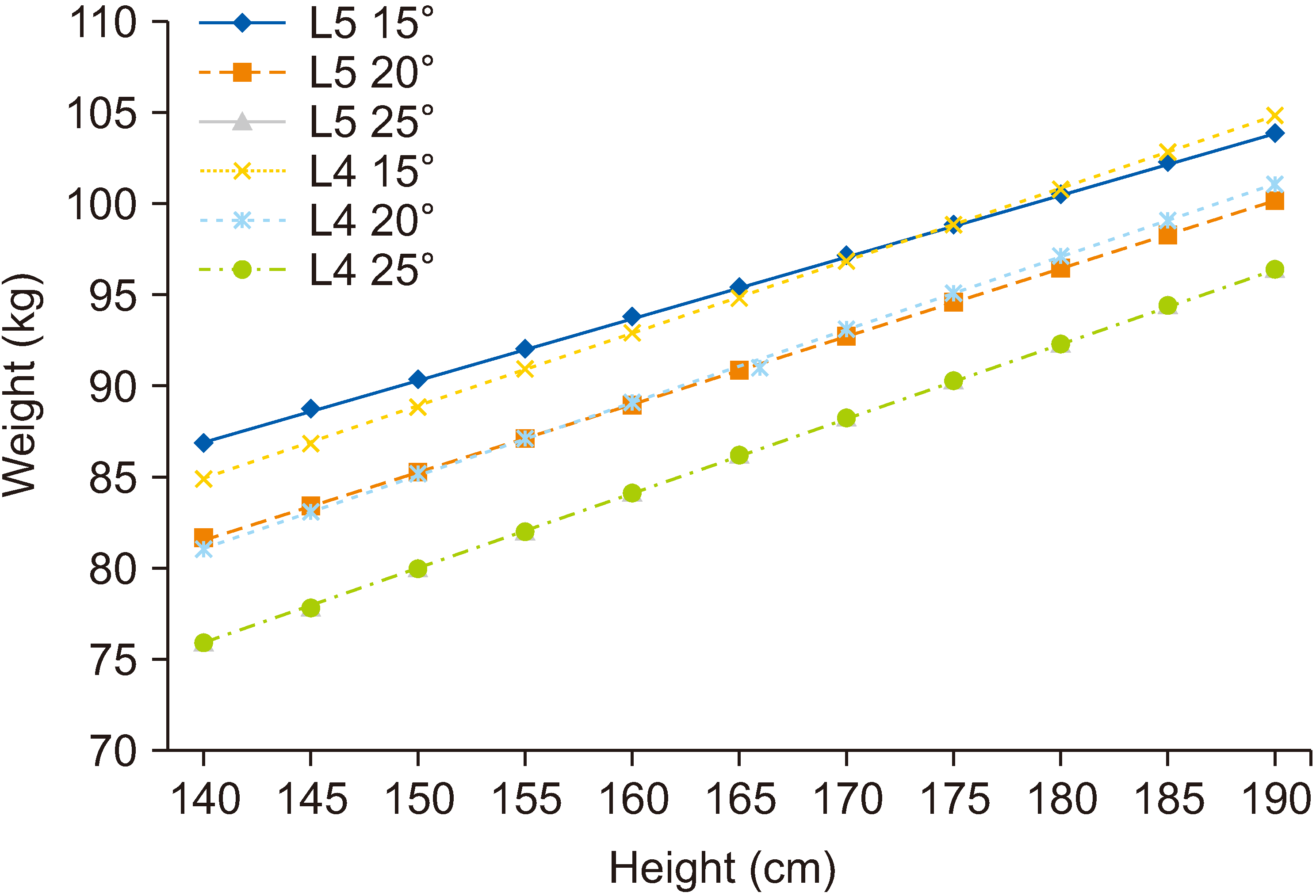
We formulated a predictive equation of needle depth to ventral transforaminal epidural space based on multiple linear regression analysis (Table 5). This correlation can be helpful for accurate needle placement in lumbar TFEI for morbid obesity. In the study by Kim et al. [1], 248 patients were evaluated, and the longest needle depth for the TFEI was 8.6 cm. In our center, the first needle choice is usually the 23G, 3.5-inch, Quincke-type point spinal needle (TaeChang Industrial Co., Gongju, Korea or Becton Dickinson, Franklin Lakes, NJ), and occasionally this needle is too short for obese patients. Longer needles are more expensive and difficult to manipulate. Therefore, a long needle should not be the preferred choice for all patients. A graph based on the equation from Table 5 could be helpful for preparation of a proper size needle (Fig. 6). If the patient’s weight is beyond the maximum value on the X-axis corresponding to the patient’s height, a needle length > 3.5-inch (88.9 mm) is recommended. Longer needles (5-inch or 6-inch, Quincke type point needle [Becton Dickinson]) could be a choice for them. Pre-analysis of needle depth using the equation can reduce unnecessary multiple needle insertions, patient discomfort, possible complications, and radiation exposure time, thus increasing both the safety of patients and clinicians.
We formulated a more practical equation of needle depth based on BMI (Table 6). This equation can minimize the preparation time in comparison to the equation based on weight and height. A graph based on the equation from Table 6 can also be a useful tool (Fig. 5). Based on the equation from Table 6, maximal BMI is listed at three different angles (15°, 20°, and 25°) at lumbar levels L4 and L5 (Table 7). This also serves to arrange the proper needle size. If a patient has a bigger BMI than the one in the table, the clinician should prepare a longer needle than usual spinal needle. For example, when you perform an L5 TFEI with a 20° angle approach on a patient with BMI over 34.97, you need to prepare a longer needle than the commonly used 3.5-inch (88.9 mm) needle.
We found the equation of needle depth in three different oblique angles. Although the C-arm fluoroscopic angle is same as the assumed angle, it is likely that the needle path is longer or shorter than the needle length obtained from this equation in actual clinical situations. The needles are often inserted slightly differently from the fluoroscopic angle, which may require a longer needle than the length we have calculated. Nevertheless, this equation can serve as a reference for the length of the needle.
One limitation of this study is that while MRI is performed in a supine position, TFEI is performed in a prone position. According to the other studies, positions of the lumbar nerve roots are not affected by prone position [31,32]. Therefore, it is considered meaningful to apply the predicted length through MRI to patients in prone position. When the patient lies supine, especially when the patient has a large BMI and subcutaneous fat, the distance from the compressed skin to the target is expected to be shorter. The obesity of patients is expected to underestimate the length of the needle. Therefore, further research comparing the actual needle length required in the procedure and the depth calculated from the equation could help confirm the usefulness of the equation for clinical practice. The authors believe that even if the positions differ, the depth of the area may be similar regardless of the posture and that the equations will help physicians determine the proper needle length for TFEI. Another limitation is that the authors only considered a few lumbar levels. Even though the L4 and L5 levels are the most commonly used in lumbar TFEI, further investigation of other spine levels should be performed to evaluate the clinical significance of this study.
In conclusion, this study showed that using both height and weight could be more helpful for predicting epidural depth in TFEI than using only BMI. A predictive length equation was formulated which may help decide on a proper needle size for TFEI. If the patient’s BMI is greater than the values shown in the Table 7, a larger needle than the conventional needle should be used.

Notes

DATA AVAILABILITY

Data files are available from Harvard Dataverse: https://doi.org/10.7910/DVN/ZFSWHM.

CONFLICT OF INTEREST

No potential conflict of interest relevant to this article was reported.

FUNDING

No funding to declare.

REFERENCES

1. Kim LK, Kim JR, Shin SS, Kim IJ, Kim BN, Hwang GT. 2011; Analysis of influencing factors to depth of epidural space for lumbar transforaminal epidural block in Korean. Korean J Pain. 24:216–20. DOI: 10.3344/kjp.2011.24.4.216. PMID: 22220243. PMCID: PMC3248585.
crossref
2. Manchikanti L, Pampati V, Falco FJ, Hirsch JA. 2013; Growth of spinal interventional pain management techniques: analysis of utilization trends and Medicare expenditures 2000 to 2008. Spine (Phila Pa 1976). 38:157–68. DOI: 10.1097/BRS.0b013e318267f463. PMID: 22781007.
3. Sencan S, Edipoglu IS, Celenlioglu AE, Yolcu G, Gunduz OH. 2020; Comparison of treatment outcomes in lumbar central stenosis patients treated with epidural steroid injections: interlaminar versus bilateral transforaminal approach. Korean J Pain. 33:226–33. DOI: 10.3344/kjp.2020.33.3.226. PMID: 32606267. PMCID: PMC7336349.
crossref
4. Helm S, Harmon PC, Noe C, Calodney AK, Abd-Elsayed A, Knezevic NN, et al. 2021; Transforaminal epidural steroid injections: a systematic review and meta-analysis of efficacy and safety. Pain Physician. 24(S1):S209–32. DOI: 10.36076/ppj.2021.24.S209-S232. PMID: 33492919.
5. Bensler S, Sutter R, Pfirrmann CWA, Peterson CK. 2018; Particulate versus non-particulate corticosteroids for transforaminal nerve root blocks: comparison of outcomes in 494 patients with lumbar radiculopathy. Eur Radiol. 28:946–52. DOI: 10.1007/s00330-017-5045-z. PMID: 28894933.
crossref
6. Vad VB, Bhat AL, Lutz GE, Cammisa F. 2002; Transforaminal epidural steroid injections in lumbosacral radiculopathy: a prospective randomized study. Spine (Phila Pa 1976). 27:11–6. DOI: 10.1097/00007632-200201010-00005. PMID: 11805628.
7. Donohue NK, Tarima SS, Durand MJ, Wu H. 2020; Comparing pain relief and functional improvement between methylprednisolone and dexamethasone lumbosacral transforaminal epidural steroid injections: a self-controlled study. Korean J Pain. 33:192–8. DOI: 10.3344/kjp.2020.33.2.192. PMID: 32235020. PMCID: PMC7136301.
crossref
8. Rezende R, Jacob Júnior C, da Silva CK, de Barcellos Zanon I, Cardoso IM, Batista Júnior JL. 2015; Comparison of the efficacy of transforaminal and interlaminar radicular block techniques for treating lumbar disk hernia. Rev Bras Ortop. 50:220–5. DOI: 10.1016/j.rbo.2013.12.007. PMID: 26229920. PMCID: PMC4519619.
crossref
9. Chang-Chien GC, Knezevic NN, McCormick Z, Chu SK, Trescot AM, Candido KD. 2014; Transforaminal versus interlaminar approaches to epidural steroid injections: a systematic review of comparative studies for lumbosacral radicular pain. Pain Physician. 17:E509–24. DOI: 10.36076/ppj.2014/17/E509. PMID: 25054401.
10. Park JM, Jung H, Kwon OD, Hong SW, Kwak KH. 2013; Analysis of dispersion of lumbar interlaminar epidural injectates. Korean J Anesthesiol. 65(6 Suppl):S139–40. DOI: 10.4097/kjae.2013.65.6S.S139. PMID: 24478848. PMCID: PMC3903836. PMID: 58f3ce699fdf4d1ea71c18a6b35dda04.
crossref
11. Lutz GE, Vad VB, Wisneski RJ. 1998; Fluoroscopic transforaminal lumbar epidural steroids: an outcome study. Arch Phys Med Rehabil. 79:1362–6. DOI: 10.1016/S0003-9993(98)90228-3. PMID: 9821894.
crossref
12. Jeong JS, Shim JC, Woo JP, Shim JH. 2013; Fluoroscopic analysis of lumbar epidural contrast spread after retrograde interlaminar ventral epidural injection (RIVEI). Korean J Anesthesiol. 65:431–7. DOI: 10.4097/kjae.2013.65.5.431. PMID: 24363846. PMCID: PMC3866339. PMID: 08463409403c4b3b9f500a27e3bd3cd9.
crossref
13. Schaufele MK, Hatch L, Jones W. 2006; Interlaminar versus transforaminal epidural injections for the treatment of symptomatic lumbar intervertebral disc herniations. Pain Physician. 9:361–6. PMID: 17066121.
14. Lee SC, Kim MW. 2004; The evaluation of epidural depth at L3-4 and L4-5 using magnetic resonance imaging and its relationship to BMI. Korean J Anesthesiol. 47:34–7. DOI: 10.4097/kjae.2004.47.1.34.
crossref
15. Ra IH, Min WK. 2015; Optimal angle of needle insertion for fluoroscopy-guided transforaminal epidural injection of L5. Pain Pract. 15:393–9. DOI: 10.1111/papr.12187. PMID: 24690186.
crossref
16. Hong JH, Lee YC, Lee HM, Kang CH. 2008; An analysis of the outcome of transforaminal epidural steroid injections in patients with spinal stenosis or herniated intervertebral discs. Korean J Pain. 21:38–43. DOI: 10.3344/kjp.2008.21.1.38.
crossref
17. Goodman BS, Posecion LW, Mallempati S, Bayazitoglu M. 2008; Complications and pitfalls of lumbar interlaminar and transforaminal epidural injections. Curr Rev Musculoskelet Med. 1:212–22. DOI: 10.1007/s12178-008-9035-2. PMID: 19468908. PMCID: PMC2682416.
crossref
18. Chang A, Ng AT. 2020; Complications associated with lumbar transforaminal epidural steroid injections. Curr Pain Headache Rep. 24:67. DOI: 10.1007/s11916-020-00900-9. PMID: 32990823.
crossref
19. Husseini JS, Simeone FJ, Staffa SJ, Palmer WE, Chang CY. 2020; Fluoroscopically guided lumbar spine interlaminar and transforaminal epidural injections: inadvertent intravascular injection. Acta Radiol. 61:1534–40. DOI: 10.1177/0284185120903450. PMID: 32050772.
crossref
20. Algrain H, Liu A, Singh S, Vu TN, Cohen SP. 2018; Cervical epidural depth: correlation between cervical MRI measurements of the skin-to-cervical epidural space and the actual needle depth during interlaminar cervical epidural injections. Pain Med. 19:1015–22. DOI: 10.1093/pm/pnx066. PMID: 28482062.
crossref
21. Galbraith AS, Wallace E, Devitt A. 2019; Examining the association of body mass index and the depth of epidural space, radiation dose exposure and fluoroscopic screening time during transforaminal nerve block injection: a retrospective cohort study. Ir J Med Sci. 188:295–302. DOI: 10.1007/s11845-018-1845-7. PMID: 29911292.
crossref
22. Jones JH, Singh N, Nidecker A, Li CS, Fishman S. 2017; Assessing the agreement between radiologic and clinical measurements of lumbar and cervical epidural depths in patients undergoing prone interlaminar epidural steroid injection. Anesth Analg. 124:1678–85. DOI: 10.1213/ANE.0000000000001839. PMID: 28099288. PMCID: PMC5398916.
crossref
23. Hong JH, Lee MY, Jung SW, Lee SY. 2015; Does spinal stenosis correlate with MRI findings and pain, psychologic factor and quality of life? Korean J Anesthesiol. 68:481–7. DOI: 10.4097/kjae.2015.68.5.481. PMID: 26495059. PMCID: PMC4610928. PMID: 140d6b1309054adfbbbc5f0ec53f594b.
crossref
24. Brunner P, Amoretti N, Soares F, Brunner E, Cazaux E, Brocq O, et al. 2012; Approaches in injections for radicular pain: the transforaminal, epidural and transfacet approaches. Diagn Interv Imaging. 93:711–22. DOI: 10.1016/j.diii.2012.07.005. PMID: 22925594.
crossref
25. Bhatia A, Flamer D, Shah PS, Cohen SP. 2016; Transforaminal epidural steroid injections for treating lumbosacral radicular pain from herniated intervertebral discs: a systematic review and meta-analysis. Anesth Analg. 122:857–70. DOI: 10.1213/ANE.0000000000001155. PMID: 26891397.
26. Botwin KP, Gruber RD, Bouchlas CG, Torres-Ramos FM, Sanelli JT, Freeman ED, et al. 2002; Fluoroscopically guided lumbar transformational epidural steroid injections in degenerative lumbar stenosis: an outcome study. Am J Phys Med Rehabil. 81:898–905. DOI: 10.1097/00002060-200212000-00003. PMID: 12447088.
crossref
27. Botwin K, Natalicchio J, Brown LA. 2004; Epidurography contrast patterns with fluoroscopic guided lumbar transforaminal epidural injections:a prospective evaluation. Pain Physician. 7:211–5. DOI: 10.36076/ppj.2004/7/211. PMID: 16868594.
28. Lee JH, Shin KH, Park SJ, Lee GJ, Lee CH, Kim DH, et al. 2018; Comparison of clinical efficacy between transforaminal and interlaminar epidural injections in lumbosacral disc herniation: a systematic review and meta-analysis. Pain Physician. 21:433–48. DOI: 10.36076/ppj.2018.5.433. PMID: 30282389.
29. Carnie J, Boden J, Gao Smith F. 2002; Prediction by computerised tomography of distance from skin to epidural space during thoracic epidural insertion. Anaesthesia. 57:701–4. DOI: 10.1046/j.1365-2044.2002.02572_4.x. PMID: 12059829.
crossref
30. Brummett CM, Williams BS, Hurley RW, Erdek MA. 2009; A prospective, observational study of the relationship between body mass index and depth of the epidural space during lumbar transforaminal epidural steroid injection. Reg Anesth Pain Med. 34:100–5. DOI: 10.1097/AAP.0b013e31819a12ba. PMID: 19282707. PMCID: PMC2715548.
crossref
31. Yingsakmongkol W, Poriswanich K, Kotheeranurak V, Numkarunarunrote N, Limthongkul W, Singhatanadgige W. 2022; How prone position affects the anatomy of lumbar nerve roots and psoas morphology for prone transpsoas lumbar interbody fusion. World Neurosurg. 160:e628–35. DOI: 10.1016/j.wneu.2022.01.104. PMID: 35108649.
crossref
32. Amaral R, Daher MT, Pratali R, Arnoni D, Pokorny G, Rodrigues R, et al. 2021; The effect of patient position on psoas morphology and in lumbar lordosis. World Neurosurg. 153:e131–40. DOI: 10.1016/j.wneu.2021.06.067. PMID: 34166827.
crossref

Fig. 1
A virtual dotted line vertically crosses the pedicle of lumbar vertebra 4, which is the needle target point of left L4 transforaminal epidural injection.
kjp-35-3-345-f1.tif
Fig. 2
(A) For measurement of left L4 transforaminal epidural injection, move the transverse plane slightly upward as shown to check the L3/4 facet point. Then, draw a virtual line at the center of the upper material entrenchment of the facet joint (dotted line). (B) The sagittal plane image of magnetic resonance imaging. The transverse plane image of (A) is where the yellow line is located.
kjp-35-3-345-f2.tif
Fig. 3
(A) Lower the transverse plane to a view directly below the pedicle of the lumbar 4 vertebra body and measure the length (red arrow) from skin to the contact point (arrow tip) of the vertebral body and the imaginary line (dotted line). (B) The sagittal plane image of magnetic resonance imaging. The transverse plane image of (A) is where the yellow line is located.
kjp-35-3-345-f3.tif
Fig. 4
(A) Measurement of length of L4 transforaminal epidural injection at 15° was performed. (B) The sagittal plane image of magnetic resonance imaging. The transverse plane image of (A) is where the yellow line is located.
kjp-35-3-345-f4.tif
Fig. 5
Estimated needle depth based on body mass index (BMI) is shown on the graph at three different angles (15°, 20°, and 25°) at lumbar levels L4 and L5.
kjp-35-3-345-f5.tif
Fig. 6
Minimal weight ranges for injection with a 88.9 mm spinal needle at three different angles (15°, 20°, and 25°) at lumbar levels L4 and L5. The needle depth for transforaminal epidural injection is positively correlated with weight and height. A 3.5-inch (88.9 mm) needle will suffice for patients with weight lighter than the weight of the corresponding height on the x-axis of the graph, while a longer than 3.5-inch needle will be required for patients with weight heavier than the weight of the corresponding height on the x-axis of the graph.
kjp-35-3-345-f6.tif
Table 1
Descriptive statistics for study participants
Variable Value (n = 386)
Sex
Male 156 (40.4)
Female 230 (59.6)
Age (yr) 63.2 ± 15.9
Height (cm) 161.3 ± 9.2
Weight (kg) 63.1 ± 10.9
Body mass index (kg/m2) 24.2 ± 3.4

Values are presented as number (%) or mean ± standard deviation.

Table 2
The correlation between the obtained lengths of each TFEI procedure side at the same level and oblique angle
Vertebral level, side and oblique angle t P value
Rt L5 TFEI and Lt L5 TFEI at 15° –0.306 0.760
Rt L5 TFEI and Lt L5 TFEI at 20° –1.206 0.229
Rt L5 TFEI and Lt L5 TFEI at 25° –0.810 0.418
Rt L4 TFEI and Lt L4 TFEI at 15° 0.891 0.374
Rt L4 TFEI and Lt L4 TFEI at 20° 0.767 0.444
Rt L4 TFEI and Lt L4 TFEI at 25° 0.825 0.410

TFEI: transforaminal epidural injection, Rt: right, Lt: left.

Table 3
The R-squared values of height, weight, BMI, and combined height and weight
Vertebral level and oblique angle Height (cm) Weight (kg) BMI
(kg/m2)
Height and weight
Bilateral L5 TFEI at 15° 0.029 0.202 0.174 0.218
Bilateral L5 TFEI at 20° 0.024 0.199 0.179 0.218
Bilateral L5 TFEI at 25° 0.020 0.190 0.179 0.213
Bilateral L4 TFEI at 15° 0.025 0.234 0.220 0.260
Bilateral L4 TFEI at 20° 0.025 0.227 0.212 0.253
Bilateral L4 TFEI at 25° 0.023 0.223 0.211 0.251

TFEI: transforaminal epidural injection, BMI: body mass index.

Table 4
Weight and height coefficients for epidural needle depth at bilateral L5 transforaminal epidural injection considering a 15° angle
Model Unstandardized coefficients Standardized coefficients t P value
B Standard error Beta
Constant 68.11 8.904 7.649 < 0.001
Height –0.18 0.065 –0.156 –2.765 0.006
Weight 0.53 0.055 0.543 9.622 < 0.001

Dependent variable: needle depth at bilateral L5 transforaminal epidural injection (15°).

Table 5
The equation of epidural needle depth using weight and height
Vertebral level and oblique angle Epidural needle depth (mm)
Bilateral L5 TFEI at 15° 68.11 – 0.18 × height (cm) + 0.53 × weight (kg)
Bilateral L5 TFEI at 20° 72.65 – 0.21 × height (cm) + 0.56 × weight (kg)
Bilateral L5 TFEI at 25° 78.00 – 0.23 × height (cm) + 0.58 × weight (kg)
Bilateral L4 TFEI at 15° 72.29 – 0.23 × height (cm) + 0.58 × weight (kg)
Bilateral L4 TFEI at 20° 74.10 – 0.23 × height (cm) + 0.58 × weight (kg)
Bilateral L4 TFEI at 25° 77.70 – 0.25 × height (cm) + 0.61 × weight (kg)

TFEI: transforaminal epidural injection.

Table 6
The equation of epidural needle depth using BMI
Vertebral level and oblique angle Epidural needle depth (mm)
Bilateral L5 TFEI at 15° 40.91 + 1.30 × BMI (kg/m²)
Bilateral L5 TFEI at 20° 41.10 + 1.37 × BMI (kg/m²)
Bilateral L5 TFEI at 25° 42.15 + 1.42 × BMI (kg/m²)
Bilateral L4 TFEI at 15° 37.09 + 1.42 × BMI (kg/m²)
Bilateral L4 TFEI at 20° 38.51 + 1.44 × BMI (kg/m²)
Bilateral L4 TFEI at 25° 39.51 + 1.50 × BMI (kg/m²)

TFEI: transforaminal epidural injection, BMI: body mass index.

Table 7
The maximal BMI that possibly be treated with commonly used 3.5-inch (88.9 mm) needle
L4 15° TFEI L5 15° TFEI L4 20° TFEI L5 20° TFEI L4 25° TFEI L5 25° TFEI
BMI 36.41 36.89 34.92 34.97 32.97 32.93

TFEI: transforaminal epidural injection, BMI: body mass index.

TOOLS
Similar articles