

 PDF
PDF Citation
 Print
Print


INTRODUCTION
Neuropathic pain (NP) is an intractable consequence of lesion or disease to the somatosensory system that results in detrimental psychosocial life of the affected patients. It has been estimated that NP affects 7 to 10% of the world population [1]. NP is characterised by hyperalgesia, allodynia, and anxiodepresive cormobidities [2,3]. Treatment of NP has proved difficult, as only about 2% of current drugs used yielded 50% pain reduction in addition to adverse side effects [1,3]. The multifaceted mechanisms involved in the development and maintenance of NP make it challenging to diagnose and treat successfully.
The roles played by elevated levels of proinflammatory mediators released by the traumatised nerve and non-neuronal cells (glial cells, infiltrating macrophages, monocytes, and T-lymphocytes) increase the neuronal sensitisation and potentiations [4,5]. Tumour necrotic factor-alpha (TNF-α) and interleukin-1-beta (IL-1β) play an active role in the advent of NP by mediating mechanical allodynia and thermal hyperalgesia [6,7]. The involvement of the reactive oxygen species (ROS) in NP has been well established. ROS and reactive nitrogen species (RNS) released from increased activities of the damaged sensory neurons and non-neuronal cells result in synaptic remodelling and functions that mediate allodynia and hyperalgesia. ROS and RNS increase the expression of sodium ion channels, excitatory receptors, and neuronal disinhibition. All this mediates increased sensitisation and a reduced neuronal threshold to nociceptive stimuli [8].
Acetaminophen (N-Acetyl-para-aminophenol) is a derivative of p-aminophenol from the metabolite of acetanilide and phenacetin. It is a multimodal analgesic agent with reduced opioid-related side effects [9] and an antipyretic function [10]. L-carnosine is a histidine-containing dipeptide molecule synthesised naturally in the liver from β-alanine and L-histidine [11]. It is widely distributed in various tissues of the body, including the nervous tissue. It is has been reported that L-carnosine possesses a proton buffering effect [12] and acted as an antioxidant via scavenging of free radicals and singlet oxygen and chelating with heavy metals [11,13].
There is limited information on the role of acetaminophen in NP. Likewise, the combined therapeutic effect of both acetaminophen and L-carnosine in painful neuropathy is yet to be investigated. Hence, this study is undertaken to investigate the combined potential antinociceptive effects of co-administration of acetaminophen and L-carnosine in chronic constriction injury (CCI)-induced peripheral neuropathy. The authors further assessed the anti-inflammatory properties and neuroprotective benefits of this combined treatment with acetaminophen and L-carnosine.
Go to : 
MATERIALS AND METHODS
1. Ethical approval
This study was consonant with the ethical guideline in animal experimentation outlined in the 2019 updated ARRIVE (Animal Research: Reporting of In Vivo Experiments) [14]. All experimental procedures were approved by the University Ethical Review Committee (UERC) of the University of Ilorin (approval number: UERC/ASN/2016/369).
2. Animals
Fifty-six male Wistar rats weighing 150–200 g were used for the study. The sample size was estimated with the aid of SigmaPlot version 12 software (Systat software, Inc., Chicago, IL). The statistical power of 80% at an alpha level of 0.05 was used for the estimation. Based on a previous study on the paw-withdrawal latency to the hotplate, a sample size of eight per group was derived. The animals were randomly hand-picked and grouped into seven. They were housed and acclimatised in the animal facilities of the Faculty of Basic Medical Sciences with free access to food and water.
3. CCI
Peripheral neuropathy was induced using the CCI model as described by Bennett and Xie [15]. Briefly, the animal was completely anaesthetised with xylazine and ketamine hydrochloric (100 mg/kg intraperitoneal injection [i.p]). The proximal back of the left hind limb was shaved, sterilised, and dissected to expose the main sciatic nerve. Four loose ligatures (4-0 ligating silk) were placed around the sciatic nerve, approximately 0.5 mm apart before the trifurcation. The muscle and skin were subsequently sutured back in layers, and penicillin powder was applied to the wound after sutured to prevent infection. The rats were returned to their respective home cage after recovery with free access to food and water. The animals were only dissected and sutured back without ligatures on the sciatic nerve in the sham group operation.
4. Grouping and treatment
Animals were treated with oral administration of either normal saline/acetaminophen/acetaminophen + L-carnosine once daily for twenty-one consecutive days. Post-treated groups were administered their respective treatment three days after surgical induction of NP and continued for the next twenty-one days. Pretreated groups were treated for seven consecutive days before induction of NP and continued subsequently. The study groups include:
Group A (Control): 10 mL/kg of body weight of normal saline
Group B (Sham): 10 mL/kg of body weight of normal saline
Group C (Ligated control): 10 mL/kg of body weight of normal saline
Group D (Pre-treated acetaminophen): 200 mg/kg of body weight
Group E (Post-treated acetaminophen): 200 mg/kg of body weight
Group F (Pre-treated co-treatment): acetaminophen (200 mg/kg of body weight) + L-carnosine (100 mg/kg of body weight)
Group G (Post-treated co-treatment): acetaminophen (200 mg/kg of body weight) + L-carnosine (100 mg/kg of body weight)
L-carnosine was a product of Hubei Hongpeptide Biotechnology Co., Ltd. China while acetaminophen was a product of Anhui BBCA pharmaceutical Co., Ltd. China. The dosage of 200 mg/kg of body weight of L-carnosine [16] and 400 mg/kg of body weight of acetaminophen [17] were used based on previously study.
5. Pain behavioral tests
Pain behavioral assessment was done to investigate the antinociceptive effects of the treatment on thermal hyperalgesia and mechanical allodynia. Baseline tests were carried out before CCI and three days after CCI. Pain behaviour was further monitored on the 3rd, 7th, 14th, and 21st day of treatments.
6. Thermal hyperalgesia
A hotplate test [18] was used to evaluate the thermal hyperalgesia in each treatment group. Briefly, a hotplate with a restricting pyrex cage, 30 cm high, with the temperature set and maintained at 55°C ± 0.5°C was used. The rats were dropped gently on the surface of the hotplate. The time taken by each rat to either flinch, lick, or jump out of the pyrex enclosure was recorded chronologically with a stopwatch, and taken as paw withdrawal latency (PWL).
7. Mechanical allodynia test
Responses to the mechanical bending force of various strengths of von Frey filaments were used to assess mechanical allodynia as described in the author’s previous study [18]. Animals were placed in a transparent Perspex cage with a wire mesh floor and allowed to rest for 15 minutes. Von Frey filaments grading 1.4 g, 2 g, 4 g, 6 g, 8 g, 10 g, 15 g, 26 g, 60 g, and 100 g bending forces were applied individually to the plantar surface of each ligated hind paws of rats in ascending orders. The paw withdrawal threshold was defined by the mechanical force (in gram) that produced ligated hind limb withdrawal, licking, or flinching in three consecutive trials. Von Frey filaments that produced a lifting of the whole hind limb without sudden withdrawal, licking or flinching were taken as a positive response.
8. Biochemical parameters
After twenty-one days of post-treatment with respective drugs, the rats were euthanised with a high dose of ketamine hydrochloride (180 mg/kg i.p.). The lumbar segment of the spinal cord (L4-L6) was collected via the hydraulic extrusion method. The sample was homogenised in Tris-buffer (1 M, pH 7.4) and centrifuged at 16,000 RPM for fifteen minutes. The supernatants collected were used for the assessment of inflammatory markers, calcium ions, antioxidant parameters, and lipid peroxidation. IL-1β (Abcam, Cambridge, UK), TNF-α (Abcam), nuclear factor kappa light chain enhancer B cell inhibitor (NF-κB) (Cayman Chemical, Ann Arbor, MI), were analysed with microplate reader using their ELISA manufacturer’s instructional manual. Superoxide dismutase (SOD) activities [19], reduced glutathione (GSH) concentration [20], malondialdehyde (MDA) concentration [21], and calcium ion level were estimated spectrophotometrically.
9. Histological study
Haematoxylin and eosin (H&E) stain as described by Muthuraman et al. [22] was used to investigate the neuronal architecture of the sciatic nerve. Briefly, the sciatic nerve was excised from the rat following trans-cardiac perfusion with phosphate buffer and formalin. The tissues were fixed in 10% formalin solution and blocked. They were sectioned longitudinally into 5 μm before staining with H&E and observed under a high-power light microscope. Images were acquired from the microscope by using Leica ICC50 E Digital Camera (Leica Microsystems, Wetzlar, Germany) connected to a computer and analysed with Armscope software version 3.7 (Amscope, Irvine, CA).
10. Statistical analysis
Graphpad prism software version 5 (GraphPad Software, San Diego, CA) was used for the analysis of data. All data were expressed as mean ± standard error of the mean. Two-way analysis of variance (ANOVA) was used to analyse behavioural pain tests, while one-way ANOVA was used for the analysis of biochemical parameters. The Bonferroni post hoc multiple comparison test with (P < 0.05) was used for comparison among groups.
Go to :
RESULTS
1. Hyperalgesic test
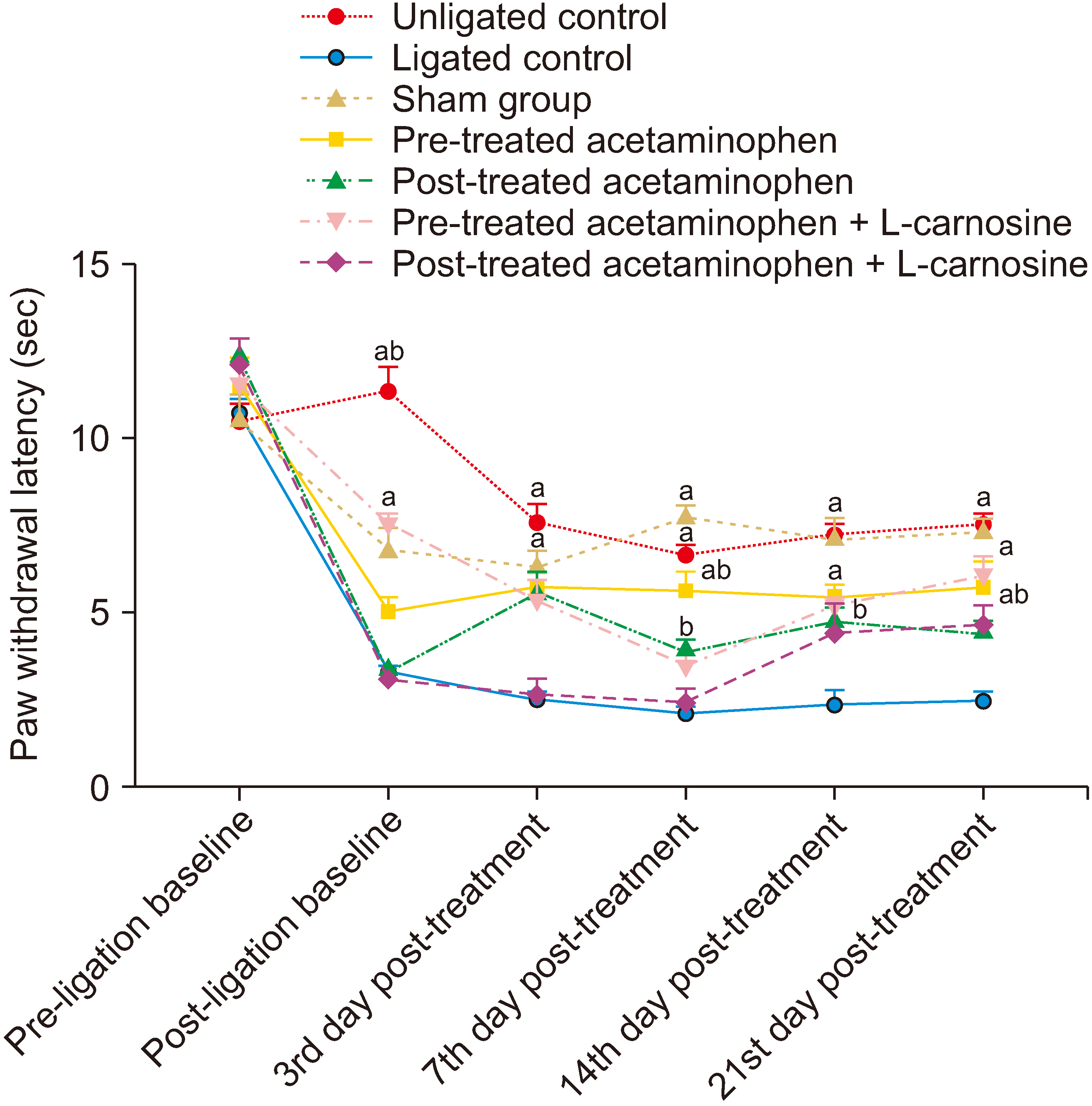
The hotplate test showed that CCI significantly (P < 0.001) reduced the PWL compared with the sham group (3.28 ± 0.16 sec vs. 6.78 ± 0.66 sec) as indicated in Fig. 1. The combined pretreatment of ligated rats with acetaminophen and L-carnosine showed a significant (P < 0.001) increase in PWL compared with the ligated control group (7.51 ± 0.36 sec vs. 3.28 ± 0.16 sec) on the post-ligation baseline test. Treatment of ligated animals with either acetaminophen or its combination with L-carnosine significantly (P < 0.001) increased the PWL compared with ligated naïve group on the third day post-treatment test except for the post-treatment co-administrated acetaminophen and L-carnosine group. There was only a significant (P < 0.001) increase in PWL of the pretreated acetaminophen group compared with the ligated group on the seventh-day post-treatment hyperalgesia test. However, both the post-treated and pretreated groups showed significantly increased PWL on the fourteenth and twenty-first day post-treatment tests. There was no significant difference between the pretreated acetaminophen and L-carnosine co-treatment group and the sham group on the twenty-first-day post-treatment test.
2. Allodynia test
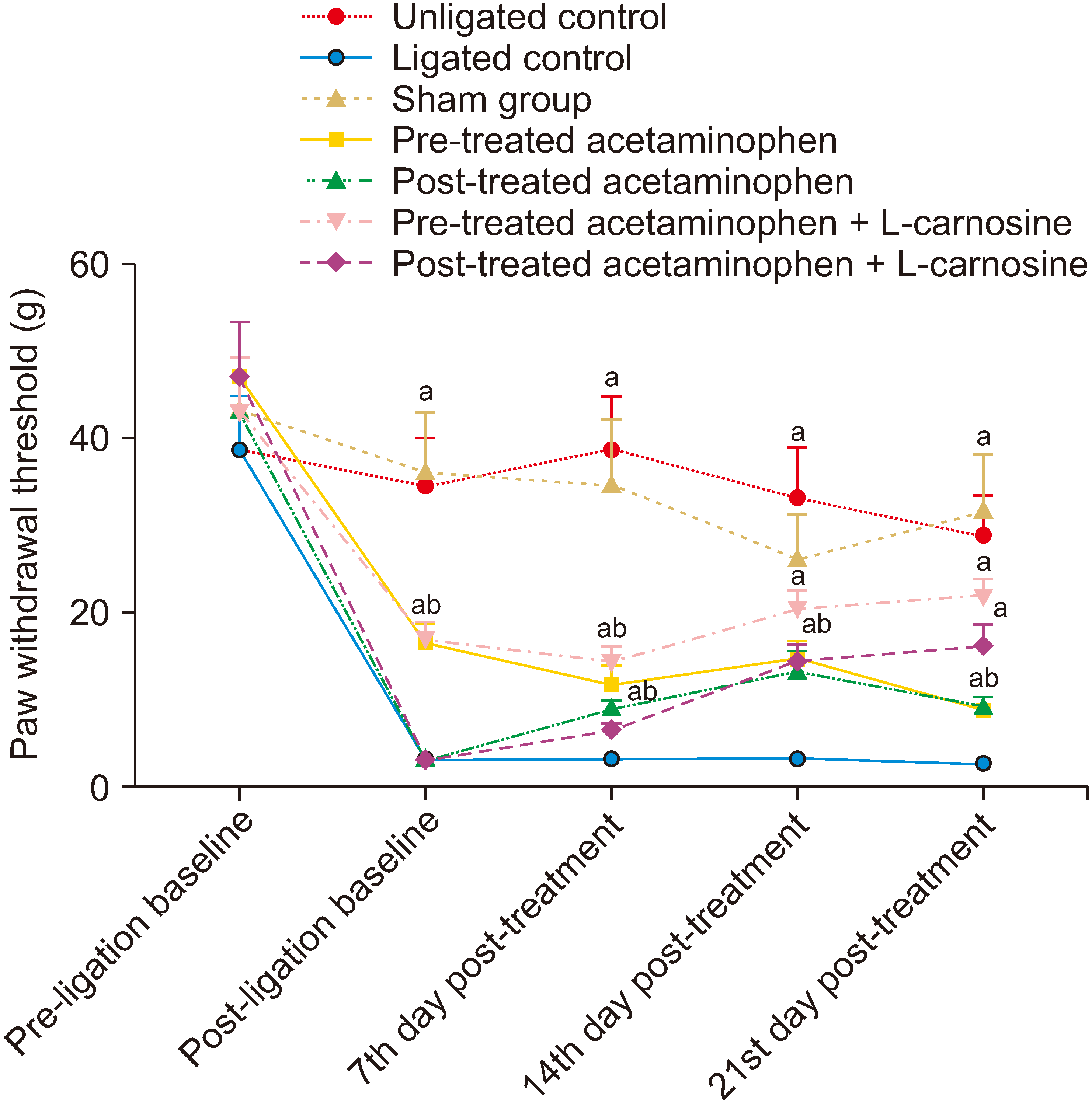
CCI significantly (P < 0.001) reduced the threshold to the mechanical bending force of von Frey filaments compared with unligated groups (3.25 ± 0.37 g vs. 36.00 ± 7.21 g) as represented in Fig. 2. Pretreatment with either acetaminophen or co-administration of acetaminophen and L-carnosine showed a significantly increased threshold to von Frey bending force after the CCI in animals on the post-ligation baseline test. There were significant increases in the threshold to von Frey filament bending force following treatments with acetaminophen, and a progressive threshold increment with co-administration of acetaminophen and L-carnosine on the seventh, fourteenth, and twenty-first post-treatment day tests. There were no significant differences between the combined acetaminophen and L-carnosine treatment groups and the sham group on the twenty-first post-treatment day test.
3. Biochemical parameters
CCI significantly (P < 0.001) increased the concentration of TNF-α (36.92 ± 0.63 pg/mL vs. 22.10 ± 0.80 pg/mL, Fig. 3A), IL-1β (290.60 ± 4.27 pg/mL vs. 230.60 ± 8.90 pg/mL, Fig. 3B), NF-κB (135.2 ± 2.78 pg/mL vs. 87.49 ± 5.88 pg/mL, Fig. 3C), and calcium ion (Ca2+, 6.11 ± 0.07 ppm vs. 5.24 ± 0.04 ppm, Fig. 3D) compared with the unligated control group. Co-administration of acetaminophen and L-carnosine led to a significant reduction in the spinal level of the proinflammatory markers (TNF-α, IL-1β, NF-κB, and Ca2+). There was a significant decrease between the combined acetaminophen and L-carnosine treatment groups and unligated control groups on IL-1β and NF-κB concentration. It was observed that there were no significant differences in the NF-κB concentration between the ligated control group and the acetaminophen treated group.
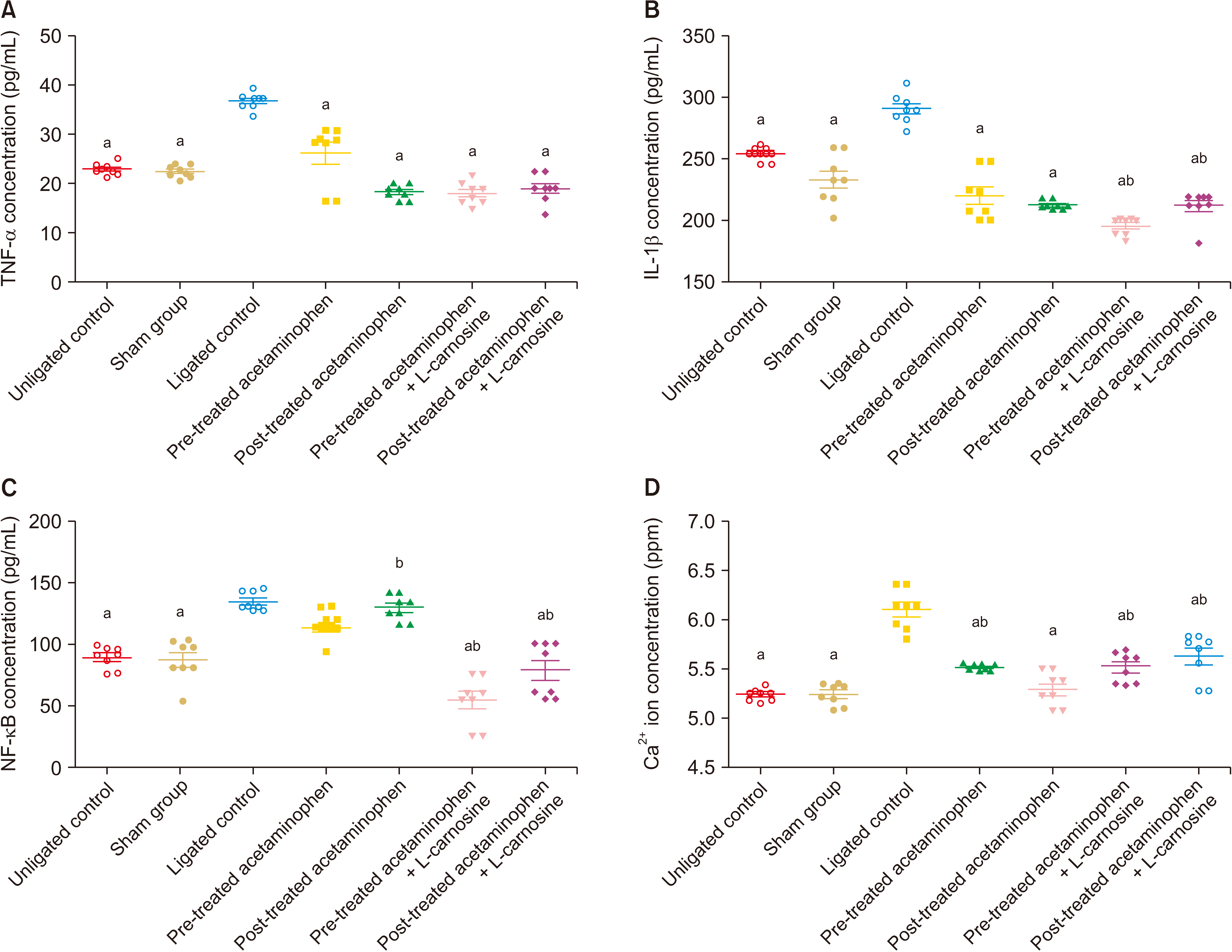 | Fig. 3Acetaminophen + L-carnosine mitigated proinflammatory mediators. Acetaminophen and L-carnosine significantly reduced proinflammatory mediators in the spinal lumbar neurons. (A) Acetaminophen + L-carnosine significantly inhibit tumour necrotic factor-alpha (TNF-α) concentration. (B) Acetaminophen + L-carnosine significantly reduced interleukin-1-beta (IL-1β) concentration. (C) Acetaminophen + L-carnosine significantly attenuated nuclear factor kappa light chain enhancer B cell inhibitor (NF-κB) concentration. (D) Acetaminophen + L-carnosine significantly inhibit calcium (Ca2+) ion concentration. Data were represented as mean ± standard error of the mean (n = 8). aP < 0.05 vs. ligated control, bP < 0.05 vs. sham control.
|
There was a significant (P < 0.001) elevation in the concentration of MDA and decreases in the reduced GSH concentration as well as SOD activities in the ligated naïve group compared with the unligated control groups as indicated in Fig. 4. Treatment with a single administration of acetaminophen for twenty-one consecutive days showed significant increases in GSH concentration and SOD activities but no significant (P < 0.001) reduction in MDA level. However, combined treatment with acetaminophen and L-carnosine showed significantly (P < 0.001) reduced MDA concentration (0.80 ± 0.03 U/mg protein vs. 1.05 ± 0.03 U/mg protein) as well as increased GSH concentration (116.20 ± 3.35 U/mg protein vs. 42.91 ± 1.50 U/mg protein) and SOD activities (133.80 ± 7.13 U/I vs. 39.11 ± 2.96 U/I). There were significant differences (P < 0.001) in the SOD activities between the combined treated acetaminophen + L-carnosine group and singly administered acetaminophen group. However, only the pretreated acetaminophen + L-carnosine group showed a significant (P < 0.001) increase in GSH concentration and decreased MDA concentration compared with pretreated and post-treated acetaminophen groups.
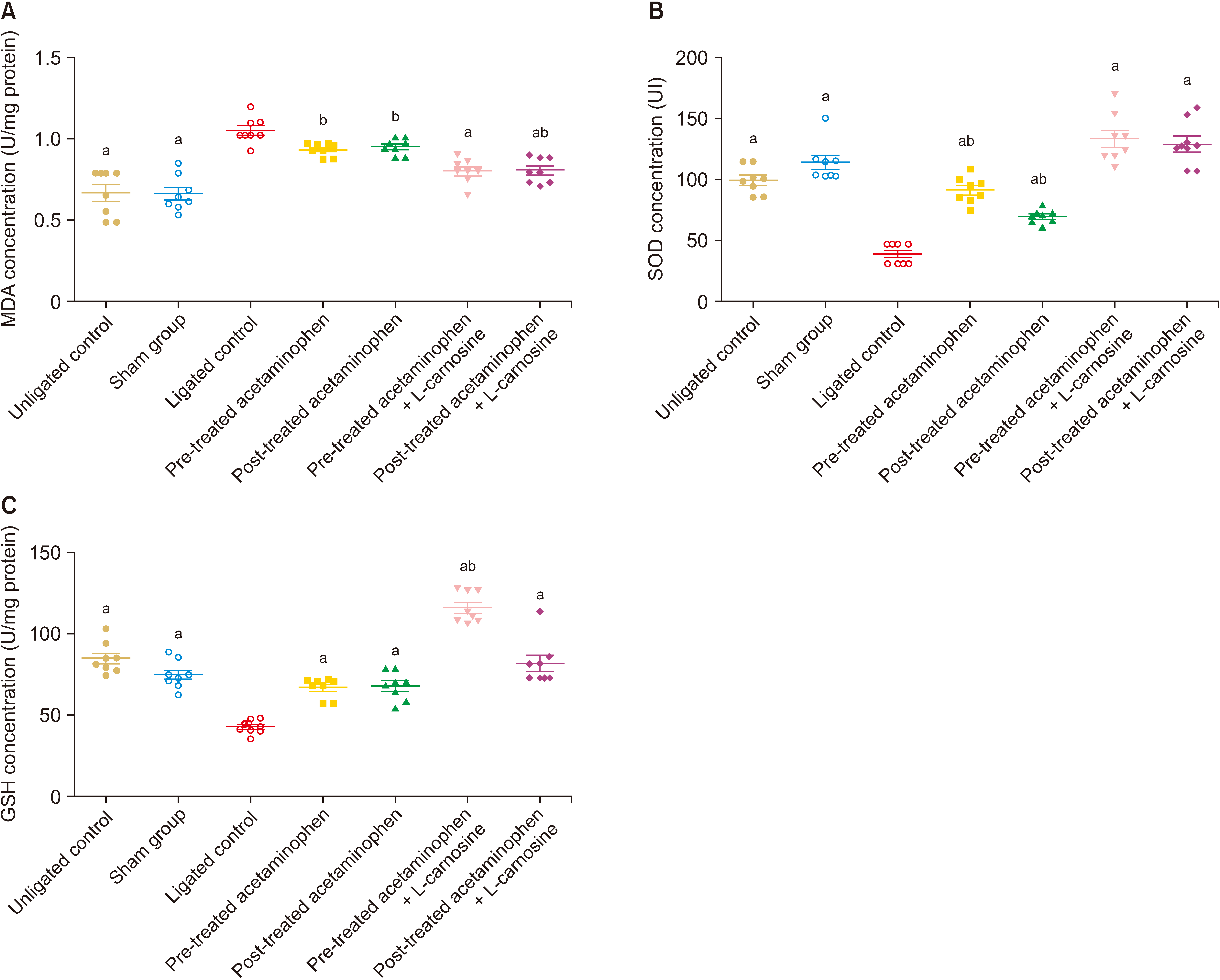 | Fig. 4Acetaminophen + L-carnosine improved neuroprotection of the neurons against oxidative stress. Combined treatment with acetaminophen and L-carnosine significantly increased antioxidant enzyme activities and reduced lipid peroxidation. (A) Acetaminophen + L-carnosine significantly reduced malondialdehyde (MDA) concentration. (B) Acetaminophen + L-carnosine significantly increased superoxide dismutase (SOD) activities. (C) Acetaminophen + L-carnosine significantly increased glutathione (GSH) concentration. Data were represented as mean ± standard error of the mean (n = 8). aP < 0.05 vs. ligated control, bP < 0.05 vs. sham control.
|
4. Histological analysis
1) Sciatic nerve
Fig. 5A and 5B shows the normal cytoarchitecture of the sciatic nervein the unligated naïve group, and sham group groups respectively. CCI induces derangement in the cytoarchitecture of the sciatic nerve, as indicated in Fig. 5C. There is wide demyelination and degeneration of the neuron fibres as well as shrinkage and extinction of the nuclei of Schwann cell. CCI results in pronounced vacuolisation of the sciatic nerves. Post-treatment with acetaminophen showed preservation of myelinated and unmyelinated neurons. However, there is the presence of vacuolation and a significant reduction in Schwann cells (Fig. 5D). Animals pretreated with acetaminophen have improved myelinated neurons, Schwann cell, and reduced vacuolation, as indicated in Fig. 5E. The combined treatment with acetaminophen + L-carnosine showed a well improved normal cytoarchitecture of the sciatic nerve. There is increased Schwann cells, myelinated and unmyelinated neurons, as well as drastically reduced vacuolation (Fig. 5F, G).
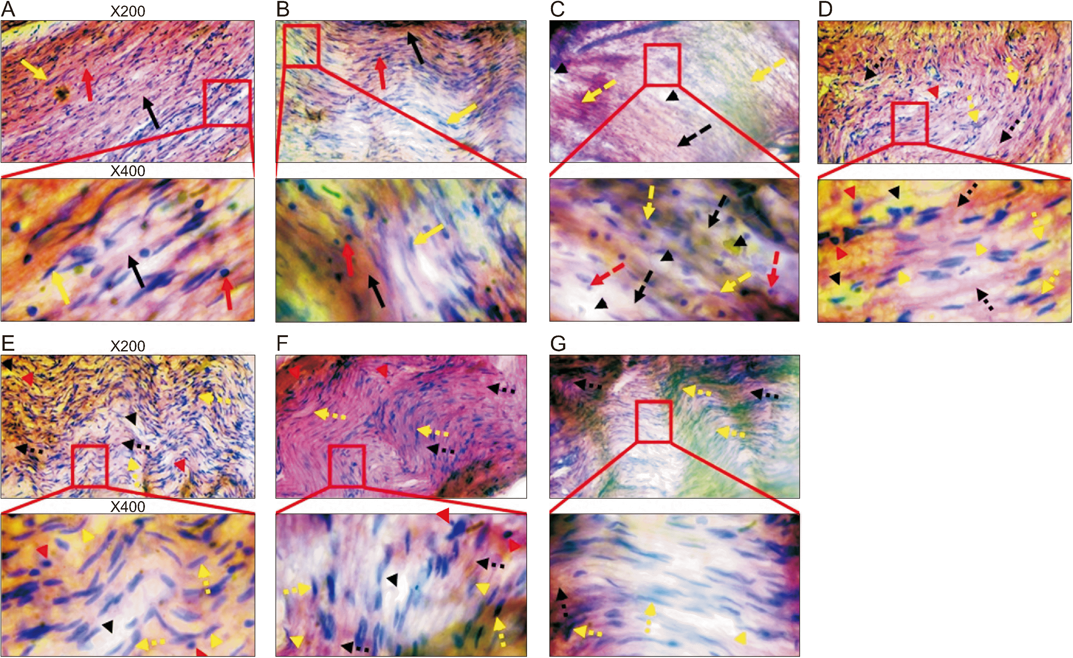 | Fig. 5Micrograph showing haematoxyline and eosin stained longitudinal section of the left sciatic nerve. Sciatic nerve ligation induces lost of myelinated (single broken yellow arrow) and unmyelinated (single broken black arrow), shrinkage and lost of nuclei of Schwann cell (single broken red arrow), vacuolarization (black arrow head). Acetaminophen + L-carnosine improved cytoarchitecture of the sciatic nerve by preserving the myelinated (double-broken yellow arrow) and unmyelinated (double-broken black arrow) fibres, nuclei of Schwann cell (red arrow head), regeneration of myelinated neuron (yellow arrow head), and reduces vacuolarization. Red arrow indicate normal Schwann cell, normal myelinated nerve fibre (yellow arrow), and normal unmyelinated nerve fibre (black arrow). (A) Unligated naïve group, (B) Sham group, (C) Ligated naïve group, (D) Post-treated acetaminophen group, (E) Pre-treated acetaminophen group, (F) Post-treated acetaminophen + L-carnosine group, (G) Pre-treated acetaminophen + L-carnosine group.
|
Go to :
DISCUSSION
The treatment of NP remains a challenge worldwide. This study reports the antinociceptive effects of combined administration of acetaminophen and L-carnosine in chronic constricted injury-induced peripheral neuropathy.
CCI is a well-established procedure in modelling peripheral neuropathy in laboratory animals. This study showed that animals developed thermal hyperalgesia and mechanical allodynia following CCI. This is evidenced by a widely reduced threshold to both mechanical and thermal stimuli. Treatment with either acetaminophen or its combination with L-carnosine effectively mitigated both thermal hyperalgesia and mechanical allodynia. Pretreatment of rats with acetaminophen only is more beneficial in preventing the development of allodynia than thermal hyperalgesia. However, combined pretreatment with acetaminophen with L-carnosine effectively slowed down the onset of thermal hyperalgesia. It ameliorated mechanical allodynia, as evidenced by increased paw response threshold to thermal and mechanical stimuli. Post-treatment with a combined dose of acetaminophen and L-carnosine has delayed therapeutic onset, unlike post-treatment with acetaminophen alone. Hence, it was deduced that the antinociceptive properties of acetaminophen are strengthened by co-treatment with L-carnosine.
Hyperalgesia and allodynia have been reported to be mediated by the increased advent of proinflammatory cytokines [4,7]. Excercabation of NF-κB promotes the production of TNF-α and IL-β. Coadministered acetaminophen and L-carnosine effectively reversed increased TNF-α and IL-β in the ligated animals. This may be due to their effects on NF-κB and calcium ion concentration. TNF-α and IL-β have been widely reported to mediate hypersensitivities by increasing neuronal excitation and lowering the neuronal threshold [6,7]. They are involved in synaptic remodelling leading to increased neuronal hyperexcitation [6,23]. A single treatment with acetaminophen alone led to reduced TNF-α and IL-β but not via the NF-κB pathway. Enhanced NF-κB is associated with increased excitatory synapses, neuronal hyperexcitability, and disinhhibition [24,25]. Hence the improved antinociceptive effects of combined acetaminophen and L-carnosine are mediated via anti-inflammatory activities in mitigating NF-κB.
ROS play a pivotal role in the development of hyperalgesia and allodynia [8,26]. Agents that possess antioxidant properties have been reported to be effective in treating NP. Therapeutic agents targeting oxidative stress improve NP via scavenging the ROS [27,28]. This study was in consonant with a previous study that showed that CCI is characterised by increased oxidative stress [26]. A combined dose of acetaminophen and L-carnosine ameliorated increased oxidative stress as indicated by a marked reduction in lipid peroxidation in the spinal cord. Combined acetaminophen and L-carnosine also stimulated the production of antioxidant enzyme (SOD) and reduced GSH that mopped up ROS. This further explains the combined potential of the two therapeutic agents that mediated their antinociceptive properties. Antioxidant properties of the combined treatment are evidence of the structural integrity of the sciatic nerves. Improved structural architecture of the sciatic nerve with combined acetaminophen and L-carnosine indicated a high degree of neuroprotection that manifested the observed antinociceptive properties. Increased lipid peroxidation results in distortion in neuronal integrity and derangement in nerve conduction. Pretreatment with combined acetaminophen and L-carnosine showed a better neuroprotective effect which may be due to increased antioxidant properties via increased production of reduced GSH.
In conclusion, the antinociceptive effects of combined acetaminophen and L-carnosine are mediated by its neuroprotective effect via reduced lipid peroxidation and NF-κB. Additive antinociceptive properties of acetaminophen and L-carnosine are mediated by the inhibition of IL-1β via reductions in NF-κB and stimulation of GSH and SOD synthesis. This serves as an effective mechanism that underpins the combined antinociceptive activities of acetaminophen and L-carnosine.
Go to :
 XML Download