Journal List > Korean J Pain > v.35(3) > 1516078744

Doménech-García, Peirotén, Imaz, Palsson, Herrero, and Bellosta-López: Not just sensitization: sympathetic mechanisms contribute to expand experimental referred pain

Abstract

Background

Widespread pain partially depends upon sensitization of central pain mechanisms. However, mechanisms controlling pain distribution are not completely known. The present study sought to assess skin temperature variations in the area of experimentally-induced pain and potential sex differences.

Methods

Pressure-pain thresholds (PPTs) were measured on the right infraspinatus muscle. At the end of Day 0, all participants performed an eccentric exercise of the shoulder external rotators to induce muscle soreness 24 hours after. On Day 1, participants indicated on a body chart the area of pain induced by 60 seconds of suprathreshold pressure stimulation (STPS; PPT + 20%) on the right infraspinatus muscle. Skin temperature variations in the area of referred pain were recorded with an infrared thermography camera, immediately before and after the STPS.

Results

Twenty healthy, pain-free individuals (10 females) participated. On Day 0, the pre-STPS temperature was higher than the post-STPS temperature on the arm (P = 0.001) and forearm (P = 0.003). On Day 1, the pre-STPS temperature was higher than the post-STPS temperature on the shoulder (P = 0.015), arm (P = 0.001), and forearm (P = 0.010). On Day 0, the temperature decrease after STPS in females was greater than in males on the forearm (P = 0.039). On Day 1, a greater temperature decrease was found amongst females compared with males at the shoulder (P = 0.018), arm (P = 0.046), and forearm (P = 0.005).

Conclusions

These findings indicate that sympathetic vasomotor responses contribute to expand pressure-induced referred pain, especially among females.

INTRODUCTION

Chronic musculoskeletal pain is a major health problem, imposing a considerable socioeconomic burden affecting up to one third of males and females [1]. Even though the primary pain complaint is commonly local, widespread pain is more prevalent than localized pain only, and is associated with a poor prognosis [2]. It is believed that sensitization of central pain mechanisms is key to pain becoming widespread [3], although the mechanisms controlling the expansion of the spatial distribution of pain are not completely known [4]. Experimentally-induced pain can be seen to spread in both healthy populations and people suffering from chronic pain [5]. This applies both for somatic [6] and visceral structures [7], and indicates that experimental referred pain [5,8] can be a valid biomarker of pain mechanisms as its expansion throughout several body regions increases when pain sensitivity increases [5,9].
Peripheral and central mechanisms have been considered to contribute to the development of expanded referred pain via e.g. sensitizing changes in the neurobiological milieu around the primary pain site and dorsal horn in addition to impaired endogenous pain inhibition [4]. Recently however, increased sympathetic activity has been found in conditions characterized by expanded pain distribution such as whiplash-associated disorders [10], fibromyalgia [11], or sore muscles [12]. Furthermore, it has been suggested that sympathetic vasomotor activity manifested as variations in skin temperature may contribute to referred pain and altered sensations [12], and that the changes in local blood flow correlate with pain intensity in conditions such as trapezius myalgia [13]. Interestingly, a nociceptive stimulation of both a healthy [5,8] and a sore muscle can induce a composite of sensations, for example aching, numbness, pressure, and heaviness, in addition to referred pain which likewise can be modulated by increased sympathetic activity [14]. Although sympathetic vasomotor activity could be a significant contributor to these altered sensations, there does not seem to be a consensus on whether these factors are related. On the one hand, it has been demonstrated that there is a temporal relationship between experimentally induced central sensitization and skin temperature decrease within segmentally linked dermatomes in healthy individuals [15], whereas, on the other hand, no changes have been found in the skin temperature after glutamate injection into either sore or asymptomatic muscles [12]. Additionally, given the differences between males and females in the regulation of the sympathetic nervous system [1618], and the higher sensitivity to experimental pain among females compared to males [19], sympathetic mechanisms could partially explain the sex differences in referred pain distribution seen in acute and chronic pain conditions [3].
Infrared thermography is a non-invasive, non-painful, and non-ionizing technique that can capture sympathetic vasomotor activity in real time via variations in skin temperature [2022]. This technique has been used in the assessment and diagnosis of musculoskeletal pain conditions [21] and has been shown to have good inter-rater reliability [22,23]. Previously, it has been demonstrated that skin temperature variations due to sympathetic vasomotor activity were present in the area of referred pain elicited by stimulating sore muscles [24]. However, it is unclear whether such variations are related to the stimulation intensity per se, a response to stimulation of a sore structure, or the sensitivity of central pain mechanisms. Additionally, despite the known sex differences in pain perception and pain sensitivity [18,19], it is also unclear whether sex might contribute to those changes in sympathetic vasomotor activity. Knowledge regarding this is important to understand if and to what extent the specific sympathetic vasomotor responses contribute to referred pain.
Despite different authors highlighting the potential of thermography for clinical use [21], it is necessary first to develop experimental studies using a controlled environment to have a broader mechanistic understanding of clinical phenomena such as skin temperature changes and their relationship with referred pain and pain sensitization. This study sought to assess whether sympathetic activation in the referred pain area of experimentally induced muscle pain would result in skin temperature changes, and if males and females would react differently. The hypotheses were that i) a sympathetic activation in the area of pressure-induced referred pain would produce a skin temperature decrease, ii) which would be further enhanced in the presence of muscle soreness, and iii) that females would demonstrate larger temperature decreases than males.

MATERIALS AND METHODS

1. Study design and setting

This study was conducted in two sessions spaced 24 hours apart. All measurements were conducted in the same university laboratory. Skin temperature variations following a suprathreshold pressure stimulation (STPS) at the infraspinatus muscle were assessed using an infrared thermography camera at baseline (Day 0) and 24 hours after (Day 1) where the participants had exercise-induced muscle pain (Fig. 1).
The protocol adhered to the guidelines established by the American Academy of Thermology [25]. The two sessions were identical except at the end of Day 0, where participants performed an eccentric exercise for the external rotators of the shoulder with the aim of generating exercise-induced muscle pain the following day. This has been demonstrated to be a good experimental pain model for mimicking clinical pain and provoking temporary pain sensitization [5,9,26].
The study complied with the Declarations of the Helsinki World Medical Association and reported following the STROBE (Strengthening the Reporting of Observational Studies in Epidemiology) statement for observational studies. All participants signed written informed consent. The local Ethics Committee for Clinical Research approved the study (Act No. CP15/2015) which was registered at ClinicalTrials.gov (NCT04097249).

2. Participants

The study sample consisted of healthy participants of both sexes recruited through advertisements in a university population and on social media. Participants were included if they were in the age range of 18 to 65 years and reported being pain-free (in the shoulder and/or in general), without a history of pain in the last six months, or previous history of fracture or surgery in the upper limbs or neck. Individuals with any systemic or regional pathology (e.g., spinal stenosis, nerve lesions, Raynaud’s syndrome) that might interfere with the evaluation or presented a psychological disorder, pregnancy, or were under pharmacological treatment were not included in the study. In addition, participants were excluded if they presented wounds, tattoos, or scars over the upper limb. Participants were instructed not to use nasal decongestants, analgesics, or anti-inflammatory medication prior to the experimental sessions. Likewise, participants were instructed not to use any substances that might affect the function of the sympathetic nervous system such as caffeine, alcohol, or smoking two hours before the assessment in each session.

3. Assessment of pressure-induced pain referral

At the beginning of each session, the pressure pain threshold (PPT) at the infraspinatus muscle on the right side was determined using a digital algometer (Somedic SenseLab AB, Hörby, Sweden) with a flat 1 cm2 probe area (covered with a latex sheath). Determining PPT has proven to be a highly reliable method to test pain sensitivity [27] and, as such, can be used to determine the amount of mechanical stimulus needed to evoke pain. PPT measurements were conducted in sitting, with the contralateral (left) forearm supported and elbow flexed at 90º. The assessed limb was in a neutral position, avoiding contact against any surface (as this could bias thermographic measurements). The PPT was assessed at the midpoint of the infraspinatus muscle belly using the midpoint of the spine of the scapula as a reference [5,9]. The point was marked to ensure that the following measurements were made at the same site. The infraspinatus muscle was selected, as previous studies have demonstrated a characteristic extensive referred pain following a nociceptive stimulus to the muscle of the shoulder, upper arm and forearm [5,24,28]. To determine the PPT, the pressure was gradually increased at a rate of 30 kPa/s, where the participant was instructed to press a button at the first instance the pressure became slightly painful. Three measurements were performed with a 30-second interval between measurements, and the mean value was calculated and extracted for data analysis.
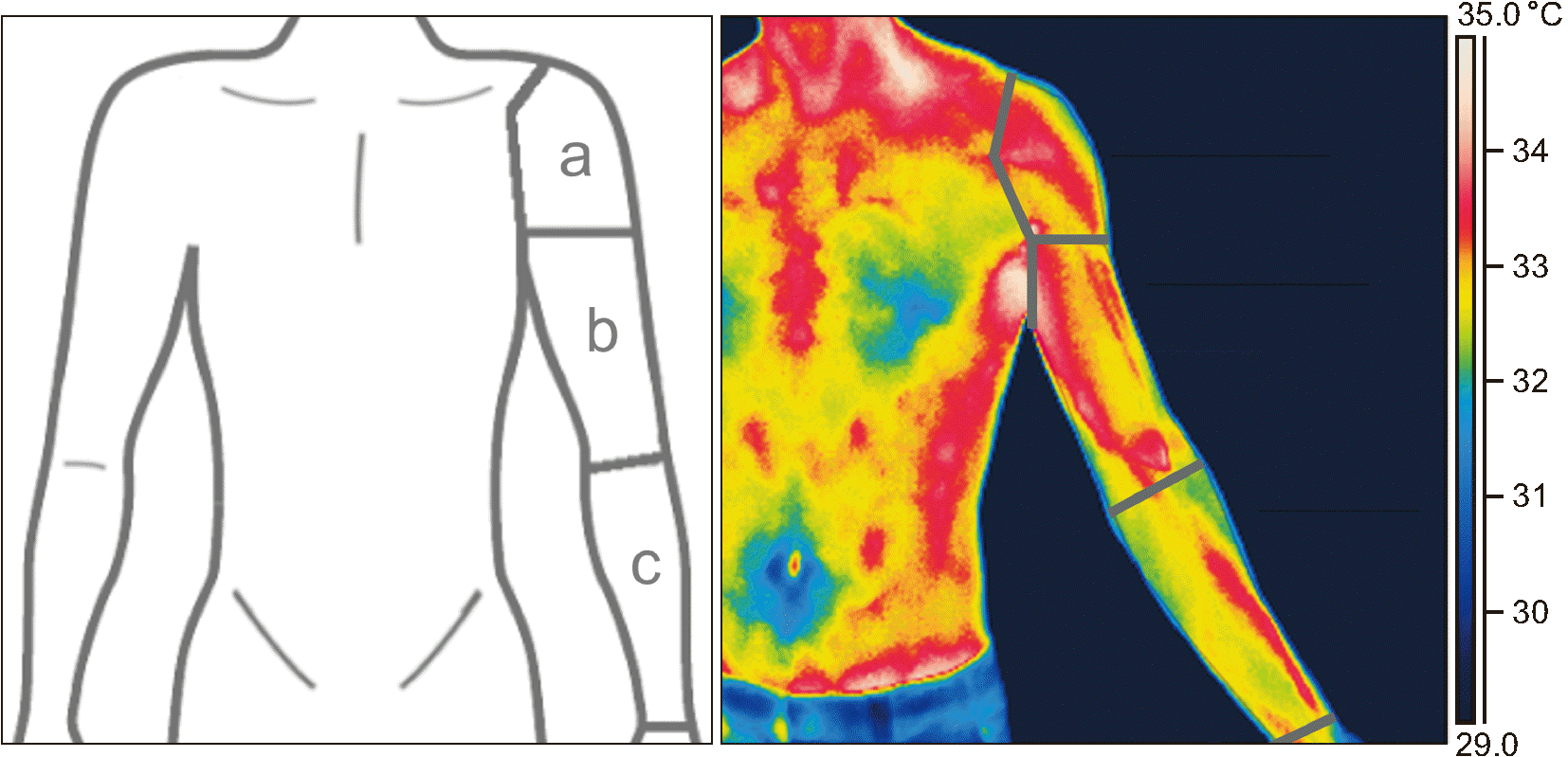
The mean PPT value was used to calculate the amount of force needed to apply during the STPS, which was done at 20% over the PPT and was used to evoke referred pain from the infraspinatus muscle. The STPS was performed with the algometer and sustained for 60 seconds in accordance with previous studies [5,9]. Immediately after STPS, the participants were asked to indicate pain distribution by shading the area of experimental pain on a printed body chart. For data analysis, the upper limb was divided into three regions: 1) the shoulder region comprising the area between the stimulation site and the insertion of the deltoid muscle on the deltoid tuberosity; 2) the arm region, from the insertion of the deltoid muscle to the axis of the elbow joint; and 3) the forearm region, from the axis of the elbow joint to the fingertips (Fig. 2a). In line with previous studies, referred pain was considered to be any pain occurring outside the stimulation area [5].

4. Assessment of temperature variations

To assess sympathetic vasomotor activity in relation to STPS, an infrared thermography camera (FLIR Thermacam E60; FLIR Systems, Boston, MA) with a specific software (FLIR Tools-Software; FLIR Systems) was used to assess the temperature variations in the area of the experimental referred pain. The room temperature ranged from 24–25°C and the humidity was between 45% and 50% in both sessions. Before starting data collection, the participants stood quietly for 15 minutes in the room to obtain a stable body temperature and to ensure correct camera set-up [25] before initiating the assessments. All thermography recordings were performed by a person with extensive experience in using the technique (CVC). Thermography was performed by taking two snapshots in each session, before (pre-STPS) and immediately after the STPS stimulation ceased (post-STPS). On Day 0, a thermographic picture was taken of both upper limbs to confirm that no side differences were present. All pictures were taken in a standing position at a one-meter distance from the participant. All participants were assessed with the shoulder girdle and the upper arm exposed and were instructed not to touch their upper limbs throughout the entire duration of the assessment.
For analysis of thermographic images, the same division of the upper limb was used as for the distribution of referred pain: shoulder region, arm region, and forearm regions (Fig. 2b). The mean temperature of each area of interest was extracted, taking into account that 0.3°C was the margin of error for the thermography equipment [20,22].

5. Model of exercise-induced muscle pain: delayed onset muscle soreness (DOMS)

At the end of Day 0, all participants performed an eccentric exercise of the external rotators of the shoulder to provoke exercise-induced muscle pain in a similar manner as described previously [5,9], by performing 4 sets of 10 repetitions of eccentric contractions against the resistance of an elastic band. The participants stood in a stable position with a straight back and the elbow flexed at 90°. Twenty-four hours later (Day 1), the participants completed a 7-item Likert scale to evaluate the level of exercise-induced muscle pain (0 = no soreness; 1 = dull feeling of soreness; 2 = light, continuous soreness; 3 = more than light soreness; 4 = annoying soreness; 5 = severe soreness; 6 = intolerable soreness). This scale has been frequently used in studies to assess DOMS [29].

6. Sample size

A sample size calculation was conducted using G*Power (v3.1.9.2; Heinrich-Heine-University, Dusseldorf, Germany) with a significance level of 0.05, a power of 90% and a desired medium effect size (Cohen’s f of 0.25) to detect a difference of 0.3°C [22,23]. As the correlation between repeated measures of infrared thermography image analysis is not lower than 0.75 with a conservative non-sphericity correction of 0.7 [30], a minimum of 20 participants were required.

7. Statistics

The statistical analysis was performed with SPSS Statistics (ver.25; IBM Co., Armonk, NY) by a person blinded to the study hypothesis and collected data (PBL). Data are presented as mean and standard deviation (SD) or median and interquartile range (IQR) depending on the distribution of the data which was evaluated using the Shapiro–Wilk tests. Greenhouse–Geisser correction was applied when the assumption of sphericity was violated. The significance level for all statistical tests was set at P ≤ 0.05.
A paired sample t-test was used to ensure that no side differences in temperature were present at baseline. A paired sample t-test was used to analyze changes in PPT at the infraspinatus muscle between Day 0 and Day 1. Chi-square tests were used to assess differences in the frequencies of the regions affected by referred pain after STPS between Day 0 and Day 1. The Wilcoxon signed-rank test was used to compare the total number of regions affected by referred pain on Day 0 and Day 1. The Mann–Whitney U-test (MWU) was used to compare the total number of regions affected by referred pain between males and females at Day 0 and Day 1. Furthermore, the MWU was also used to compare the Likert scale scores for exercise-induced pain (DOMS intensity).
A repeated-measures analysis of covariance (RM-ANCOVA) was used to investigate temperature values with time (pre-STPS and post-STPS), site (shoulder, arm, and forearm regions), and day (Day 0, Day 1) as within factors. A RM-ANCOVA was used to evaluate variations in temperature before and after STPS (“temperature value post-STPS” minus “temperature value pre-STPS”) with site and day as repeated factors. Besides, the effect of sex was analyzed in both previous models by introducing sex as a covariate. Additionally, a mixed model analysis of variance (ANOVA) was used to explore specific sex interaction in temperature variations and PPTs with sex (male and female) set as a fixed factor and site and day as repeated factors. Bonferroni correction was used as a post hoc test to account for multiple comparisons.
Spearman’s rank correlation coefficient was calculated to display the relationship between binomial temperature variations according the margin of error of the thermography equipment (“No”: a variation lower than 0.3°C; “Yes”: a variation equal to or higher than 0.3°C) and the number of body regions affected by referred pain following STPS.

RESULTS

The sample consisted of 20 healthy participants (31.7 ± 9.1 years, 10 females). The skin temperature of both limbs was similar at baseline on Day 0. Twenty-four hours after the eccentric exercise protocol (Day 1), the median exercise-induced pain value (DOMS intensity) on the Likert-scale was 2 (2–3 IQR) with no differences between males and females (MWU = 41.0; Z = –0.77; P = 0.529). The mean PPT value used for the STPS was not significantly different between sessions (Day 0, 341 ± 121 kPa; Day 1, 314 ± 140 kPa). Additionally, the mixed model ANOVA for the PPTs revealed no interaction between sex and time for the PPTs, although females showed lower PPTs than males (ANOVA, F(1,18) = 7.85, P = 0.012) at Day 0 (Bonferroni: P = 0.014; CI, –226 to –29) and Day 1 (Bonferroni: P = 0.016; CI, –259 to –30).

1. Temperature variations

A significant interaction between site, time, and day was detected (RM-ANCOVA, F(2,38) = 3.35, P = 0.046) in absolute temperature values, whilst no effect of sex was detected (P = 0.350). On Day 0, the values of temperature pre-STPS were higher than temperature post-STPS on the arm (Bonferroni: P = 0.001; CI, 0.13 to 0.45) and forearm (Bonferroni: P = 0.003; CI, 0.11 to 0.50). On Day 1, pre-STPS temperature was higher than post-STPS temperature on the shoulder (Bonferroni: P = 0.015; CI, 0.05 to 0.37), arm (Bonferroni: P = 0.001; CI, 0.15 to 0.50) and forearm (Bonferroni: P = 0.010; CI, 0.07 to 0.46). Temperature values on the shoulder region were higher than temperature values on the arm (Bonferroni: P < 0.001; CI, 0.29 to 0.76) and forearm (Bonferroni: P = 0.010; CI, 0.13 to 0.74) on both days (Table 1).
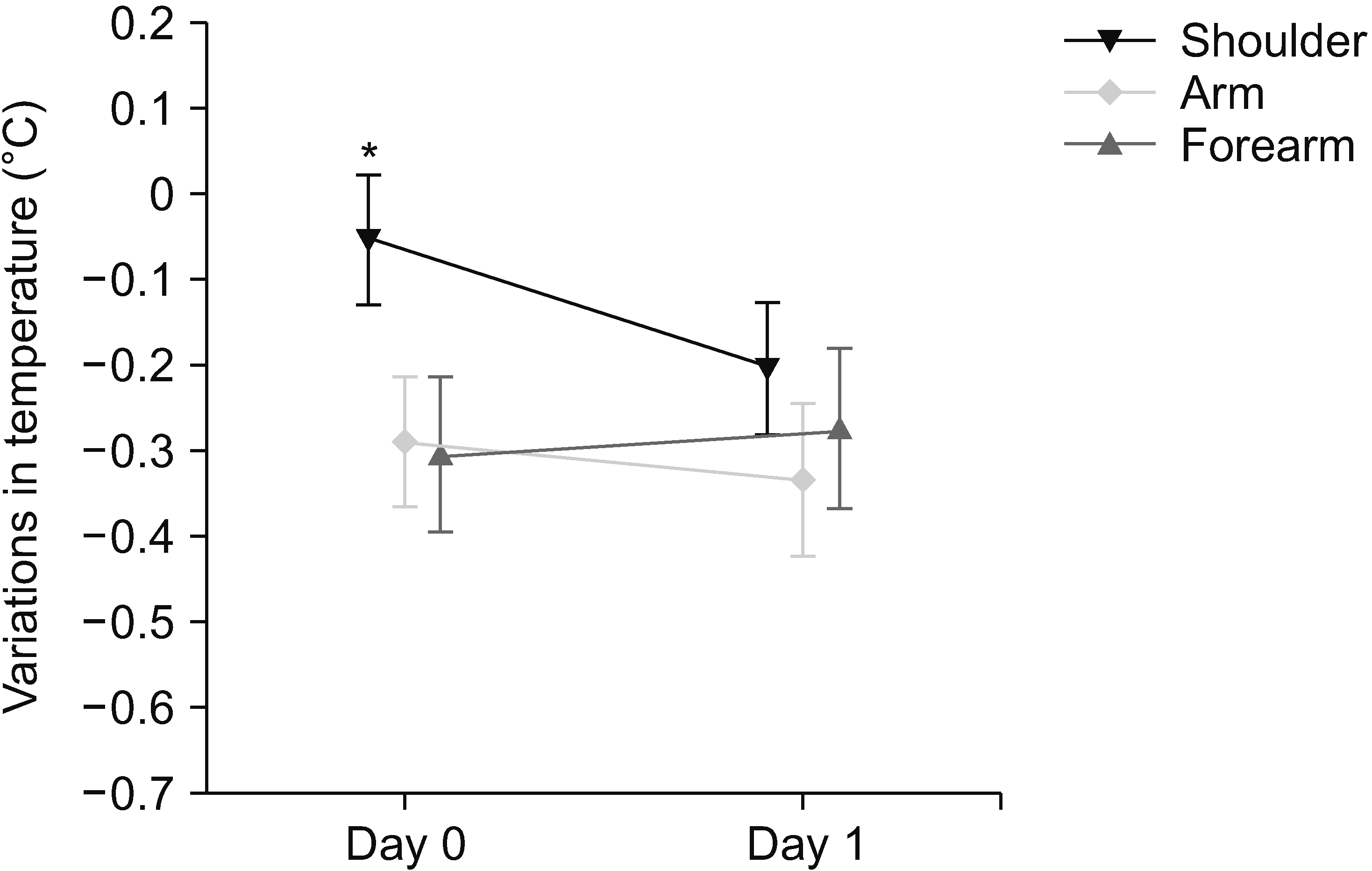
For temperature variations, a significant interaction was detected between site and day (RM-ANCOVA, F(2,38) = 3.35, P = 0.046). On Day 0, the decrease in temperature at the shoulder was lower compared to the arm (Bonferroni: P = 0.001; CI, 0.11 to 0.37) and forearm (Bonferroni: P = 0.018; CI, 0.04 to 0.46). On Day 1, no significant differences were found in the decrease in temperature among the body regions (Fig. 3). Additionally, a sex interaction was detected (P = 0.010), suggesting that male and female present different variations in temperature.
Fig. 4 shows variations in temperature before and after STPS on Day 0 and Day 1, divided by sex. The mixed model ANOVA revealed a significant interaction between site and sex (ANOVA, F(2,36) = 3.44, P = 0.043). On Day 0, temperature decrease after STPS in females was greater than in males in the forearm (Bonferroni: P = 0.039; CI, 0.02 to 0.72). On Day 1, a greater temperature decrease was found amongst females compared with males at the shoulder (Bonferroni: P = 0.018; CI, 0.07 to 0.63), arm (Bonferroni: P = 0.046; CI, 0.01 to 0.69) and forearm (Bonferroni: P = 0.005; CI, 0.17 to 0.81). A greater decrease in temperature was likewise found in females at the shoulder on Day 1 compared to Day 0 (Bonferroni: P = 0.050; CI, 0.00 to 0.58).

2. Pressure-induced referred pain

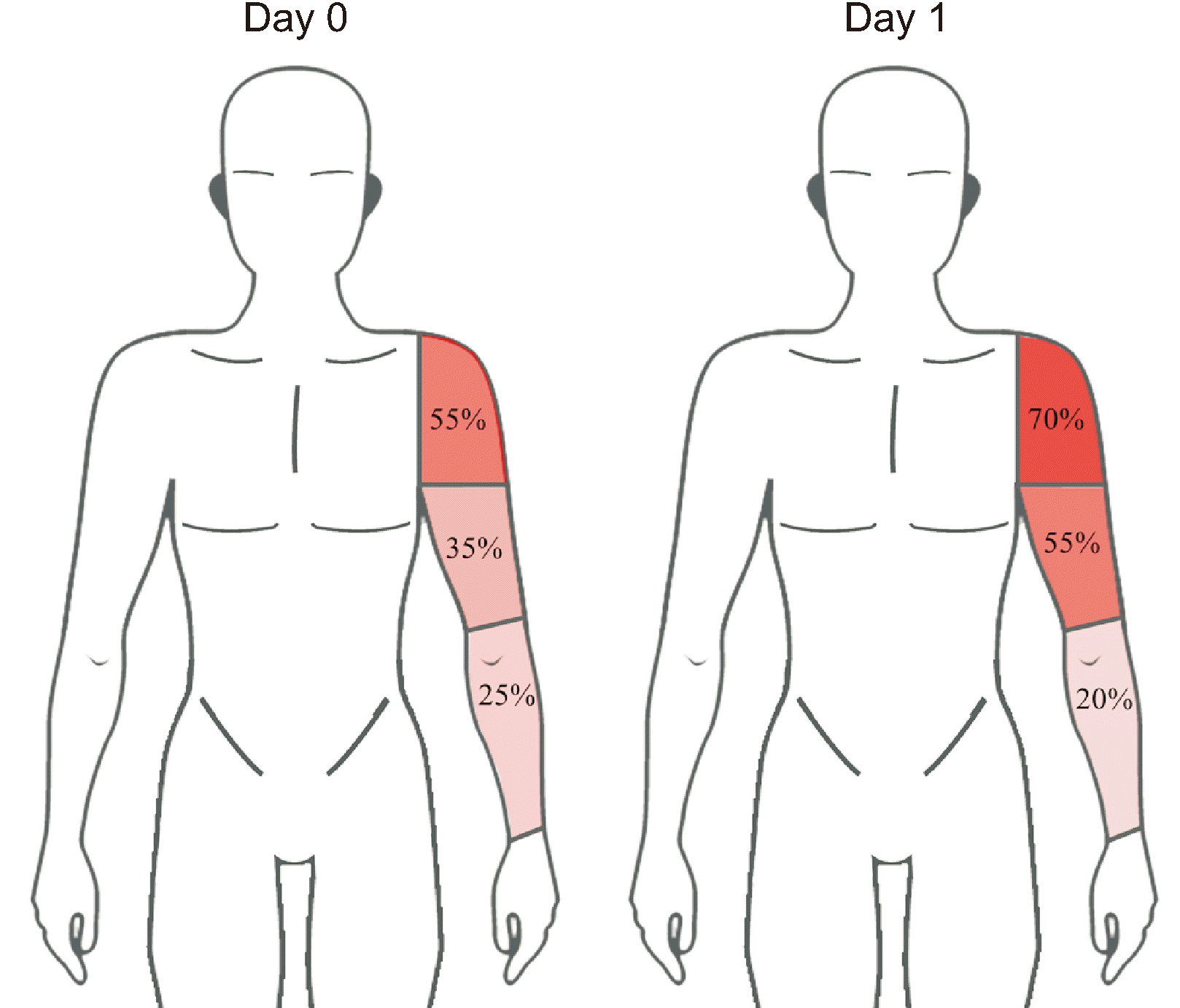
Following STPS on Day 0, 55% of participants experienced referred pain in the shoulder region, 35% in the arm region, and 25% in the forearm region. On Day 1, 70% of participants experienced referred pain in the shoulder region, 55% in the arm region, and the 20% in the forearm region. The frequencies of referred pain in each region did not differ between days (Fig. 5). However, the total number of body regions affected by pain on Day 1 (1; 1–2 IQR) was larger compared to Day 0 (1; 1–1 IQR) (T = 21.0; z = –2.45; P = 0.014) in the total group. The number of regions affected by experimental referred pain was higher in females than in males on Day 0 (MWU = 28.0; z = –2.19; P = 0.029), while there were no differences between the sexes on Day 1 (MWU = 49.0; z = –0.90; P = 0.929). A negative correlation between the number of body regions affected by referred pain and binomial temperature changes after STPS was found in the arm and forearm regions on Day 1, revealing that participants who displayed a temperature reduction greater than 0.3°C were more likely to experience a higher number of referred pain areas. No significant correlation was found on Day 0 (Table 2).

DISCUSSION

The present study used infrared thermography for investigating skin temperature variations after experimental referred pain in healthy individuals. Following STPS, pain was consistently referred to the upper limb in parallel with a decrease in the arm region temperature at baseline and during exercise-induced muscle pain. However, for the shoulder area, the STPS only showed significant differences in temperature between Days 0 and 1, in contrast to the arm and forearm regions which showed changes in the temperature already at Day 0. This could be explained by different factors such as the degree of DOMS or that the arm and forearm regions of referred pain are segmentally linked with the infraspinatus muscle, whereas the shoulder region has only partial links where the C4 segment also includes the shoulder region. Furthermore, exercise-induced muscle pain resulted in a higher number of body regions affected by experimental referred pain and a negative correlation between the number of body regions affected by experimental referred pain and skin temperature variations. These findings were more prominent in females than males at baseline.

1. Sympathetic vasomotor activity and experimental referred pain

The participants in this study demonstrated a decrease in skin temperature following STPS of the infraspinatus muscle (Fig. 2, Table 1). In a previous study investigating referred pain and thermography, Kruse and Christiansen [31] observed that a 60 sec pressure stimulation at tolerance level caused a significant temperature decrease in the areas of referred pain across the entire upper limb. The greater temperature decrease and higher number of limb regions with temperature change, found by Kruse and Christiansen [31], may relate to differences in stimulation intensity where this study only applied a stimulus 20% above PPT. Later studies using similar methods have shown both a skin temperature decrease in the referral area [32] or no change [12], and that painful stimuli could affect sympathetic vasoconstrictive activity and prevent primary neural vasodilation. In line with our results, Srbely et al. [15] found a temporal relationship between experimentally induced central sensitization and skin temperature decrease within segmentally linked dermatomes in healthy individuals. Furthermore, the present study induced referred pain in both basal and sensitized (DOMS) conditions, which is an important novelty compared to previous results since pain sensitization has been linked to larger areas of experimentally induced referred pain [5,26]. The authors believe that their findings support central sensitization possibly being a contributing mechanism in the clinical expression of sympathetic responses in humans. Overall, these findings can be interpreted as strong nociceptive stimuli causing a global change in vasomotor activity [18], which may relate to a general response to threat. The present study, however, demonstrated a unilateral change in cutaneous blood flow, although the relatively low stimulation intensity used here might have been so low that the entire variations are not significant. However, it is worth noting that the intensity of the muscle stimulation as a factor partially explaining these diverse results is a hypothesis grounded in previous studies showing larger pain durations following continuous versus bolus injections of hypertonic saline [4].
In contrast, recent studies have shown that a vasodilation occurs in the area of referred pain when applying a nociceptive stimulus to sore muscle [33,34] and that increases in overlying skin blood flow occur at the stimulation site, but not in distant regions in healthy individuals [35,36] and fibromyalgia patients [37]. From there, it was hypothesized that the variations in cutaneous blood flow might be related to the increased pain sensitivity and increased sympathetic activity [37,38]. To potentially better understand this discrepancy, it is worth noting the dynamic of a normal vasomotor response following noxious stimulation of a healthy muscle. Here, an immediate decrease in skin blood flow is seen, which normalizes approximately 15 minutes after the stimulation and is followed by an increase in such skin blood flow [18,39].
However, despite the present study showing temperature decreases after STPS at baseline and in sore conditions in the area of the referred pain, the temperature variations at baseline and in male participants at all timepoints were below the margin of error of the thermography camera, while the variations in the sore condition were above that margin. Furthermore, the negative correlation between the temperature variations and the number of regions affected by experimental referred pain was found only in the exercise-induced muscle pain condition.
In summary, the current and previous findings indicate that the degree of sympathetic vasomotor responses are related to the stimulation intensity, where more intense stimuli result in the greatest temperature changes [18]. Importantly, the level of pain sensitization due to peripheral and central mechanisms produced by DOMS [5,40] may further amplify the response; not only resulting in expanded referred pain [5] but also in greater vasomotor responses.

2. Sex differences in referred pain and sympathetic mechanisms

At baseline, and between days, females displayed greater temperature variations following the pressure stimulation (Fig. 3). These sex differences in vasomotor activity suggest a greater interaction between mechanisms related to sympathetic regulation of vasomotor responses and nociception, which is in line with previous studies showing sex differences in sympathetic nervous system regulation [16-18]. These sex differences have been attributed to sympathoadrenal processes regulating blood pressure and vascular resistance, although this relationship is not fully understood [41]. Interestingly, while greater temperature variations were seen in females compared to males in both sessions, females showed a higher number of upper limb regions affected by referred pain only at baseline (Table 2). Besides, it is necessary to take into account the aforementioned interpretation of the data in the context of the thermography margin of error, which leads to the conclusion that real changes only occurred in the female group. These differences might be related to females being more sensitive to mechanical pressure [42] and having a less efficient endogenous pain modulation than males [43]. Both factors can contribute to the larger referred pain areas found in females at baseline but not on Day 1, when the participants were assessed in a sensitized condition. In fact, original studies indicate that female facilitation of pain perception could be observed through signs of sympathetic activity such as pupil constriction [44]. Furthermore, it has been suggested that sex-differences in perceived muscle pain may be due to supraspinal rather than peripheral or spinal mechanisms [45]. This has further been supported in a study where the peripheral anesthetic block of nociceptive tissue afferents, after the injection of hypertonic saline, only prevented increases in skin blood flow in males [18]. Furthermore, the possibility of females showing higher temperature variations and more expanded pain, due to receiving a more intense exercise than males, can be ruled out, as no differences were found in perceived DOMS on Day 1 between females and males.
Taken together, these findings suggest that sympathetic vasomotor activity is likely related to the facilitated referred pain response found in females compared with males, and that sympathetic vasomotor activity may partly be accountable for the higher prevalence of clinical widespread pain in females [46]. Studies carried out in healthy individuals found either a decrease [15] or no changes [32] of temperature [47] when a chemical model of sensitization was used through infiltration of capsaicin [15] or glutamate [12,32]. In this study a decrease of temperature was found, although using a DOMS model instead of a chemical one, which the authors think clinical pain (prolonged movement-related pain) can be mimicked more successfully. In the case of studies regarding painful conditions, Skorupska et al. [34] found an increase in temperature following treatment of sore muscles in individuals with sciatica, but a decrease in temperature following treatment of individuals with sciatica and without sore muscles. Moreover, this study also showed a higher decrease of temperature in distal body regions, similarly to what the authors of the present study found, although the reason for this is unknown and should be explored in future studies.

3. Strengths and limitations

The software used to analyze the infrared thermographic image did not allow for accurately superimposing the thermographic image with the area of referred pain. This has previously been considered important when assessing small regions with thermograms [48], as the results obtained tend to be more reliable. Additionally, pain drawings were done on a paper-body chart, but if an electronic body chart would have been used, it would have been possible to calculate the total area of self-perceived pain in a more detailed manner [49] than simply calculating the number of body regions affected by referred pain. Another limitation is that although the DOMS model can mimic clinical pain [50], the study was conducted in a small sample of healthy individuals, and therefore the results cannot be generalized or extrapolated to painful conditions. Moreover, the observational nature of this study and the absence of a control group and/or control side does not allow an evaluation of causality. Future studies should take into account these limitations and also include other outcome measures of sympathetic flow and vasomotor activity to better analyze potential associations with referred pain.
This is the first study to demonstrate temperature decreases in the area of experimental referred pain which correlate with the expansion of referred pain in a sensitized condition based on DOMS. Moreover, these findings seem to be more pronounced in females than males. Collectively, the study findings indicate that sympathetic vasomotor responses, as measured by infrared thermography, could be considered a valuable objective biomarker to assess pain conditions manifesting as expanded pain distribution.

ACKNOWLEDGMENTS

We thank Carlos Valero del Campo for his technical assistance and support with thermographic assessments.

Notes

DATA AVAILABILITY

Data files are available from Harvard Dataverse: https://doi.org/10.7910/DVN/VYIKLK.

CONFLICT OF INTEREST

No potential conflict of interest relevant to this article was reported.

FUNDING

Pablo Bellosta-López has been supported during the elaboration of the present work by the Grant CPB09/18 from “Gobierno de Aragón” and co-financed by “Programa Operativo FSE Aragón 2014-2020, Construyendo Europa desde Aragón” and the Grant FPU19/05237. The funders did not have any role in this study.

REFERENCES

1. Steingrímsdóttir ÓA, Landmark T, Macfarlane GJ, Nielsen CS. 2017; Defining chronic pain in epidemiological studies: a systematic review and meta-analysis. Pain. 158:2092–107. DOI: 10.1097/j.pain.0000000000001009. PMID: 28767506.
crossref
2. Gerhardt A, Hartmann M, Blumenstiel K, Tesarz J, Eich W. 2014; The prevalence rate and the role of the spatial extent of pain in nonspecific chronic back pain--a population-based study in the south-west of Germany. Pain Med. 15:1200–10. DOI: 10.1111/pme.12286. PMID: 24341931.
crossref
3. Vaegter HB, Palsson TS, Graven-Nielsen T. 2017; Facilitated pronociceptive pain mechanisms in radiating back pain compared with localized back pain. J Pain. 18:973–83. DOI: 10.1016/j.jpain.2017.03.002. PMID: 28344100.
crossref
4. Graven-Nielsen T. 2006; Fundamentals of muscle pain, referred pain, and deep tissue hyperalgesia. Scand J Rheumatol Suppl. 122:1–43. DOI: 10.1080/03009740600865980. PMID: 16997767.
5. Doménech-García V, Palsson TS, Herrero P, Graven-Nielsen T. 2016; Pressure-induced referred pain is expanded by persistent soreness. Pain. 157:1164–72. DOI: 10.1097/j.pain.0000000000000497. PMID: 26808146.
crossref
6. van der Wurff P, Buijs EJ, Groen GJ. 2006; Intensity mapping of pain referral areas in sacroiliac joint pain patients. J Manipulative Physiol Ther. 29:190–5. DOI: 10.1016/j.jmpt.2006.01.007. PMID: 16584942.
crossref
7. Dimcevski G, Staahl C, Andersen SD, Thorsgaard N, Funch-Jensen P, Arendt-Nielsen L, et al. 2007; Assessment of experimental pain from skin, muscle, and esophagus in patients with chronic pancreatitis. Pancreas. 35:22–9. DOI: 10.1097/mpa.0b013e31805c1762. PMID: 17575541.
crossref
8. Masuda M, Iida T, Exposto FG, Baad-Hansen L, Kawara M, Komiyama O, et al. 2018; Referred pain and sensations evoked by standardized palpation of the masseter muscle in healthy participants. J Oral Facial Pain Headache. 32:159–66. DOI: 10.11607/ofph.2019. PMID: 29561916.
crossref
9. Doménech-García V, Skuli Palsson T, Boudreau SA, Herrero P, Graven-Nielsen T. 2018; Pressure-induced referred pain areas are more expansive in individuals with a recovered fracture. Pain. 159:1972–9. DOI: 10.1097/j.pain.0000000000001234. PMID: 29608510.
crossref
10. Passatore M, Roatta S. 2006; Influence of sympathetic nervous system on sensorimotor function: whiplash associated disorders (WAD) as a model. Eur J Appl Physiol. 98:423–49. DOI: 10.1007/s00421-006-0312-8. PMID: 17036216.
crossref
11. Martinez-Lavin M. 2007; Biology and therapy of fibromyalgia. Stress, the stress response system, and fibromyalgia. Arthritis Res Ther. 9:216. DOI: 10.1186/ar2146. PMID: 17626613. PMCID: PMC2206360.
crossref
12. Zhang Y, Ge HY, Yue SW, Kimura Y, Arendt-Nielsen L. 2009; Attenuated skin blood flow response to nociceptive stimulation of latent myofascial trigger points. Arch Phys Med Rehabil. 90:325–32. DOI: 10.1016/j.apmr.2008.06.037. PMID: 19236988.
crossref
13. Larsson SE, Bodegård L, Henriksson KG, Oberg PA. 1990; Chronic trapezius myalgia. Morphology and blood flow studied in 17 patients. Acta Orthop Scand. 61:394–8. DOI: 10.3109/17453679008993548. PMID: 2239160.
14. Morikawa Y, Takamoto K, Nishimaru H, Taguchi T, Urakawa S, Sakai S, et al. 2017; Compression at myofascial trigger point on chronic neck pain provides pain relief through the prefrontal cortex and autonomic nervous system: a pilot study. Front Neurosci. 11:186. DOI: 10.3389/fnins.2017.00186. PMID: 28442987. PMCID: PMC5386976.
crossref
15. bely JZ Sr, Dickey JP, Montaholi Y. 2017; Experimentally induced central sensitization evokes segmental autonomic responses in humans. Int Phys Med Rehabil J. 1:47–53. https://medcraveonline.com/IPMRJ/experimentally-induced-central-sensitization-evokes-segmental-autonomic-responses-in-humans.html. DOI: 10.15406/ipmrj.2017.01.00013.
16. Hinojosa-Laborde C, Chapa I, Lange D, Haywood JR. 1999; Gender differences in sympathetic nervous system regulation. Clin Exp Pharmacol Physiol. 26:122–6. DOI: 10.1046/j.1440-1681.1999.02995.x. PMID: 10065332.
crossref
17. Hogarth AJ, Mackintosh AF, Mary DA. 2007; Gender-related differences in the sympathetic vasoconstrictor drive of normal subjects. Clin Sci (Lond). 112:353–61. DOI: 10.1042/CS20060288. PMID: 17129210.
crossref
18. Lei J, You HJ. 2012; Variation of pain and vasomotor responses evoked by intramuscular infusion of hypertonic saline in human subjects: influence of gender and its potential neural mechanisms. Brain Res Bull. 87:564–70. DOI: 10.1016/j.brainresbull.2011.11.003. PMID: 22100333.
crossref
19. Fillingim RB, King CD, Ribeiro-Dasilva MC, Rahim-Williams B, Riley JL 3rd. 2009; Sex, gender, and pain: a review of recent clinical and experimental findings. J Pain. 10:447–85. DOI: 10.1016/j.jpain.2008.12.001. PMID: 19411059. PMCID: PMC2677686.
crossref
20. Hildebrandt C, Raschner C, Ammer K. 2010; An overview of recent application of medical infrared thermography in sports medicine in Austria. Sensors (Basel). 10:4700–15. DOI: 10.3390/s100504700. PMID: 22399901. PMCID: PMC3292141.
crossref
21. Nahm FS. 2013; Infrared thermography in pain medicine. Korean J Pain. 26:219–22. DOI: 10.3344/kjp.2013.26.3.219. PMID: 23861995. PMCID: PMC3710935.
crossref
22. Ring EF, Ammer K. 2012; Infrared thermal imaging in medicine. Physiol Meas. 33:R33–46. DOI: 10.1088/0967-3334/33/3/R33. PMID: 22370242.
crossref
23. Dibai-Filho AV, Guirro RRJ. 2015; Evaluation of myofascial trigger points using infrared thermography: a critical review of the literature. J Manipulative Physiol Ther. 38:86–92. DOI: 10.1016/j.jmpt.2014.10.010. PMID: 25467609.
crossref
24. Simons DG, Travell JG, Simons LS. 2001. Dolor y disfunción miofascial: el manual de los puntos gatillo. 2nd ed. Vol. 1:Médica Panamericana;Madrid: Spanish. https://books.google.es/books?id=30IRTsoFvqcC&printsec=copyright&hl=es#v=onepage&q&f=false. DOI: 10.1016/j.jmpt.2014.10.010.
25. RG S. 2015. Guidelines for neuro-musculoskeletal thermography [Internet]. American Academy of Thermology;Greenville (SC): Available at: https://aathermology.org/wp-content/uploads/2018/04/AAT-NMSK-Guidelines-2021.pdf.
26. Gibson W, Arendt-Nielsen L, Graven-Nielsen T. 2006; Delayed onset muscle soreness at tendon-bone junction and muscle tissue is associated with facilitated referred pain. Exp Brain Res. 174:351–60. DOI: 10.1007/s00221-006-0466-y. PMID: 16642316.
crossref
27. Chesterton LS, Sim J, Wright CC, Foster NE. 2007; Interrater reliability of algometry in measuring pressure pain thresholds in healthy humans, using multiple raters. Clin J Pain. 23:760–6. DOI: 10.1097/AJP.0b013e318154b6ae. PMID: 18075402.
crossref
28. Madeleine P, Mathiassen SE, Arendt-Nielsen L. 2008; Changes in the degree of motor variability associated with experimental and chronic neck-shoulder pain during a standardised repetitive arm movement. Exp Brain Res. 185:689–98. DOI: 10.1007/s00221-007-1199-2. PMID: 18030457.
crossref
29. Impellizzeri FM, Maffiuletti NA. 2007; Convergent evidence for construct validity of a 7-point likert scale of lower limb muscle soreness. Clin J Sport Med. 17:494–6. DOI: 10.1097/JSM.0b013e31815aed57. PMID: 17993794.
crossref
30. Choi E, Lee PB, Nahm FS. 2013; Interexaminer reliability of infrared thermography for the diagnosis of complex regional pain syndrome. Skin Res Technol. 19:189–93. DOI: 10.1111/srt.12032. PMID: 23331254.
crossref
31. Kruse RA Jr, Christiansen JA. 1992; Thermographic imaging of myofascial trigger points: a follow-up study. Arch Phys Med Rehabil. 73:819–23. PMID: 1514891.
32. Kimura Y, Ge HY, Zhang Y, Kimura M, Sumikura H, Arendt-Nielsen L. 2009; Evaluation of sympathetic vasoconstrictor response following nociceptive stimulation of latent myofascial trigger points in humans. Acta Physiol (Oxf). 196:411–7. DOI: 10.1111/j.1748-1716.2009.01960.x. PMID: 19210492.
crossref
33. Skorupska E, Rychlik M, Pawelec W, Samborski W. 2015; Dry needling related short-term vasodilation in chronic sciatica under infrared thermovision. Evid Based Complement Alternat Med. 2015:214374. DOI: 10.1155/2015/214374. PMID: 25821479. PMCID: PMC4363707.
crossref
34. Skorupska E, Rychlik M, Samborski W. 2015; Intensive vasodilatation in the sciatic pain area after dry needling. BMC Complement Altern Med. 15:72. DOI: 10.1186/s12906-015-0587-6. PMID: 25888420. PMCID: PMC4426539.
crossref
35. Cagnie B, Barbe T, De Ridder E, Van Oosterwijck J, Cools A, Danneels L. 2012; The influence of dry needling of the trapezius muscle on muscle blood flow and oxygenation. J Manipulative Physiol Ther. 35:685–91. DOI: 10.1016/j.jmpt.2012.10.005. PMID: 23206963.
crossref
36. Sandberg M, Lundeberg T, Lindberg LG, Gerdle B. 2003; Effects of acupuncture on skin and muscle blood flow in healthy subjects. Eur J Appl Physiol. 90:114–9. DOI: 10.1007/s00421-003-0825-3. PMID: 12827364.
crossref
37. Sandberg M, Lindberg LG, Gerdle B. 2004; Peripheral effects of needle stimulation (acupuncture) on skin and muscle blood flow in fibromyalgia. Eur J Pain. 8:163–71. DOI: 10.1016/S1090-3801(03)00090-9. PMID: 14987626.
crossref
38. Sandberg M, Larsson B, Lindberg LG, Gerdle B. 2005; Different patterns of blood flow response in the trapezius muscle following needle stimulation (acupuncture) between healthy subjects and patients with fibromyalgia and work-related trapezius myalgia. Eur J Pain. 9:497–510. DOI: 10.1016/j.ejpain.2004.11.002. PMID: 16139178.
crossref
39. Lei J, You HJ, Andersen OK, Graven-Nielsen T, Arendt-Nielsen L. 2008; Homotopic and heterotopic variation in skin blood flow and temperature following experimental muscle pain in humans. Brain Res. 1232:85–93. DOI: 10.1016/j.brainres.2008.07.056. PMID: 18680731.
crossref
40. Nie H, Arendt-Nielsen L, Madeleine P, Graven-Nielsen T. 2006; Enhanced temporal summation of pressure pain in the trapezius muscle after delayed onset muscle soreness. Exp Brain Res. 170:182–90. DOI: 10.1007/s00221-005-0196-6. PMID: 16328284.
crossref
41. Just TP, DeLorey DS. 2017; Sex differences in sympathetic vasoconstrictor responsiveness and sympatholysis. J Appl Physiol (1985). 123:128–35. DOI: 10.1152/japplphysiol.00139.2017. PMID: 28473610. PMCID: PMC5538817.
crossref
42. Petrini L, Matthiesen ST, Arendt-Nielsen L. 2015; The effect of age and gender on pressure pain thresholds and suprathreshold stimuli. Perception. 44:587–96. DOI: 10.1068/p7847. PMID: 26422905.
crossref
43. Riley JL 3rd, Cruz-Almeida Y, Staud R, Fillingim RB. 2020; Age does not affect sex effect of conditioned pain modulation of pressure and thermal pain across 2 conditioning stimuli. Pain Rep. 5:e796. DOI: 10.1097/PR9.0000000000000796. PMID: 32072094. PMCID: PMC7004505.
crossref
44. Ellermeier W, Westphal W. 1995; Gender differences in pain ratings and pupil reactions to painful pressure stimuli. Pain. 61:435–9. DOI: 10.1016/0304-3959(94)00203-Q. PMID: 7478686.
crossref
45. Lei J, Jin L, Zhao Y, Sui MY, Huang L, Tan YX, et al. 2011; Sex-related differences in descending norepinephrine and serotonin controls of spinal withdrawal reflex during intramuscular saline induced muscle nociception in rats. Exp Neurol. 228:206–14. DOI: 10.1016/j.expneurol.2011.01.004. PMID: 21238453.
crossref
46. Mansfield KE, Sim J, Jordan JL, Jordan KP. 2016; A systematic review and meta-analysis of the prevalence of chronic widespread pain in the general population. Pain. 157:55–64. DOI: 10.1097/j.pain.0000000000000314. PMID: 26270591. PMCID: PMC4711387.
crossref
47. Skorupska E, Rychlik M, Samborski W. 2015; Validation and test-retest reliability of new thermographic technique called Thermovision Technique of Dry Needling for gluteus minimus trigger points in sciatica subjects and TrPs-negative healthy volunteers. Biomed Res Int. 2015:546497. DOI: 10.1155/2015/546497. PMID: 26137486. PMCID: PMC4475557.
crossref
48. Haddad DS, Brioschi ML, Arita ES. 2012; Thermographic and clinical correlation of myofascial trigger points in the masticatory muscles. Dentomaxillofac Radiol. 41:621–9. DOI: 10.1259/dmfr/98504520. PMID: 23166359. PMCID: PMC3528192.
crossref
49. Boudreau SA, Badsberg S, Christensen SW, Egsgaard LL. 2016; Digital pain drawings: assessing touch-screen technology and 3D body schemas. Clin J Pain. 32:139–45. DOI: 10.1097/AJP.0000000000000230. PMID: 25756558.
50. Slater H, Arendt-Nielsen L, Wright A, Graven-Nielsen T. 2005; Sensory and motor effects of experimental muscle pain in patients with lateral epicondylalgia and controls with delayed onset muscle soreness. Pain. 114:118–30. DOI: 10.1016/j.pain.2004.12.003. PMID: 15733637.
crossref

Fig. 1
Two-day study protocol. PPTs: pressure pain thresholds, STPS: suprathreshold pressure stimulation, DOMS: delayed-onset muscular soreness.
kjp-35-3-240-f1.tif
Fig. 2
Body divisions show the areas of interest to quantify temperature variations in thermography images following the infraspinatus suprathreshold pressure stimulation: shoulder (a), arm (b), and forearm (c).
kjp-35-3-240-f2.tif
Fig. 3
Mean temperature variations before and after suprathreshold pressure stimulation on Day 0 and Day 1 for the shoulder, arm, and forearm regions. Error bars indicate standard error of the mean. *Significantly different compared to arm and forearm regions after Bonferroni post hoc test, P < 0.05.
kjp-35-3-240-f3.tif
Fig. 4
Mean temperature variations before and after suprathreshold pressure stimulation on Day 0 and Day 1. Error bars indicate standard error of the mean. *Significant sex differences after Bonferroni post hoc test, P < 0.05. #Significantly different compared to Day 0 after Bonferroni post hoc test, P < 0.05.
kjp-35-3-240-f4.tif
Fig. 5
Proportion of participants experiencing pressure-induced referred pain at the shoulder, arm, and forearm regions following suprathreshold pressure stimulation on Day 0 and Day 1.
kjp-35-3-240-f5.tif
Table 1
Temperature values at Day 0 and Day 1 before and after a suprathreshold pressure stimulation
Temperature (ºC) Pre-STPS
(n = 20)
Post-STPS
(n = 20)
Pre-post STPS differences
[CI 95%]
Shoulder Day 0 33.20 (0.84) 33.15 (0.75) [–0.10 to 0.21]
Day 1 33.37 (0.63) 33.16 (0.71)* [0.05 to 0.37]
Arm Day 0 32.88 (0.83) 32.59 (0.79)* [0.13 to 0.45]
Day 1 32.83 (0.63) 32.49 (0.78)* [0.15 to 0.52]
Forearm Day 0 32.97 (0.74) 32.66 (0.84)* [0.11 to 0.50]
Day 1 32.89 (0.53) 32.62 (0.66)* [0.07 to 0.46]

Values are presented as mean (standard deviation).

STPS: suprathreshold pain stimulation, CI: confidence interval.

*P < 0.05, significant differences compared to pre-STPS after Bonferroni post hoc test.

Table 2
Correlation between number of body regions affected by referred pain and relevant temperature variations after STPS on Day 0 and Day 1
No. of regions
affected by
referred pain
Regions ρ P value
Binomial temperature variations Day 0 Shoulder –0.138 0.563
Arm –0.333 0.152
Forearm –0.184 0.436
Day 1 Shoulder –0.213 0.367
Arm –0.523* 0.018
Forearm –0.523* 0.018

STPS: suprathreshold pain stimulation, ρ: Spearman’s Rho.

*Significant correlation at P < 0.05 (two-tailed).

TOOLS
Similar articles