

 PDF
PDF Citation
 Print
Print


INTRODUCTION
Rheumatoid arthritis (RA) is a chronic autoimmune inflammatory disease characterized by synovial inflammation and bone erosion, which can eventually lead to structural and functional impairments and a decrease in the quality of life [1]. The pathogenesis of RA is still under investigation, but the interplay between genetic predisposition and environmental triggers plays a key role in the development of RA. Human leukocyte antigen (HLA)-DRB1 is a renowned genetic risk factor that is strongly associated with the development and progression of RA [2-5]. Hypervariable region encoded by particular HLA-DRB1 alleles, the so-called “shared epitope (SE)” is responsible for the presentation of self-antigen to T lymphocytes [6]. A previous study has also shown that the interaction between SEs and the aryl-hydrocarbon receptor accounts for the influence of environmental pollutants, such as tobacco smoking in SE-positive RA patients [7].
HLA-DRB1 SEs bind to citrullinated peptides with higher affinity than unmodified peptides, leading to T-cell activation and the subsequent generation of antibodies to citrullinated peptides [8]. Anti-cyclic citrullinated peptide antibody (ACPA) is the key clinical index for determining the diagnosis, classification, and prognosis of RA [9]. Radiographic progression tends to be more severe as the ACPA titer become higher [10-12]. ACPAs are frequently found in SE-positive RA patients, but rarely in SE-negative patients [13]. Alternatively, SEs are present in 70%~80% of ACPA-positive patients and are strongly associated with joint destruction [14-17]. Of note, a SE gene–dose effect on RA risk has been documented in ACPA-positive patients [14].
Given that SE and ACPA are clinically significant in the characterization of RA, SE and ACPA are expected to be involved in the treatment response to disease-modifying anti-rheumatic drugs (DMARDs). The paradigm of RA treatment has greatly shifted with the introduction of biologic DMARDs (bDMARDs). Although, biologic therapy has greatly improved RA management, some needs remain unmet. About 30% of patients show inadequate response to the first biologic therapy and the response may decrease over time in some patients [18]. Only a subset of patients achieves long-term remission or low disease activity. This disappointing reality is similar to that of most biologics [18]. To spare the expense of time and money, predictive indicator for specific drug response would be of great clinical benefit. In the current studies, ACPA positivity was associated with a better response to abatacept in RA patients and patients with the highest ACPA concentrations showed better clinical outcomes [19-23]. However, disease manifestation and treatment response can differ in patients of varing races and ethnicity with RA [24,25]. The clinical implications of SE and ACPA have not been fully evaluated in Korean patients with RA.
In this study, we compared the clinical characteristics of SE-positive patients to those of SE-negative patients and investigated the clinical significance of SE and ACPA in terms of bDMARD therapy in Korean patients with RA.
Go to : 
MATERIALS AND METHODS
Patients
A total of 1,639 patients with RA who fulfilled the 2010 RA classification criteria [9] and had received care at St. Vincent’s Hospital, the Catholic University of Korea (Suwon, Republic of Korea) between 2003 and 2020 were identified. Clinical, laboratory and radiographic data at the time of initial diagnostic evaluation were retrieved from the hospital’s medical records. Of the 1,639 patients, HLA-DRB1 alleles were identified in 533 patients, including 216 patients who had been treated or were currently being treated with bDMARDs or targeted synthetic DMARDs (tsDMARDs). The bDMARDs include tumor necrosis factor inhibitors (etanercept, adalimumab, infliximab, and golimumab), tocilizumab, abatacept, and rituximab. The tsDMARDs include tofacitinib and baricitinib [26]. Disease activity was assessed using the Disease Activity Score in 28 joints (DAS28) using the C-reactive protein (CRP) level [27]. The study was carried out in accordance with the Helsinki Declaration and was approved by the Institutional Review Board of St. Vincent’s Hospital, the Catholic University of Korea (no. VC19OISI0252).
Assay of RA-associated antibodies
ACPA was analyzed by chemiluminescent microparticle immunoassay (Abbott Laboratories, North Chicago, IL, USA) and a positive reading was defined as a cut-off value of 5 U/mL. The maximum antibody concentration was defined as 340 U/mL. For statistical calculations, a value of 340 U/mL was assigned to all measurements >340 U/mL. ACPA was divided into three categories: <5 U/mL (negative), 5~200 U/mL (low to moderate level), and >200 U/mL (high level) in agreement with a previous study [11]. Rheumatoid factor (RF) titers were measured with a latex agglutination test (Beckman Coulter, Brea, CA, USA) with a cut-off value of 14 U/mL. RF levels were also divided into three categories: <14 U/mL (negative), 14~100 U/mL (low to moderate level) and >100 U/mL (high level) in agreement with a previous study [30].
Radiographic evaluation
Anteroposterior radiographs of the hands were scored by two experienced readers using van der Heijde modified Sharp score (SHS) [31]. The films were scored in chronological order, and the readers were blinded to the patient data. The potential maximum total score for both hands is 280 (16 areas scored for erosions [score 0~5] and 15 areas for joint space narrowing [score 0~4] in each hand). Interobserver reliability was assessed by calculating the intraclass correlation coefficient, which was 0.860 (95% confidence interval [CI], 0.779 to 0.922).
Statistical analyses
For continuous data, the results were presented as means with standard deviations or medians with interquartile ranges. The between-group comparisons were performed using Student’s t-test or the Mann–Whitney U-test. Categorical or dichotomous variables were expressed as frequencies and percentages, and were compared using the Chi-squared test or Fisher’s exact test. The initiation of b- or tsDMARDs with hazard ratios (HRs) and corresponding 95% CIs were assessed via Kaplan–Meier analysis and compared using log-rank tests. To identify significant predictors of b- or tsDMARD initiation, clinically relevant variables were entered into a multivariable Cox proportional hazards regression model. A two-sided p-value of less than 0.05 was considered statistically significant. All statistical analyses were performed using R (version 4.0.4; R Project for Statistical Computing, www.r-project.org).
Go to :
RESULTS
Characteristics of the study population
The baseline characteristics of the study subjects (n=533) and separate baseline values for patients with and without SE alleles are presented in Table 1. A total of 329 patients (61.7%) were identified as having a SE allele. The median age at diagnosis was 50 years (41, 59), and 80.3% (n=428) of the patients were women. At baseline, 88.9% (n=474) were seropositive for RF and 91.0% (n=485) for ACPA. Erosive disease was identified in 32.8% (n=175) of patients, and the median SHS was 0 (0, 7).
Table 1
Baseline characteristics of the study subjects with respect to the shared epitope
| Variable | All (n=533) | Shared epitope | p-value | |
|---|---|---|---|---|
| Negative (n=204) | Positive (n=329) | |||
| Female | 428 (80.3) | 173 (84.8) | 255 (77.5) | 0.052 |
| Age at diagnosis (yr) | 50 (41, 59) | 48 (40, 60) | 50 (42, 59) | 0.536 |
| BMI | 0.203 | |||
| Underweight | 36 (6.8) | 11 (5.4) | 25 (7.6) | |
| Normal | 379 (71.1) | 148 (72.5) | 231 (70.2) | |
| Overweight | 101 (18.9) | 35 (17.2) | 66 (20.1) | |
| Obese | 17 (3.2) | 10 (4.9) | 7 (2.1) | |
| Smoking* | 117 (22.0) | 40 (19.6) | 77 (23.4) | 0.357 |
| Diabetes | 70 (13.1) | 28 (13.7) | 42 (12.8) | 0.852 |
| Hypertension | 197 (37.0) | 70 (34.3) | 127 (38.6) | 0.366 |
| Dyslipidemia | 274 (51.4) | 107 (52.5) | 167 (50.8) | 0.771 |
| ESR (mm/h) | 45 (26, 69) | 42 (26, 68) | 46 (26, 70) | 0.575 |
| CRP (mg/dL) | 0.8 (0.2, 2.3) | 0.6 (0.2, 2.0) | 1.0 (0.3, 2.9) | 0.005 |
| IgM RF | ||||
| Positive | 474 (88.9) | 180 (88.2) | 294 (89.4) | 0.794 |
| Titer (IU/mL) | 70.9 (29.7, 184.0) | 77.9 (30.8, 199.8) | 70.7 (28.8, 175.7) | 0.811 |
| Subgroup | 0.899 | |||
| Negative | 59 (11.1) | 24 (11.1) | 35 (10.6) | |
| Low to moderate | 348 (65.3) | 93 (45.6) | 155 (47.1) | |
| High | 137 (25.7) | 87 (42.6) | 139 (42.2) | |
| ACPA | ||||
| Positive | 485 (91.0) | 179 (87.7) | 306 (93.0) | 0.056 |
| Titer (IU/mL) | 100.0 (30.9, 200.0) | 95.2 (22.1, 200.0) | 100.0 (35.2, 200.0) | 0.177 |
| Subgroup | 0.105 | |||
| Negative | 48 (9.0) | 25 (12.3) | 23 (7.0) | |
| Low to moderate | 348 (65.3) | 126 (61.8) | 222 (67.5) | |
| High | 137 (25.7) | 52 (26.0) | 84 (25.5) | |
| SHS (units) | 0 (0, 7) | 0 (0, 4) | 0 (0, 10) | 0.020‡ |
| Erosion | 72 (13.5) | 19 (9.3) | 53 (16.1) | 0.035 |
| DAS28-CRP (units) | 3.9 (3.4, 4.3) | 3.9 (3.3, 4.3) | 3.9 (3.5, 4.3) | 0.220 |
| Bone mineral density | ||||
| L-spine | 0.132 | |||
| Normal | 286 (53.7) | 119 (58.3) | 167 (50.8) | |
| Osteopenia | 116 (21.8) | 44 (21.6) | 72 (21.9) | |
| Osteoporosis | 131 (24.6) | 41 (20.1) | 90 (27.4) | |
| Femur | 0.361 | |||
| Normal | 224 (42.0) | 93 (45.6) | 131 (39.8) | |
| Osteopenia | 248 (46.5) | 91 (44.6) | 157 (47.7) | |
| Osteoporosis | 61 (11.4) | 20 (9.8) | 41 (12.5) | |
| Osteoporosis | 148 (27.8) | 46 (22.5) | 102 (31.0) | 0.044 |
| bDMARDs or tsDMARDs† | 216 (40.5) | 71 (34.8) | 145 (44.1) | 0.043 |
Values are presented as number (%) or median (interquartile range). ACPA: anti-cyclic citrullinated protein antibody, BMI: body mass index, CRP: C-reactive protein, DMARD: disease-modifying anti-rheumatic disease, ESR: erythrocyte sedimentation rate, RF: rheumatoid factor, SHS: van der Heijde modified Sharp score, DAS28: disease activity score in 28 joints. *Include ex- and current smokers. †Biologic DMARDs (bDMARDs) include tumor necrosis factor inhibitors (etanercept, adalimumab, infliximab, golimumab), tocilizumab, abatacept, and rituximab. Targeted synthetic DMARDs (tsDMARDs) include tofacitinib and baricitinib. ‡When adjusted by sex and onset age, p-value was 0.194 (odds ratio [95% confidence interval] 1.0052 [0.9972~1.0134]).
![]()
As compared with the patients without SE alleles, the patients with SE alleles had a smaller proportion of females (77.5% vs. 84.8%, p=0.052) and a higher level of CRP (1.0 [0.3, 2.9] vs. 0.6 [0.2, 2.0] mg/dL, p=0.005). There was a trend toward higher DAS28-CRP in SE-positive patients (4.0 [3.4, 4.5] vs. 3.9 [3.3, 4.3], p=0.057). At baseline, the proportion of erosive disease was higher in SE-positive patients (16.1% vs. 9.3%, p=0.035) and osteoporosis (lumbar spine or femur) was also more frequent in SE-positive patients (31.2% vs. 22.5%, p=0.044).
Association between the SE and ACPA
ACPA was not significantly associated with SE-positive patient (93.0% vs. 87.7%, p=0.056) (Figure 1A). When the ACPA was categorized into three subgroups by titer (<5 U/mL [negative], 5~200 U/mL [low to moderate level], and >200 U/mL [high level]), high titer of ACPA was found in about a quarter of RA patients (n=137, 25.7%). The proportion of patients with high levels of ACPA did not differ between SE-positive and SE-negative patients (25.5% vs. 26.0%, p=0.990) (Figure 1B). Given that smoking interacts with SE in the development of ACPA-positive RA [5,29,32,33], the association between ACPA positivity and smoking was evaluated in SE-positive and SE-negative patients separately. In SE-positive patients, the proportion of ACPA positivity was marginally higher in smokers than in non-smokers (97.4% vs. 91.7%, p=0.141). In contrast, there was no difference in ACPA positivity between smokers and non-smokers in SE-negative patients (87.5% vs. 87.8%, p=1.000).
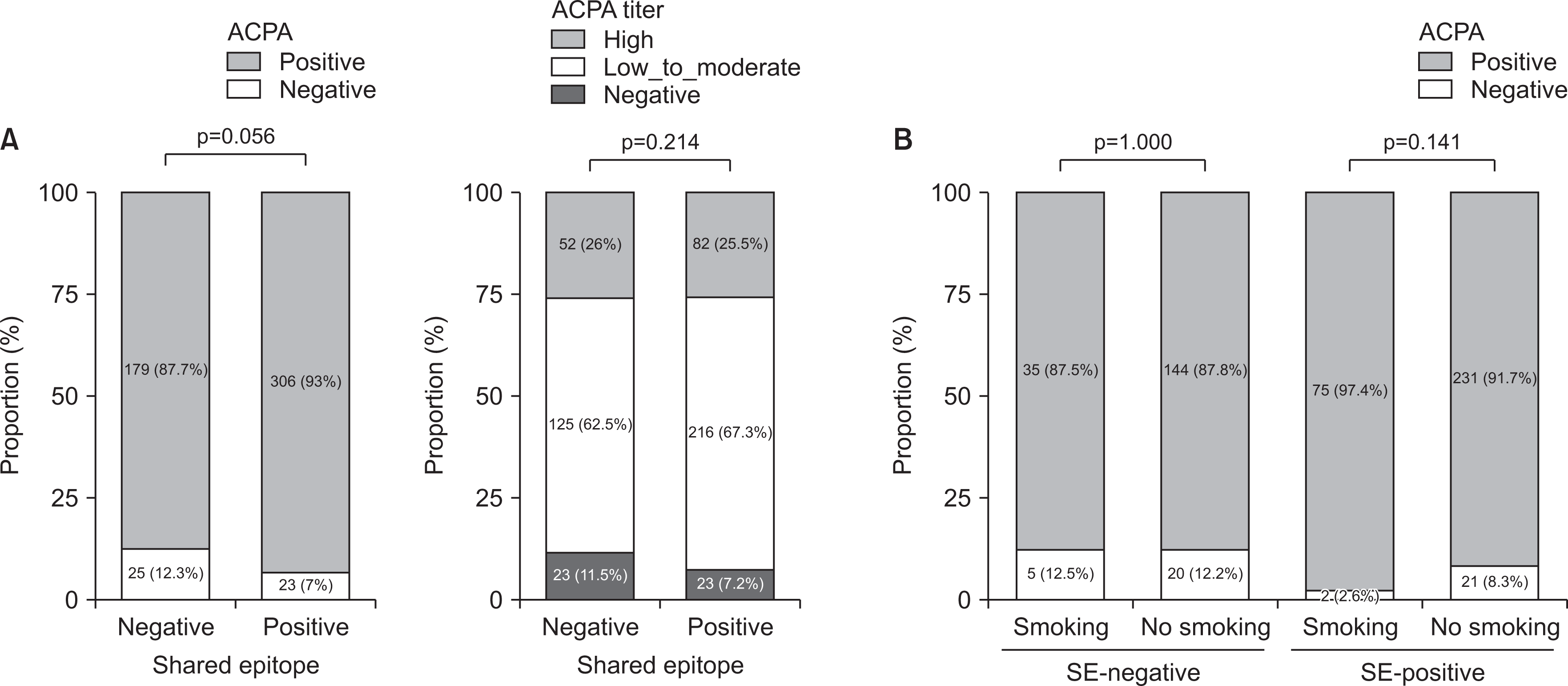 | Fig. 1(A) ACPA positivity and the three ACPA subgroups with respect to SE status. ACPA was divided into three categories: <5 U/mL (negative), 5~200 U/mL (low to moderate level), and >200 U/mL (high level). (B) ACPA positivity for different combination of smoking and SE status. Categorical comparison between the two groups was verified using the Chi-squared test. The number (percentage) is marked in the middle of each bar. ACPA: anti-cyclic citrullinated protein antibody, SE: shared epitope.
|
Risk of initiation of bDMARDs or tsDMARDs
Methotrexate (MTX)-based conventional synthetic DMARD (csDMARD) combinations comprise the initial therapy for RA. The b- or tsDMARDs are administered only if disease activity is not sufficiently controlled despite the use of MTX-based double or triple combinations of csDMARDs for over 6 months, according to the National Health Insurance guidelines in South Korea. Under the assumption that the lack of response to csDMARDs and the initiation of b- or tsDMARDs are surrogate indices for poor long-term outcomes [34], we evaluated the use of b- or tsDMARDs according to the presence or absence of SE. During the follow-up period, b- or tsDMARDs were more often used in SE-positive patients than in SE-negative patients (44.1% vs. 34.8%, p=0.046) (Figure 2A). The cumulative incidence of b- or tsDMARD initiation within the first 5 years was also significantly higher in the SE-positive subgroup (log-rank test, p=0.020) (Figure 2B). The HR of b- or tsDMARD initiation was 1.48 (95% CI, 1.063~2.022) in patients with SE alleles compared with patients without SE alleles.
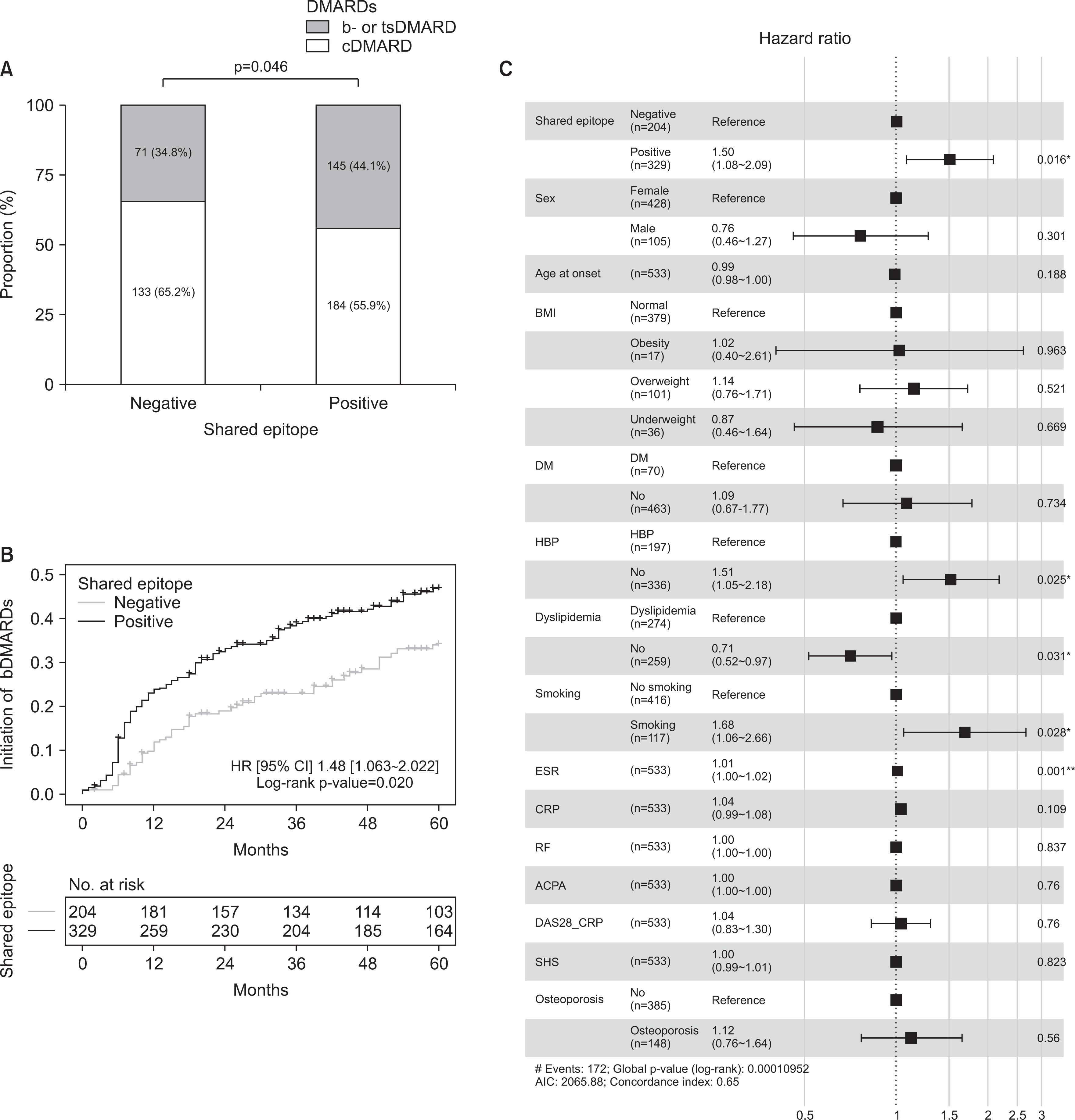 | Fig. 2(A) Proportion of b- or ts-DMARDs use with respect to the SE status. The number (percentage) is marked in the middle of each bar. (B) Cumulative incidence of the initiation of b- or ts-DMARDs based on the SE status over 5 years. (C) Forest plots showing hazard ratios of b- or tsDMARD initiation in patients with RA. The Cox proportional hazards model was used to estimate hazard ratios and associated 95% confidence intervals. ACPA: anti-citrullinated protein antibody, bDMARDs: biologic disease-modifying anti-rheumatic drugs, BMI: body mass index, cDMARDs: conventional disease-modifying anti-rheumatic drugs, CRP: C-reactive protein, DM: diabetes mellitus, ESR: erythrocyte sedimentation rate, HBP: hypertension, HR: hazard ratio, RF: rheumatoid factor, SE: shared epitope, SHS: van der Heijde modified Sharp score, tsDMARDs: targeted synthetic disease-modifying anti-rheumatic drugs. *p<0.05, **p<0.01.
|
To identify significant predictors of b- or tsDMARD initiation, clinical variables were entered inesrto the multivariable Cox proportional hazards regression model (Figure 2C). The presence of SE (HR [95% CI] 1.50 [1.08~2.09], p=0.016), smoking (HR [95% CI] 1.68 [1.06~2.66], p=0.028), dyslipidemia (HR [95% CI] 1.412 [1.031~1.934], p=0.031), and erythrocyte sedimentation rate (HR [95% CI] 1.009 [1.004~1.016], p=0.001) were significantly associated with the initiation of b- or tsDMARDs. However, ACPA and RF positivity were not independently associated.
Drug survival of abatacepts and TNF inhibitors by SE or ACPA positivity
Of the 533 patients, 59 patients received abatacept and 146 patients received TNF inhibitors as the first bDMARD. The drug survival rate of abatacept and TNF inhibitors within 24 months was shown in Figure 3A. The drug survival rate of abatacept did not differ according to the presence of SE (HR [95% CI] 1.608 [0.686~3.764], p=0.274), and this was the same for TNF inhibitors (HR [95% CI] 1.087 [0.599~1.959], p=0.784). When compared by the presence or absence of ACPA (Figure 3B), the drug survival rate of abatacept in the ACPA-positive group was higher than that in the ACPA-negative group (HR [95% CI] 2.799 [1.147~6.829], p=0.024). The drug survival rate of TNF inhibitors did not depend on the presence of ACPA (HR [95% CI] 1.879 [0.748~4.714], p=0.179). To identify significant predictors of abatacept (ABTC) survival, clinical variables were entered into the multivariable Cox proportional hazards regression model. In this model, ACPA positivity (HR [95% CI] 8.166 [1.892~35.200], p=0.005) was significantly associated with ABTC survival and the presence of SE (HR [95% CI] 3.148 [0.782~12.700], p=0.107) was not.
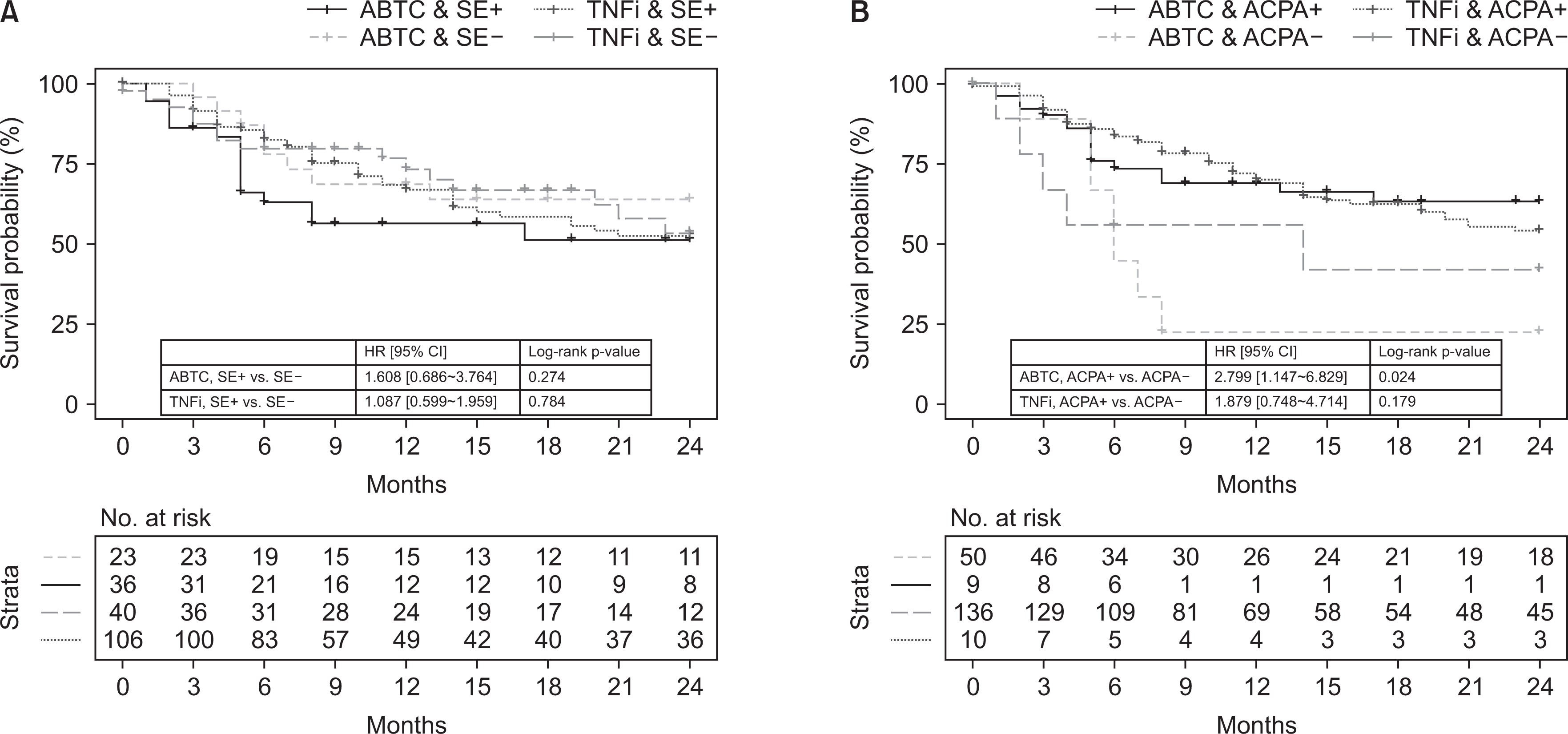 | Fig. 3The drug survival rate of TNF inhibitors and abatacept over 24 months by Kaplan–Meier estimates. (A) Plot with respect to SE status. (B) Plot with respect to ACPA status. ABTC: abatacept, ACPA: anti-citrullinated protein antibody, SE: shared epitope, TNFi: tumor necrosis factor inhibitors, HR: hazard ratio, CI: confidence interval.
|
Go to :
DISCUSSION
In this study, we investigated the relationship between SE and ACPA and their clinical implications regarding disease severity and treatment response to abatacept and TNF inhibitors in RA. Patients with SE had higher CRP levels and more erosive changes at baseline. There was also a strong trend toward ACPA positivity in SE-positive patients, particularly in smokers. Administration of b- or tsDMARDs was more frequent in SE-positive patients and SE positivity was a significant predictor for early introduction of b- or tsDMARDs. The drug survival rate of abatacept or TNF inhibitors in the 24 months did not differ according to SE positivity. However, the drug survival rate of abatacept was better in patients with ACPA than in patients without ACPA, while those of TNF inhibitors did not differ between the ACPA-positive and ACPA-negative groups.
HLA-DRB1 SE is strongly associated with the development of ACPA, particularly in the presence of smoking [5,13,14,29,32,33]. We also confirmed that the ACPA positivity rate was higher in SE-positive patients than in SE-negative patients, and this was more prominent in smokers. However, this was not statistically significant, probably owing to the small proportion of ACPA-negative patients (9.0%) in this study. HLA-DRB1 SE not only confers a higher risk for RA development, but also increases the likelihood of more severe bone erosions [3,4,35]. High acute-phase reactant levels, presence of ACPA, and early erosions were poor prognostic factors in the management of RA [26]. In our results, the SE-positive group showed higher levels of CRP, higher ACPA positivity rate, and higher frequency of erosive joint damage. These results indicate that SE-positive patients may benefit from closer monitoring and more aggressive therapy. Early administration of b- or tsDMARDs was more frequent in SE-positive patients than in SE-negative patients. The multivariate regression analysis confirmed that SE positivity was independent risk factor for the use of b- or tsDMARDs.
To improve the risk-benefit ratio and cost-effectiveness in individual patients, it is important to predict which patients are more likely to respond to a specific drug. Accordingly, the clinical and genetic characteristics of patients who respond better or worse to a particular drug should be defined. Specific alleles of SEs have be reported to be associated with treatment response to TNF inhibitors [36]. For instance, HLA-DRB1*0404 and *0101 alleles, both of which encode SE, were associated with favorable responses to etanercept at 12 months [37]. In the case of abatacept, a greater effectiveness was demonstrated in the SE-positive group was demonstrated among the Japanese population [38]. However, the association between SE and drug response was not clear in this study. The drug retention rate is a composite measure of the efficacy and safety of bDMARDs [39]. In our results, there was no significant difference in the drug survival rate of TNF inhibitors and abatacept by SE positivity. This discrepancy may arise from differences in the study design and target population. The present study analyzed the association between a set of SEs and the entire class of TNF inhibitors rather than the relationship between a specific allele and a specific drug. The ethnic and racial difference may also influence the distinct SE alleles and treatment responses [24,25]. The previous Japanese study defined seven alleles as the SE of RA [38], while 10 SE alleles were considered in the Korean RA population [29].
Although SE positivity failed to demonstrate an association with drug response, ACPA status affected by the presence of SE was related to the response to abatacept, consistent with previous reports. It has been noted that RA patients with ACPA, particularly those with a high titer of ACPA, are more likely to achieve a good response to abatacept compared to ACPA-negative patients in contrast to TNF inhibitors [19-23]. We confirmed that the drug survival rate of abatacept was better in ACPA-positive patients than in ACPA-negative patients and this difference was not observed in the TNF inhibitor-treated group. The reason for the differential pattern of response to abatacept and TNF inhibitors may be in part due to their different mechanisms of action [20]. Abatacept selectively modulates T-cell co-stimulation and autoantibody production via interaction with B cells, whereas TNF inhibitors directly bind to TNF or TNF receptors. In routine clinical practice, it is almost infeasible to check SE due to high cost out of insurance coverage. In this respect, ACPA is a good biomarker because it comprises the clinical and mechanistic significance of the SE and is available at a low cost.
This study has some limitations. First, there may be selection or attrition biases because of the inherent limitations of retrospective data collection in a single center. Second, the seronegativity of ACPA was not sufficiently leveraged in the analysis due to the small proportion of ACPA-negative patients. Third, the maximal cut-off of ACPA was set at 200 U/mL. Thus, ACPA titer levels could not be accurately compared.
Go to :
CONCLUSION
The presence of HLA-DRB1 SEs is an important genetic factor closely linked with clinical features that define a severe form of RA and a useful surrogate index for predicting the need for early aggressive therapy. Although routine inspection of HLA-DR alleles is not feasible in the current clinical practice due to high costs, it can be applied to early diagnosis and risk assumptions in future medicine. The SEs were not associated with treatment response to abatacept or TNF inhibitors in this Korean RA population, but we observed that ACPA had a predictive value for abatacept retention. In ACPA-positive patients with multiple comorbidities such as interstitial lung disease, abatacept may be a good treatment choice, considering the favorable safety profile.
Go to :
 XML Download