

 PDF
PDF Citation
 Print
Print


INTRODUCTION
Hepatocellular carcinoma (HCC) is one of the leading causes of cancer-related death.1 Surgical resection and liver transplantation are considered the treatment of choice but most patients with advanced diseases are not amendable to these surgical treatments. Locoregional treatment, such as radiofrequency ablation (RFA), transcatheter arterial chemoembolization (TACE), percutaneous ethanol injection (PEI), radioembolization, and radiotherapy, can be used in inoperable patients.
Historically, radiotherapy in HCC patients was limited because of radiation-induced liver disease (RILD). On the other hand, with the advances in radiotherapy, stereotactic body radiotherapy (SBRT) has become a new treatment option. SBRT can deliver a high dose of radiation in a few fractions while minimizing the dose to the adjacent organs. Therefore, by delivering a sufficient dose to tumors without concerns for RILD, SBRT is effective in tumor control and is used widely in the treatment of HCC.2,3
The inflammatory response is important in cancer development and progression, and the prognostic value of systemic inflammatory markers has been investigated in various cancers.4-9 In particular, the strong correlation between chronic inflammation and tumorigenesis of HCC is well established, and the prognostic role of various systemic inflammatory markers has been investigated in HCC.10,11 The neutrophil to lymphocyte ratio (NLR) and the platelet to lymphocyte ratio (PLR) are markers that are readily available from the complete blood count. On the other hand, the association between these systemic inflammatory markers and the prognosis in a HCC patient treated with SBRT is unknown. This study investigated the effect of the SBRT on NLR and PLR in HCC patients treated with SBRT and evaluated the prognostic values of NLR and PLR.
Go to : 
SUBJECTS AND METHODS
1. Patients
Between 2008 and 2019, 61 patients underwent SBRT for HCC at Soonchunhyang University Seoul Hospital. Patients who were unsuitable for surgery or RFA were referred for SBRT. Patients with 1-3 lesions, Child-Pugh class A or B, and normal liver volume (total liver volume excluding tumor volume) ≥700 mL were treated with SBRT. The patients who received SBRT with radical aim were included in this study. The exclusion criteria were as follows: 1) double primary cancer; 2) palliative SBRT; 3) distant metastasis; 4) lost to follow- up within 6 months after SBRT; 5) no laboratory test or radiologic image after SBRT. Five, eight, and eight patients with double primary cancer, lost to follow-up, and treated with a palliative aim, respectively, were excluded. One patient who had a repeated infection was eliminated from the study to exclude the compounding effect of infection on the laboratory test. Finally, 39 patients were included, and none received any further treatment for HCC after SBRT until disease progression. The medical records of the included patients were reviewed retrospectively according to the protocol approved by the Institutional Review Board (IRB) of Soonchunhyang University Seoul Hospital (IRB No. 2020-12-001). Liver cirrhosis was diagnosed with liver imaging, such as sonography, CT, or MRI.
2. Treatment
SBRT was delivered using Cyberknife® (Accuray, Inc., Sunnyvale, CA, USA). For real-time motion tracking, gold fiducial markers were inserted near the lesion under image guidance before the simulation. For the simulation, the patients were immobilized with Vac-Lok® (MEDTEC, Orange City, IA, USA) in the supine position. The CT scans were performed with a 1 mm slice thickness, and a MRI was taken on the same day. The gross tumor volume (GTV) was contoured on the simulation CT registered with the MRI using fiducial markers. The planning target volume was generated by adding a 5-7 mm margin to GTV. The prescription dose was 40-60 Gy in 3-5 fractions. The dose to the normal organs was constrained, as defined in a previous prospective study.12 The treatment was performed on consecutive days (QD) or every other day (QOD) and delivered with real-time tumor tracking using a Synchrony® (Accuray, Inc., Sunnyvale, CA, USA).
3. Follow-up and calculation of NLR and PLR
The complete blood count (CBC), including a differential count, was performed before SBRT and at every follow-up. The patients were followed up 1-2 months after SBRT and every 2-3 months thereafter, depending on the risk. The CBC counts within 2 months after SBRT was selected as the post-SBRT value. NLR and PLR were calculated by dividing the neutrophil or platelet count by the lymphocyte count. The changes in the NLR and PLR were obtained by post-SBRT value minus the pre-SBRT value.
4. Statistical analysis
The differences in the NLR and PLR values before and after SBRT were compared using a paired t-test. The receiver operating characteristic curves were generated to determine the optimal cut-off values of the NLR and PLR. The overall survival (OS) was calculated from the first day of SBRT to the date of death. The progression-free survival (PFS) was calculated from the first day of SBRT to the date of disease progression detected by radiological imaging using the Response Evaluation Criteria in Solid Tumor (RECIST) criteria. Patients lost to follow-up or who died without disease progression were considered to be censored. The actuarial survival rates were calculated using Kaplan-Meier methods, and the statistical significance was evaluated by the log-rank test. The Cox proportional hazard regression model assessed the factors affecting the OS and PFS. Factors with p-values less than 0.5 were analyzed by multivariable analysis. All statistical analyses were performed using SPSS version 18.0 (SPSS Inc., Chicago, IL, USA).
Go to :
RESULTS
1. Baseline characteristics and treatment outcome
Table 1 lists the baseline patient and tumor characteristics. The median age was 62 years (range, 37-84 years), and 29 out of 39 patients had viral hepatitis. The Child-Pugh class was A in 38 patients and B in one patient. Seven patients received upfront SBRT, and the other patients underwent a surgical resection, RFA, or TACE. Nine patients had two or more lesions, and 49 lesions were evaluated. The median tumor size was 1.5 cm (range, 0.2-3.5 cm), and the median GTV in each patient was 12.9 cc (range, 2.4-74.7 cc). The median value of pre-SBRT neutrophil, lymphocyte and platelet count were 2.8×10⁹/L (range, 0.6-5.1×10⁹/L), 1.4×10⁹/L (range, 0.3-3.2×10⁹/L) and 105×109/L (range, 35-196×109/L), respectively.
Table 1
Baseline Characteristics of Hepatocellular Carcinoma Treated with Stereotactic Body Radiotherapy
![]()
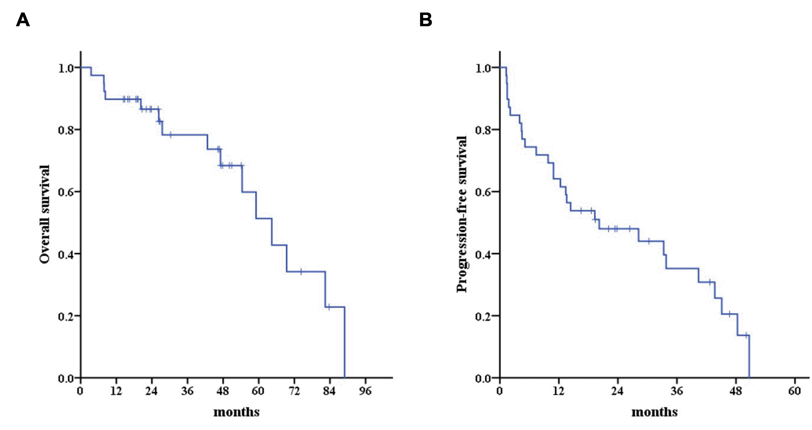
After a median follow-up of 26.8 months (range, 8.4-80.0 months), there were two local recurrences, 28 intrahepatic recurrences outside the SBRT field, and five distant metastases. All first recurrences after SBRT were intrahepatic, and the actuarial three-year local control, OS, and PFS rates were 97.4%, 78.3%, and 35.2%, respectively (Fig. 1).
2. NLR and PLR
Both NLR and PLR were significantly increased after SBRT compared to the pre-SBRT value (Fig. 2). The increase in NLR and PLR persisted until 3 months after SBRT, and statistical significances were not observed after 6 months. At the time of progression, both NLR and PLR increased again regardless of the disease-free interval after SBRT (range, 1.3-50.7 months).
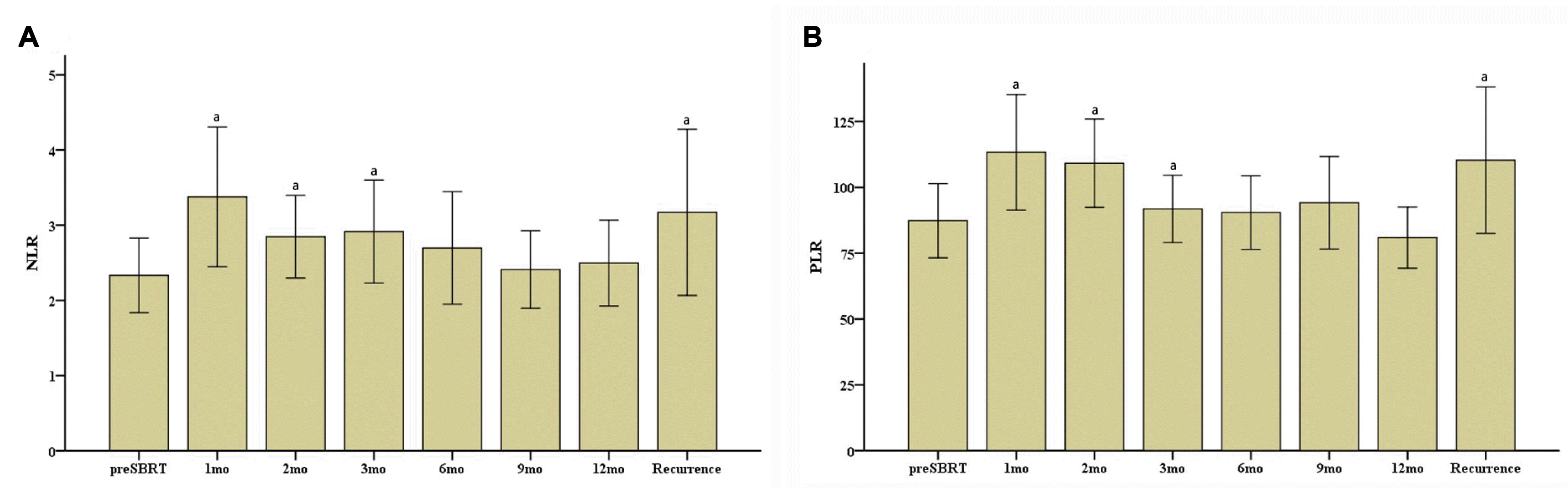
Table 2 lists the results of univariable and multivariable analysis for the OS and PFS. Among the clinical and treatment factors, only GTV was associated with OS (p=0.002). Pre-SBRT NLR, post-SBRT NLR, and change in NLR exhibited no association with OS. On the other hand, the post-SBRT PLR >90 and PLR change >30 were associated significantly with a poor OS (p=0.011 and p=0.001, respectively). Because post-SBRT PLR value and PLR change were closely correlated (Spearman’s correlation coefficient=0.692, p<0.001), only PLR change was included in multivariable analysis. As a result, the PLR change was a significant prognostic factor predicting OS (HR, 10.09; 95% CI, 1.15-88.40, p=0.037) (Fig. 3A).
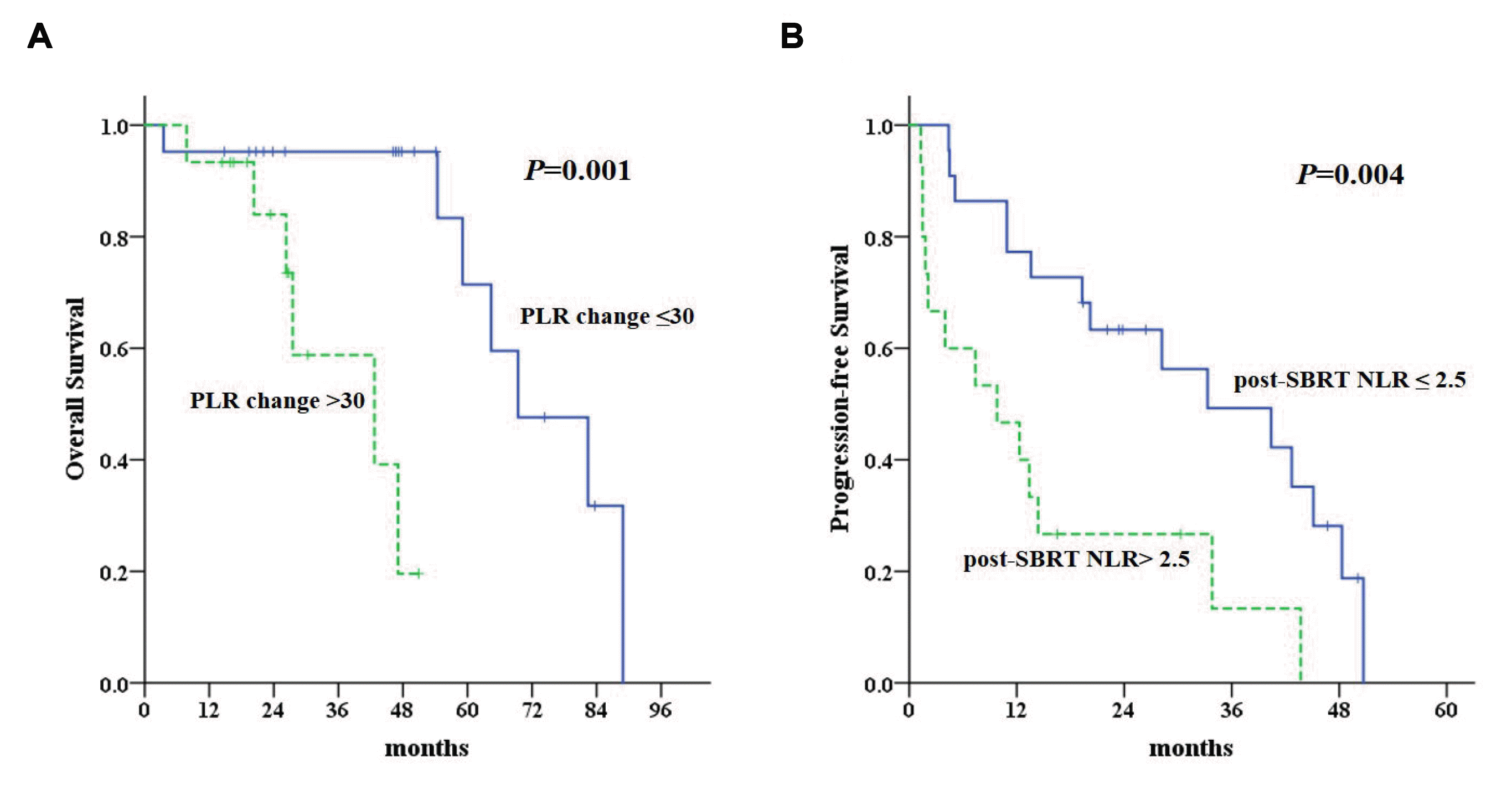 | Fig. 3Kaplan-Meier curves for the overall survival according to (A) PLR and progression-free survival according to (B) NLR. NLR, neutrophil to lymphocyte ratio; PLR, platelet to lymphocyte ratio; SBRT, stereotactic body radiotherapy.
|
Table 2
Cox Proportional Hazard Regression Analyses for Overall and Progression-free Survival
| Variable | Univariable | Multivariable | Univariable | Multivariable | ||||
|---|---|---|---|---|---|---|---|---|
|
|
|
|
|
|||||
| HR (95% CI) | p-value | HR (95% CI) | p-value | HR (95% CI) | p-value | HR (95% CI) | p-value | |
| Age ≤65 years | 1.704 (0.369-7.866) | 0.490 | 1.289 (0.540- 3.078) | 0.565 | ||||
| Male | 1.278 (0.281-5.801) | 0.750 | 1.041 (0.394-2.748) | 0.936 | ||||
| Viral hepatitis | 0.581 (0.192-1.759) | 0.331 | 1.433 (0.576-3.569) | 0.436 | ||||
| Liver cirrhosis | 22.617 (0.000-18,336,313. 720) | 0.492 | 1.722 (0.230-12.879) | 0.591 | ||||
| Portal vein thrombosis | 1.231 (0.271-5.600) | 0.788 | 2.155 (0.724-6.416) | 0.156 | ||||
| Child-Pugh Score 6 or 7a | 1.095 (0.358-3.351) | 0.874 | 1.584 (0.734-3.418) | 0.236 | ||||
| AFP >25 ng/mLb | 2.791 (0.957-8.135) | 0.500 | 2.129 (0.951-4.764) | 0.06 | ||||
| Previous treatment | 2.411 (0.530-10.958) | 0.240 | 1.150 (0.464-2.849) | 0.762 | ||||
| GTV >14 ccc | 8.059 (1.673-38.815) | 0.002 | 4.577 (0.837-25.041) | 0.079 | 2.635 (1.210-5.819) | 0.011 | 1.624 (0.696-3.791) | 0.262 |
| Pre-SBRT NLR >2.7d | 2.685 (0.929-7.761) | 0.058 | 1.418 (0.655-3.069) | 0.372 | ||||
| Post-SBRT NLR >2.5e | 2.637 (0.861-8.083) | 0.155 | 3.084 (1.384-6.872) | 0.004 | 3.084 (1.384-6.872) | 0.006 | ||
| NLR change >1.0f | 3.927 (1.032-14.952) | 0.091 | 1.988 (0.849-4.654) | 0.135 | ||||
| Pre-SBRT PLR >70g | 1.348 (0.452-4.021) | 0.591 | 1.400 (0.661-2.964) | 0.376 | 1.812 (0.787-4.173) | 0.162 | ||
| Post-SBRT PLR >90h | 9.906 (1.273-77.098) | 0.011 | 2.212 (0.986-4.960) | 0.041 | ||||
| PLR change >30i | 17.881 (2.113-151.293) | 0.001 | 10.917 (1.274 -93.512) | 0.029 | 2.074 (0.916-4.696) | 0.079 | ||
![]()
Univariable analysis for PFS, identified GTV, post-SBRT NLR, and post-SBRT PLR as statistically significant prognostic factors (p=0.011, p=0.004 and p=0.041, respectively). Among these factors, only post-SBRT NLR >2.5 remained significant on the multivariable analysis for PFS (HR, 2.44; 95% CI, 1.03-5.76; p=0.042) (Fig. 3B).
Go to :
DISCUSSION
Increasing evidence has shown that systemic inflammation and the host immune status are closely associated with the prognosis of cancer patients, and strategies to modulate the host immunity are being actively investigated.13,14 Although the overall mechanisms are not completely understood, radiotherapy is believed to have an ambivalent role in the immune system, exerting both anti-tumor immunogenic and immune- suppressive effects.15 SBRT, which delivers a very high radiation dose in a few fractions, showed higher efficacy in cancer treatment than expected, which is not fully explained by the biological model of conventional radiotherapy. The high efficacy of SBRT is believed to stem from the more pronounced effect on the immune system than conventional radiotherapy. The SBRT induces immunogenic cell death, such as necrosis and senescence, recruits immune cells to the tumor microenvironment, and increases the secretion of cytokines and chemokines that stimulate immune cells.16 On the other hand, most of these were evaluated at the preclinical level, and the overall clinical effects were not clearly elucidated.
The effects of SBRT on immune cells were examined by evaluating the change in the NLR and PLR after SBRT for HCC. In the present results, both NLR and PLR increased significantly after SBRT. Tajiri et al.17 reported a transient increase in NLR 1 week after RFA in HCC patients and suggested it as a treatment effect. In the present study, the patients were not given any further treatment after SBRT before disease progression. Therefore, the changes in the NLR and PLR are considered the sole effect of SBRT. Chew et al.18 reported that the local and systemic immune systems were activated after 90Y radioembolization, which was associated with a sustained tumor response. The underlying mechanism of the increased NLR and PLR after SBRT is unclear. The effect of SBRT on the immune system was suggested to be different from that of conventional radiotherapy. Although conventional radiotherapy induces apoptosis predominantly, SBRT can induce necrosis, which is considered inflammatory cell death.16,19,20 As a result, various cytokines and chemokines are increased, and immune cells are activated. Therefore, neutrophils and platelets could be increased due to the enhanced immune systems. The increased NLR and PLR after SBRT in the present study indicate the immune-modulatory effects of SBRT in a clinical setting.
Pre-SBRT NLR and PLR were not associated with the treatment outcome in the present results. Previous studies evaluating the outcome of various local treatments, such as surgical resection, RFA, and TACE, have reported the prognostic value of pretreatment NLR and PLR.21-24 On the other hand, the results of previous studies are heterogeneous, and there is no consensus. In the present study, SBRT was delivered in relatively well-selected patients with good liver functions, and patients treated for palliation were excluded. Therefore, the baseline inflammatory status of each patient might not have been different, and the pretreatment NLR and PLR could not be predictive of the prognosis.
The large increase in PLR after SBRT and the high post-SBRT NLR values predicted a poorer treatment outcome. Zhuang et al.25 reported similar results. They showed that a high post-treatment PLR and a large increase in NLR were associated with inferior outcomes in HCC patients treated with SBRT and suggested NLR and PLR as prognostic biomarkers for HCC patients. The association between the NLR change and the prognosis of patients has been reported in studies investigating other treatment modalities for HCC. Peng et al.26 reported that an increase in the NLR after a resection was predictive of a poor outcome in HCC patients treated with a curative resection. Dan et al.27 also reported that a decreased NLR after RFA was associated with a better survival in HCC patients. Recently, the predictive value of NLR and PLR were verified in advanced HCC patients treated with anti- PD-1 therapy.28 An elevated post-treatment NLR and PLR were predictive of poor survival and associated with a poor response to anti-PD-1 therapy. Therefore, this result also shows the role of NLR and PLR as prognostic factors after SBRT in HCC patients. Furthermore, both the NLR and PLR increased at the time of progression. Tajiri et al.17 also found that NLR persisted at a high level in patients with recurrence after RFA for HCC. Both studies suggest that an increase in NLR and PLR after treatment for HCC is associated with recurrence, and NLR and PLR could also be used as markers for monitoring the recurrence of HCC.
The underlying molecular mechanism of the association of the NLR and PLR with prognosis has been studied, but it is not completely understood.29,30 On the other hand, increasing evidence suggests that systemic inflammation is associated with tumor development and progression and neutrophils and platelets are crucial components of inflammation. Neutrophils are stimulated to produce various pro-angiogenic molecules and growth factors that promote tumor progression and metastasis.31 Platelets produce growth factors facilitating angiogenesis and adhesion molecules promoting tumor cell migration and metastasis.32 Therefore, increased NLR and PLR could be reflective of the systemic inflammatory status of the patients and associated with the prognosis.
Recently, immunotherapy is used widely in various cancers and actively investigated in HCC, but the efficacy is not conclusive.33 In the present study, the systemic immune-modulatory effect of SBRT was suggested by the increased NLR and PLR after treatment. Therefore, there is a possibility that SBRT could improve the efficacy of immunotherapy safely by adding a synergistic role in potentiating tumor immunity. Moreover, a subset of patients with a poor prognosis can be identified in advance using the changes in NLR and PLR after SBRT. Earlier administration of immunotherapy could improve the treatment outcome of these patients.
This study had some limitations. Because of the retrospective design, the follow-up duration of the patients was not homogeneous. As NLR and PLR were changed continuously after SBRT, small differences in the timing of blood collection could have underestimated the effects of SBRT. Furthermore, PLR could be confounded by underlying cirrhosis because the platelet count was lower in patients with liver cirrhosis. In the present study, patients had relatively good liver functions, and the effects of cirrhosis were similar in these patients. Despite these limitations, the result identified the potential effect of SBRT on the systemic immune system and the prognostic role of NLR and PLR in HCC treated with SBRT in a relatively homogeneous patient group. Further prospective studies involving a larger number of patients will be needed to define better the immune-modulatory effect of SBRT in HCC.
Go to :
 XML Download