Journal List > J Korean Foot Ankle Soc > v.26(2) > 1516078700

후과 골절은 족관절 골절에서 어떤 의미를 갖는가?

Abstract

The posterior malleolar fracture is relatively common fracture of the foot and ankle, but several aspects of this are still controversial. If the posterior malleolus is involved in the ankle fracture, the prognosis is usually poor. A computed tomography scan is essential for accurate diagnosis and treatment planning. Although indirect reduction and the anterior to posterior screw fixation technique have the advantages of a small incision with the requirement of relatively simple skills, direct open reduction and fixation from the posterior side provide a more biomechanically stable and accurate reduction. The precise reduction of the posterior malleolar fragment helps to achieve congruency of the tibia and fibula in the incisura and contributes to syndesmotic stability. It is important to determine the indications for surgical treatment by comprehensively evaluating the three-dimensional structure of the posterior malleolar fracture and all related injuries to the ankle.

서 론

족관절 삼과 골절은 정형외과 영역에서 비교적 흔하게 발생하는 외상이다. 후과 골절이 있는 경우 관절의 불안정성이 더 유발되며 관절면의 정확한 정복에 어려움을 주는 경우가 많다. 이러한 이유들로 인해 삼과 골절이 양과 골절보다 예후가 좋지 않다고 알려져 왔다.1-4) 이에 본 종설에서는 후과 골절이 족관절 골절에서 어떤 의미를 갖는지 그 역사 및 진단, 치료 방법, 예후 등에 대해 알아보고자 한다.

본 론

1. 역사

족관절 골절에서 원위 경골 후방 부위의 골절은 역사적으로 정형외과 및 외상 영역에서 꾸준한 관심의 대상이었다. 1828년 Earle5)이 족관절 골절 중 원위 경골의 후방 가장자리 골절에 대해서 최초로 기술한 것으로 알려져 있다. 그 이후 독일의 문헌들에서 이러한 골절편을 ‘Volkmannsches Dreieck (Volkmann 삼각형)’으로 기술하였고6) 이 용어는 최근까지도 후과 골절편을 지칭하는 용어로 종종 사용되었다. 그러나 사실 1875년에 출판된 von Volkmann7)의 원래의 설명과 그림은 원위 경골의 전외측 부분의 건열 골절을 묘사한 것이었다. 이후 Destot8)가 1911년 처음으로 과(malleolus)라는 표현을 원위 경골 후방에 사용하였으며 1932년 Henderson9)이 삼과 골절(trimalleolar fracture)이라는 용어를 도입하였다. 이후 1940년 Nelson과 Jensen10)이 후과 골절을 관절 표면의 1/3 이상을 침범한 경우와 1/3 미만을 침범한 경우로 분류하여 전자의 경우에는 후내측 접근으로 나사 고정을 권유하였고 이 방법은 현재까지도 때때로 사용되고 있다. 이후로도 후과 골절에 대한 관심은 점점 증가하고 있으나 최선의 치료법에 대한 합의는 아직 명확히 이루어져 있지 않다. 최근 컴퓨터 단층촬영(computed tomography, CT) 영상의 사용 빈도가 높아지면서 골절 유형에 대한 이해도가 더 높아지고 있고 이에 따른 개별화된 치료 접근 방식이 제시되고 있다.

2. 진단

기본적인 방사선 검사로 족관절 전후면, 측면, 격자면(mortise) 방사선 촬영을 시행한다. 그러나 이러한 단순 방사선 촬영으로는 후과 골절편의 크기 및 양상을 판단하기에는 어려움이 있다. Ferries 등11)이 삼과 골절 환자의 단순 방사선 촬영과 CT를 통하여 후과 골절편의 크기를 측정한 것을 서로 비교한 논문에서 단순 방사선 촬영을 측정한 경우가 매우 낮은 관찰자 간 및 관찰자 내부 신뢰도를 나타냈고, 단순 방사선 촬영 중 54%가 CT 측정과 비교하였을 때 25% 이상의 측정값 오류를 나타냈다고 보고하였다.
삼과 골절의 진단 및 치료 방침 결정에서 CT 촬영의 중요성은 점점 더 강조되고 있고, 현재는 CT 촬영의 필요성에 대해 어느 정도 합의가 이루어져 있다고 볼 수 있다. 후과 골절편의 크기 측정과 그 골절 형태, 후외측 골편인지 내과로 이어지는 골편인지, 후과 골절편과 경골 사이에 작은 골절편이 감입되어 있는지 등을 확인하기 위해서는 CT 촬영이 불가피하다고 할 수 있겠다.12-16)

3. 분류

1987년 제시된 단순 방사선 촬영을 이용한 분류법에서 후과 골절을 (1) 관절 외 골절, (2) 관절을 침범한 작은 골절편, (3) 관절을 침범한 큰 골절편으로 나누었다.17) 이후 1989년 Heim18)이 이 분류를 좀 더 세분화하여 5가지로 분류하였으며 이러한 분류들은 CT를 통한 후과 골절에 대한 분석이 보편화되기 전까지 사용되어 왔다.
2006년 Haraguchi 등19)이 CT를 기반으로 한 후과 골절의 분류를 최초로 제시하였고 이 분류는 다음과 같다(Fig. 1).
(1) 후외측 사선 유형(posterolateral-oblique type): 원위 경골의 후외측 부분에서 분리된 삼각형 모양의 골절편
(2) 내측 확장 유형(medial-extension type): 내과의 후방 부분으로 확장된 양상의 골절편
(3) 작은 껍질 유형(small-shell type): 후과의 피질골을 포함하는 하나 또는 여러 개의 얇은 껍질 모양의 골절편
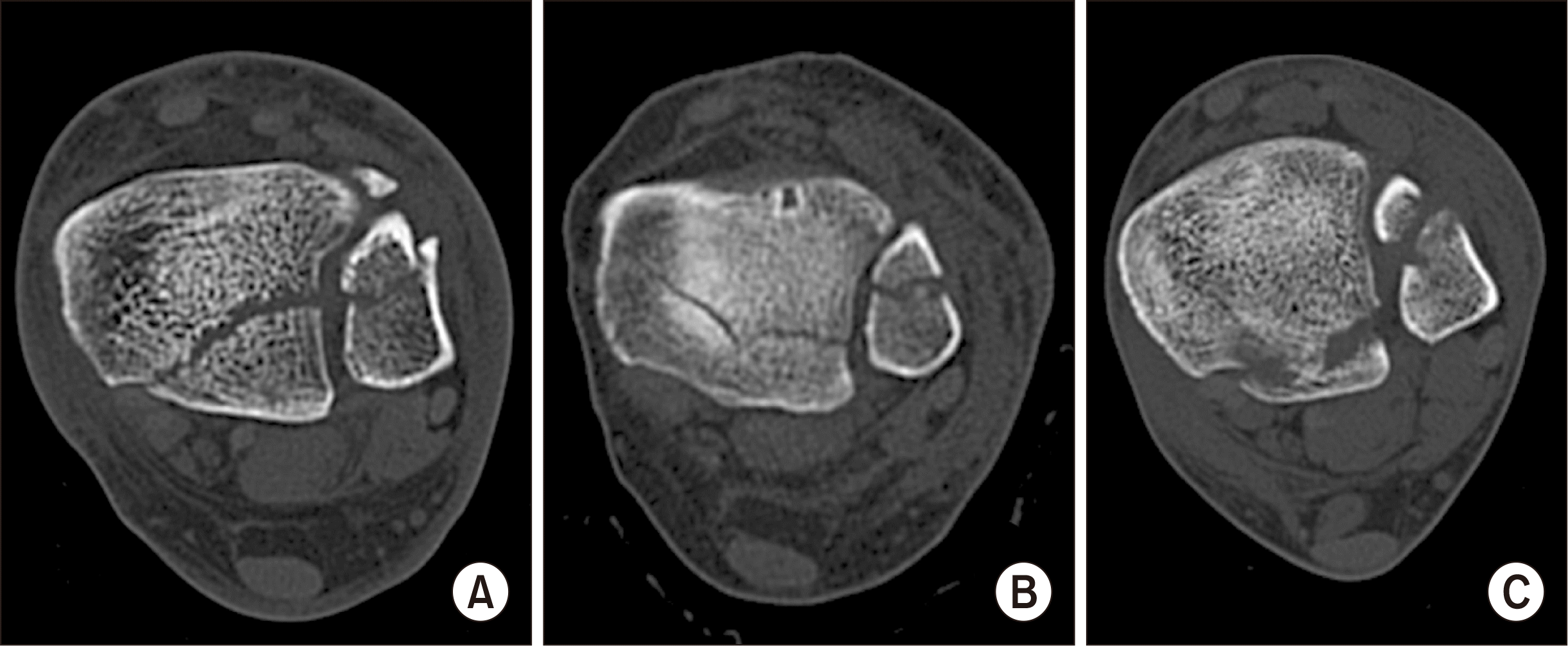
Haraguchi 등19)이 제시한 위와 같은 분류법은 수평면(axial) CT 영상을 기반으로 한 것이고 2015년 Bartoníček 등13)은 3D CT를 이용하여 시상면, 관상면, 수평면 영상에서 분석한 분류 방법을 제시하였고 이는 아래와 같다(Fig. 2).
(1) 절흔 외 골절편(extraincisural fragment)
(2) 후외측 골절편(posterolateral fragment)
(3) 후내측 2 부분 골절편(posteromedial two-part fragment)
(4) 큰 후외측 삼각형 모양 골절편(large posterolateral triangular fragment)
(5) 불규칙한 골다공증성 골절(irregular osteoporotic fracture)

4. 후방 테두리 골절(posterior rim fracture)과 후방 천정 골절(posterior pilon fracture)

후과 골절에서 후방 테두리 골절과 후방 천정 골절의 형태학적 구별 기준은 명확하지 않으나 제시된 바는 다음과 같다. 몇몇 논문에서 큰 후과 골절편이나 내측으로 연장된 후과 골절 등을 후방 전정 골절 또는 저에너지 천정 골절 등으로 묘사하였다.20-22) Haraguchi 등19)은 후과 골절과 후방 천정 골절을 구별하는 데에 경과선(transmalleolar line)을 이용하였고 Bartoníček 등13,23)은 후과 골편이 전방 둔덕(anterior colliculus)을 침범하거나 절흔(incisura)의 50% 이상을 침범할 때 부분적인 천정 골절로 분류하였다.
종합하면, 족관절 골절 및 탈구에서 인장력, 압박력, 전단력 등이 동반되나 크기가 작고 전방 둔덕 또는 비골 절흔을 거의 침범하지 않은 경우를 후방 테두리 골절에 가깝다고 볼 수 있겠고, 주로 큰 압박력에 의하여 발생하였으며 전방 둔덕을 침범하였거나 비골 절흔 절반 이상을 침범하였을 때 후방 천정 골절로 분류할 수 있겠다.

5. 수술적 치료 결정의 기준

후과 골절편의 크기가 큰 경우 수술적 치료를 하지 않았을 때 예후가 좋지 않다는 것이 알려지면서 수술적 치료를 결정함에 있어 후과 골절편이 족관절 일반 방사선 촬영 측면상에서 관절면의 25% 또는 33% 이상을 침범하였는지, 지속적인 족관절의 아탈구가 발생하는지, 관절면의 층 형성(articular step-off)이 있는지 등을 방사선적 기준으로 삼는 방법이 오랜 시간 통용돼 왔다.1,24-28) 그러나 CT를 통한 3차원적 분석이 활성화되면서 이제는 점차 다른 기준들이 강조되고 있다.
Blom 등29)은 2019년에 73명의 후과를 침범한 족관절 골절 환자를 대상으로 Haraguchi 분류법을 이용하여 유형 1, 2, 3으로 분류해 그 예후를 분석하였고 Haraguchi 유형 2 골절이 가장 예후가 나쁘다고 보고하였다. 2020년에는 다른 논문에서 회전 유형(rotational type)의 후과 골절 환자 70명을 대상으로 역시 Haraguchi 분류법을 이용하여 분석하였고 후과 골절 치료 후 임상적 예후는 골절의 형태, 치료 후 잔존하는 관절 내 간격 등과 관계가 있으며 골절편의 크기와는 관계가 없다고 보고하였다.30) 그 외 여러 연구들에서도 골절편의 크기 외에 경비인대결합(syndesmosis)의 불안정성, 골편 사이의 관절면에 감입된 작은 골편 여부, 골절의 양상이 절흔 또는 내측으로 연장되었는지 등이 수술적 치료 및 예후를 결정하는 데 중요하다고 제시하였다.20,22,26,31,32)
종합하면, 수술적 치료를 결정함에 있어 단순 방사선 측면 촬영에서 후과 골편이 25% 또는 33% 이상 관절면을 침범하였는지 보다는 CT를 통해 골절의 양상을 확인하고 경비인대결합의 불안정성이 의심되거나 관절면의 불일치(incongruity)가 있는 경우 등에서 이를 회복시키는 것을 수술의 목표로 정하고 수술을 시행하는 것이 필요하겠다.23)

6. 수술 방법

1) 전방에서 후방으로의 나사 고정(anterior to posterior screw fixation)

후과 골절의 고정 방법 중 전방에서 후방으로의 나사 고정은 오랜 세월에 걸쳐 많이 사용되어 온 방법이다.15,24,25) 이는 앙와위(supine position)에서 작은 절개를 통해 간접적인 정복으로 수행 가능하다는 장점이 있으며 때때로 측와위에서도 수행이 가능하다. 골절편의 정복은 족관절의 배굴과 척굴 움직임을 통한 조작과 정복용 집게(reduction clamp) 등을 이용하여 시행하게 되며 유관 나사(cannulated screw)나 부분 나사산 나사(partially threaded screw)를 통해 고정하는 경우가 일반적이다. 비교적 술기가 단순하고 수술 시간이 짧다는 장점이 있으나 골절의 양상에 따라 골절편의 정복이 쉽지 않은 경우가 많고 골절편이 어느 정도 큰 경우에만 사용할 수 있다는 단점이 있다. 또한 관절 내에 부유 골절편이 있거나 감입된 골절편이 있는 경우에는 이러한 골절편의 제거 또는 조작이 사실상 불가능하다.

2) 후외측 접근법(posterolateral approach)을 통한 고정

후외측 접근을 통한 후과 골절의 직접적 정복 및 고정은 널리 사용되고 있는 방법이다.20,26,32,33) 복와위(prone position) 또는 측와위로 시행하게 되며, 골절면을 직접 확인하고 정복을 시행할 수 있고, 골절편을 조정하기에 용이하며 관절 내에 감입된 골편을 제거하기 쉬운 방법이다. 고정은 골절편의 크기 및 골절 양상에 따라 나사 고정 또는 금속판 고정 모두 가능하다. Verhage 등26)이 2018년 발표한 체계적 문헌 고찰(systematic review) 논문에서 경피적 전방에서 후방으로의 나사 고정 방법보다 후외측 접근법으로 관혈적 정복 및 내고정술을 시행하는 방법이 더 우월한 방사선학적, 기능적 결과를 보이는 것으로 보고하였다. 다만 이러한 결론에 근거를 더하기 위해서는 전향적 비교 연구들이 추가로 더 필요할 것이라고 언급하였다.
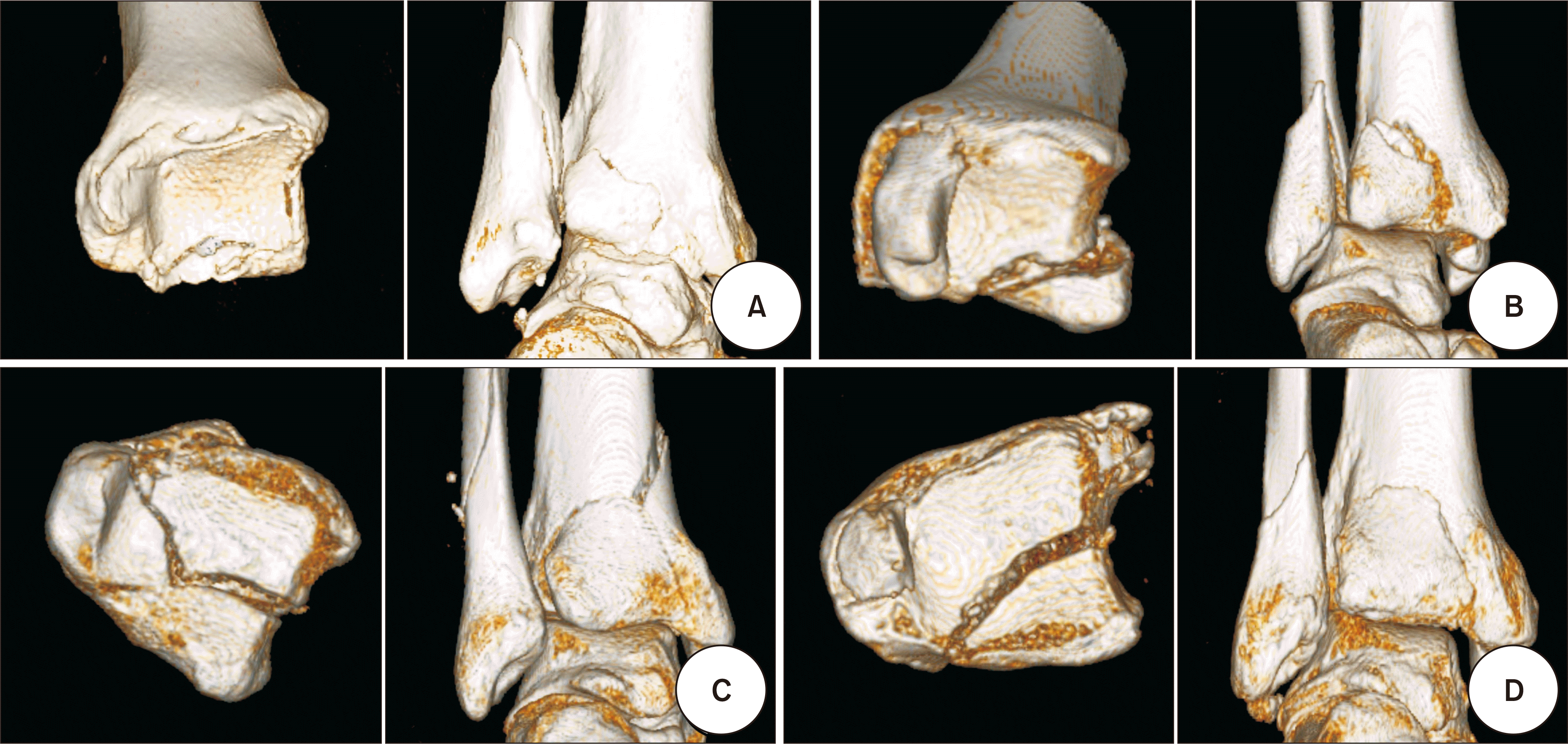
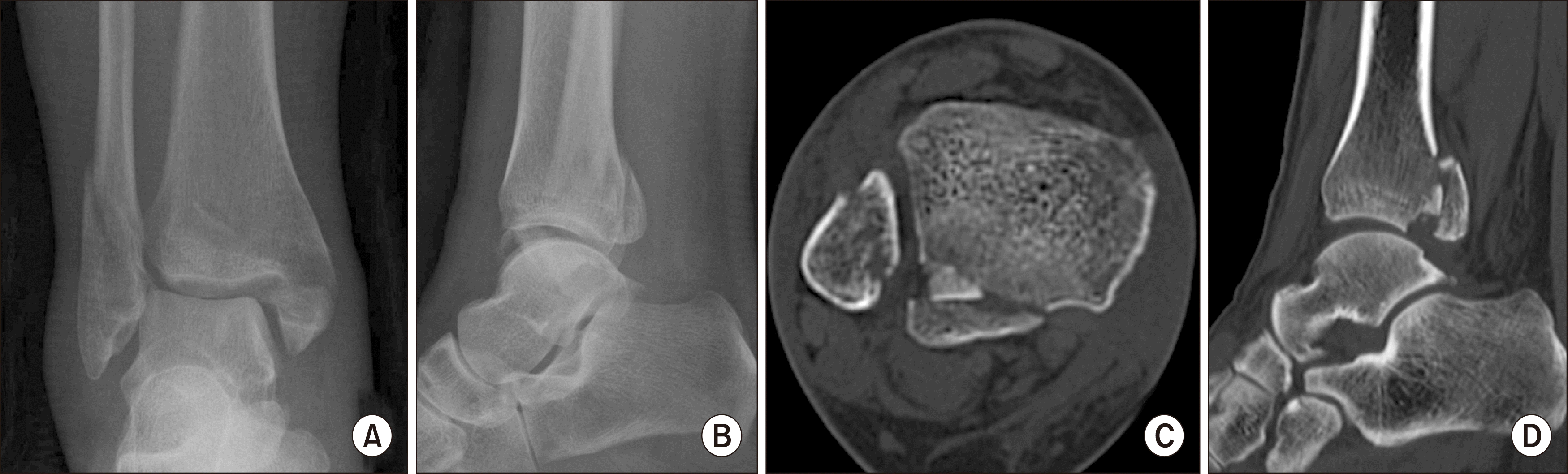
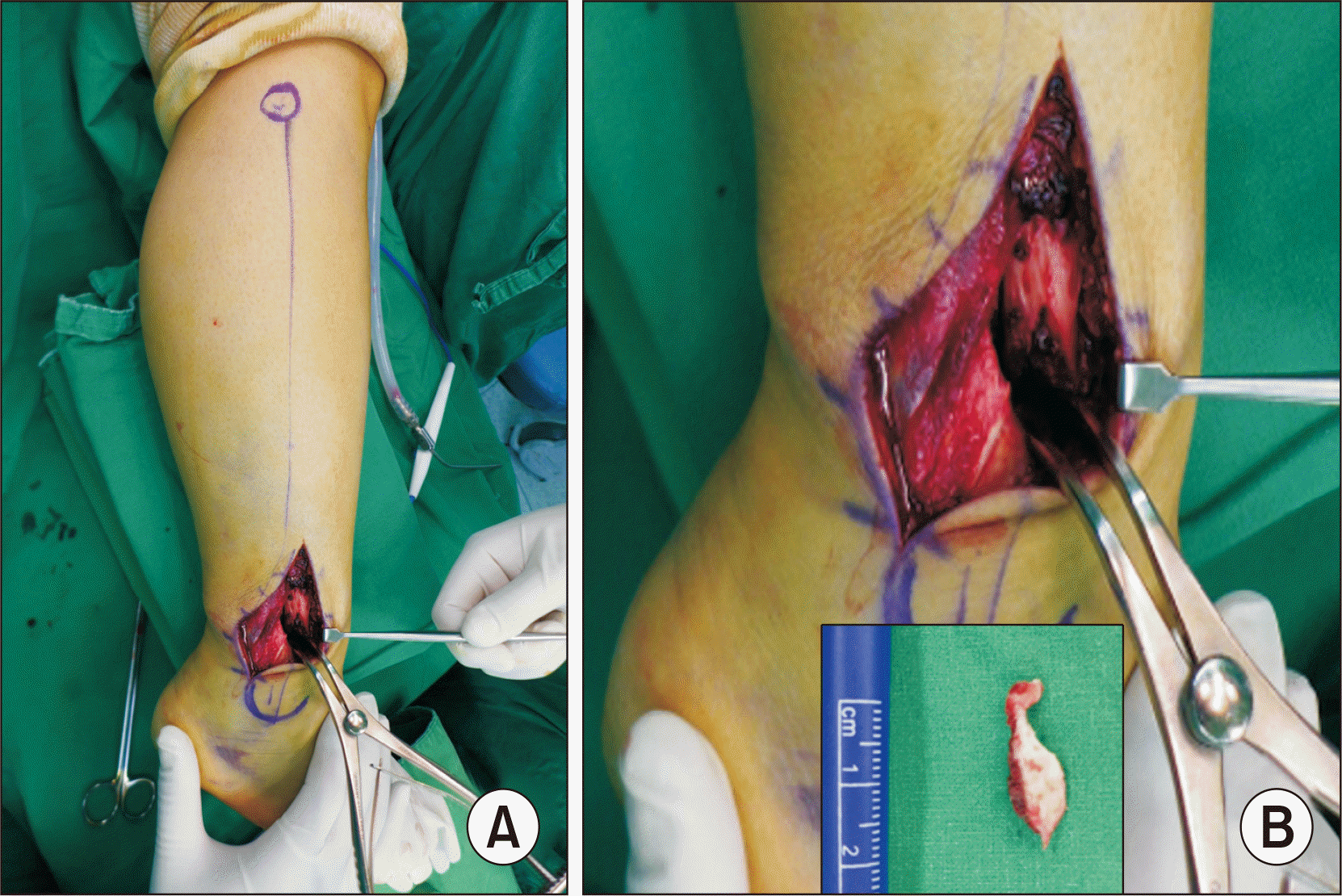
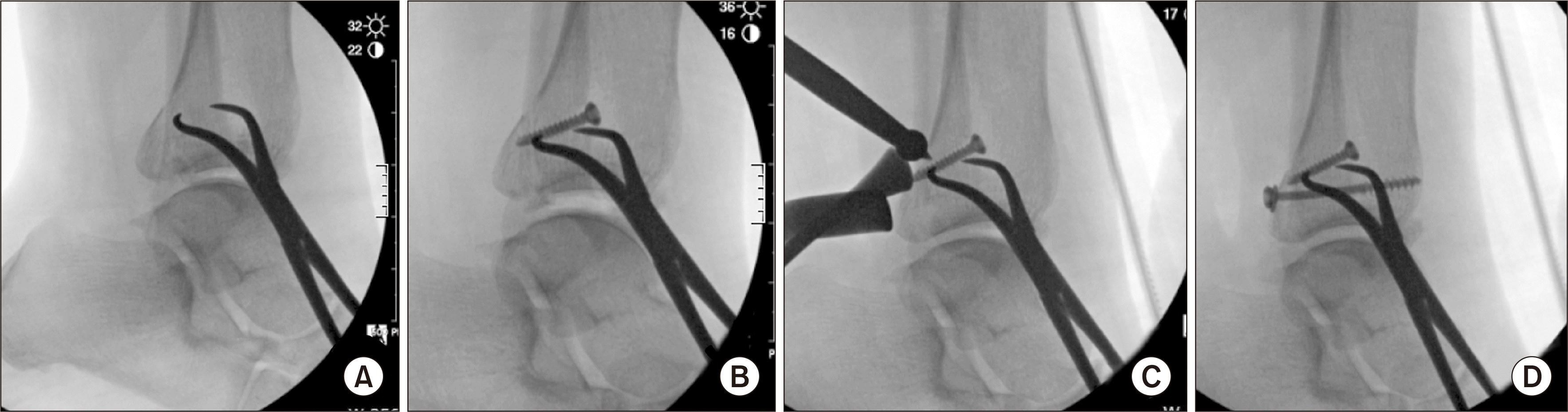
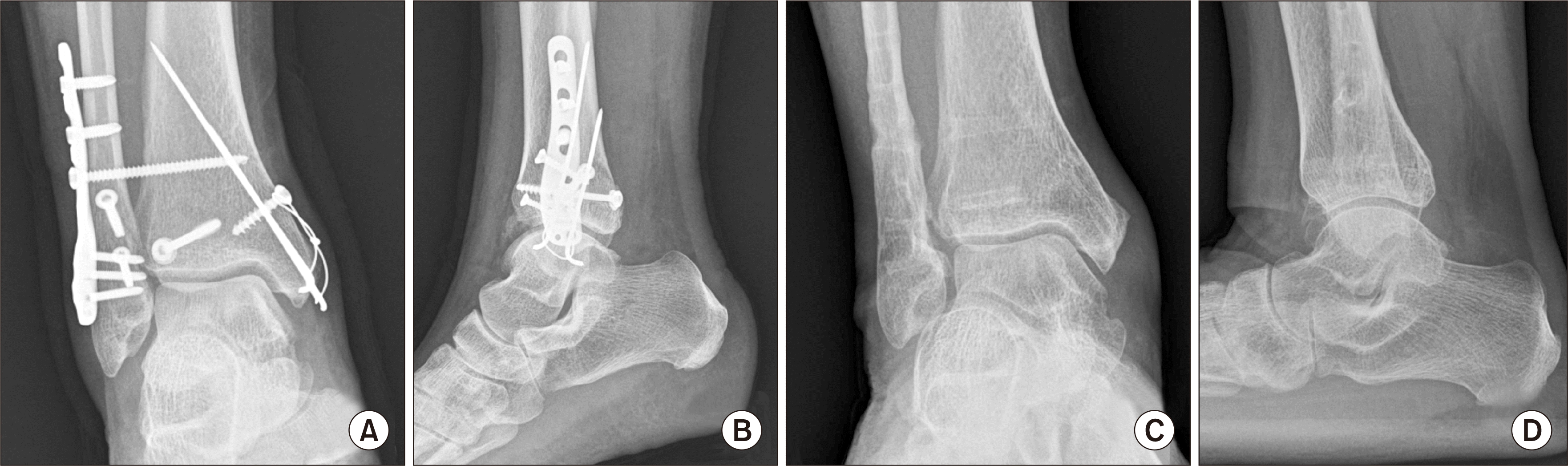
Weber B 유형의 삼과 골절(Fig. 3)에서 저자가 선호하는 접근법은 다음과 같다. 환자 자세는 측와위로 시행하며 비골의 약간 후방을 따라 종절개 또는 회전 종절개를 하여 비골 골절부를 노출시킨 후 비골 골절 부위를 라미나 스프레더(lamina spreader) 등으로 벌리면 후과 골절면을 확인할 수 있다(Fig. 4A). 골절편 사이의 혈종 등을 제거하고 관절 내 골절편 등이 있는 경우에도 비교적 쉽게 조작 및 제거할 수 있다(Fig. 4B). 이후에 먼저 비골 골절 부위를 지연 나사(lag screw)로 고정한다. 경우에 따라 나사를 삽입하지 않고 정복용 집게로 고정을 유지만 한 상태에서 진행하기도 한다. 후과 골절의 고정을 위해 비골근(peroneal muscles)과 장무지굴곡근(flexor hallucis longus muscle) 사이로 접근하여 후과 골절편의 후방면에 도달하게 되고, C-arm으로 족관절 측면상을 확인하며 ball spike pusher 등의 기구로 정복을 유지한 상태로 부분 나사산 나사를 이용하여 고정을 시행하게 된다(Fig. 5). 이후 비골에 금속판을 적용하여 비골 골절에 대한 술기를 마무리하고 내과 골절의 고정이 추가로 필요할 경우 무균 드랩을 유지한 상태로 환자 자세를 앙와위로 변경하여 내과 골절에 대한 술기를 시행한다. 수술 직후 및 1년 시점 방사선 촬영에서 관절면이 잘 보존되어 있는 것을 확인할 수 있다(Fig. 6).

3) 후내측 접근법(posteromedial approach)을 통한 고정

후내측 접근법은 내과 및 내과로 연장된 후과 골절의 접근에 용이한 방법이다.34-36) Haraguchi 분류의 유형 2 골절 등에서 후내측 접근법을 이용하면 후외측 접근법보다 골절 부위 도달 및 고정에 더 용이하다. 일반적으로 복와위에서 시행하며 경우에 따라 앙와위에서도 접근 가능하다. 내과와 아킬레스건 사이로 접근하여 신경혈관 구조물을 노출시킨 후 보호하며 수술이 진행되게 된다. 고정 방법은 후외측 접근법과 마찬가지로 골절 양상에 따라 나사 고정 또는 금속판 고정을 이용할 수 있다. Zhong 등36)은 삼과 골절 환자 48명을 대상으로 후내측 접근법과 후외측 접근법을 사용한 군으로 나누어 서로 비교하였고 임상적 및 방사선적 결과에서 두 군 간에 차이가 없었다고 보고하였다.

7. 예후

족관절 골절에서 후과 골절이 있는 경우가 후과 골절이 없는 경우에 비해 예후가 좋지 않다는 것은 여러 연구에서 알려져 왔다. Jaskulka 등2)은 142명의 족관절 골절 환자 중 후과를 침범한 군과 침범하지 않은 군으로 나누어 분석한 5년 이상의 장기 추시 연구에서 후과를 침범한 경우가 기능적 예후가 더 좋지 않았다고 보고하였고, Tejwani 등37)도 후과를 침범한 족관절 골절 54명과 후과를 침범하지 않은 255명으로 나누어 분석한 결과 후과를 침범한 군이 기능 장애 및 통증 관련 결과가 더 나쁜 것으로 보고하였다.
기능적 예후뿐만 아니라 방사선적으로 후외상성 관절염의 위험도 더 증가시키는 것으로 알려져 있다. 321명의 환자를 대상으로 한 Lindsjö4)의 전향적 연구에서 후과의 상당 부분을 침범한 삼과 골절이 그렇지 않은 경우보다 외상 후 관절염의 발병률이 더 높은 것으로 나타났다.
후과 골절편의 크기에 따른 예후에 대한 보고는 다양하다. 후과 골편이 클수록 기능적 예후 및 방사선적 예후가 좋지 않다는 보고들도 많은 반면에1,2,4,16,38) 골편의 크기는 예후와 관계가 없다는 보고들도 다수 있다.39-41)
최근 여러 논문들에서 강조되는 것은 삼과 골절에서 경비인대결합의 불안정성(syndesmotic instability)이 있을 경우 원위 비골의 절흔으로의 정확한 정복이 중요하다는 것이며23,31,42,43) 또 하나 중요한 인자로 강조되고 있는 것은 수술 후 관절면의 층 형성이 2 mm 이상인 경우 예후에 안 좋은 영향을 미치고 후외상성 관절염의 빈도를 증가시킬 가능성이 있다는 것이다.16,39,44-46)

결 론

족관절 삼과 골절에서 후과 골절은 단순히 양과 골절에 하나가 더해진 것이 아니라 족관절 격자(ankle mortise)를 이루며 족관절 전체 안정성에 중요한 역할을 하는 부위의 손상임을 이해하는 것이 중요하다. 후과 골절의 진단 및 치료 방침 결정에 있어서 전통적인 방법인 측면 방사선 촬영으로 후과 골편이 관절면의 25% 또는 33% 이상을 차지하는지를 확인하는 것은 갈수록 그 중요성이 떨어지고 있다. CT 촬영을 통해 후과 골절의 양상을 3차원적으로 확인하는 것이 필수적이며 골절편의 크기보다는 골절의 내측으로의 연장 여부나 관절의 불안정성 평가, 관절 내 부유 골절편 제거 및 정복 후 관절면의 층 형성을 줄이기 위한 노력이 중요하다고 할 수 있겠다.

Notes

Financial support

None.

Conflict of interest

None.

REFERENCES

1. Heim D, Niederhauser K, Simbrey N. 2010; The Volkmann dogma: a retrospective, long-term, single-center study. Eur J Trauma Emerg Surg. 36:515–9. doi: 10.1007/s00068-010-0061-6. DOI: 10.1007/s00068-010-0061-6. PMID: 26816305.
crossref
2. Jaskulka RA, Ittner G, Schedl R. 1989; Fractures of the posterior tibial margin: their role in the prognosis of malleolar fractures. J Trauma. 29:1565–70. doi: 10.1097/00005373-198911000-00018. DOI: 10.1097/00005373-198911000-00018. PMID: 2585569.
3. Broos PL, Bisschop AP. 1991; Operative treatment of ankle fractures in adults: correlation between types of fracture and final results. Injury. 22:403–6. doi: 10.1016/0020-1383(91)90106-o. DOI: 10.1016/0020-1383(91)90106-o. PMID: 1806506.
crossref
4. Lindsjö U. Operative treatment of ankle fracture-dislocations. A follow-up study of 306/321 consecutive cases. Clin Orthop Relat Res. 1985; (199):28–38. doi: 10.1097/00003086-198510000-00005. DOI: 10.1097/00003086-198510000-00005. PMID: 3930122.
5. Earle H. 1828; Simple, succeeded by compound dislocation forwards, of the inferior extremity of the tibia, with fracture of its posterior edge, comminuted fracture of the fibula, amputation of the leg, and death. Lancet. 29:346–8.
6. Ludloff K. 1926; [Zur Frage der Knöchelbrüche mit Heraussprengung eines hinteren Volkmannschen Dreiecks]. Zentralbl Chir. 53:390–1. German.
7. von Volkmann R. [Beiträge zur Chirurgie: anschliessend an einen Bericht über die Thätigkeit der chirurgischen Universitäts-klinik zu Halle im Jahre 1873]. Breitkopf und Härtel;Leipzig: 1875. German.
8. Destot E. [Traumatismes du pied et rayons x: malléoles, astragale, calcanéum, avant-pied]. Masson;Paris: 1911. French.
9. Henderson M. 1932; Trimalleolar fracture of the ankle. Surg Clin North Am. 12:867–72.
10. Nelson M, Jensen N. 1940; The treatment of trimalleolar fractures of the ankle. Surg Gynecol Obstet. 71:509–14.
11. Ferries JS, DeCoster TA, Firoozbakhsh KK, Garcia JF, Miller RA. 1994; Plain radiographic interpretation in trimalleolar ankle fractures poorly assesses posterior fragment size. J Orthop Trauma. 8:328–31. doi: 10.1097/00005131-199408000-00009. DOI: 10.1097/00005131-199408000-00009. PMID: 7965295.
crossref
12. Bartoníček J, Rammelt S, Tuček M, Naňka O. 2015; Posterior malleolar fractures of the ankle. Eur J Trauma Emerg Surg. 41:587–600. doi: 10.1007/s00068-015-0560-6. DOI: 10.1007/s00068-015-0560-6. PMID: 26253884.
crossref
13. Bartoníček J, Rammelt S, Kostlivý K, Vaněček V, Klika D, Trešl I. 2015; Anatomy and classification of the posterior tibial fragment in ankle fractures. Arch Orthop Trauma Surg. 135:505–16. doi: 10.1007/s00402-015-2171-4. DOI: 10.1007/s00402-015-2171-4. PMID: 25708027.
crossref
14. Büchler L, Tannast M, Bonel HM, Weber M. 2009; Reliability of radiologic assessment of the fracture anatomy at the posterior tibial plafond in malleolar fractures. J Orthop Trauma. 23:208–12. doi: 10.1097/BOT.0b013e31819b0b23. DOI: 10.1097/BOT.0b013e31819b0b23. PMID: 19516096.
crossref
15. Rammelt S, Heim D, Hofbauer LC, Grass R, Zwipp H. 2011; [Problems and controversies in the treatment of ankle fractures]. Unfallchirurg. 114:847–60. German. doi: 10.1007/s00113-011-1978-x. DOI: 10.1007/s00113-011-1978-x. PMID: 21779900.
16. Xu HL, Li X, Zhang DY, Fu ZG, Wang TB, Zhang PX, et al. 2012; A retrospective study of posterior malleolus fractures. Int Orthop. 36:1929–36. doi: 10.1007/s00264-012-1591-9. DOI: 10.1007/s00264-012-1591-9. PMID: 22777382. PMCID: PMC3427438.
crossref
17. Müller ME, Nazarian S, Koch P. [Classification AO des fractures]. Vol. 1, Les os longs:Springer-Verlag;Berlin: 1987. French. DOI: 10.1007/978-3-662-06263-0_1.
18. Heim UF. 1989; Trimalleolar fractures: late results after fixation of the posterior fragment. Orthopedics. 12:1053–9. doi: 10.3928/0147-7447-19890801-05. DOI: 10.3928/0147-7447-19890801-05. PMID: 2771825.
crossref
19. Haraguchi N, Haruyama H, Toga H, Kato F. 2006; Pathoanatomy of posterior malleolar fractures of the ankle. J Bone Joint Surg Am. 88:1085–92. doi: 10.2106/JBJS.E.00856. Erratum in: J Bone Joint Surg Am. 2006;88:1835. DOI: 10.2106/JBJS.E.00856. PMID: 16651584.
crossref
20. Klammer G, Kadakia AR, Joos DA, Seybold JD, Espinosa N. 2013; Posterior pilon fractures: a retrospective case series and proposed classification system. Foot Ankle Int. 34:189–99. doi: 10.1177/1071100712469334. DOI: 10.1177/1071100712469334. PMID: 23413057.
21. Switaj PJ, Weatherford B, Fuchs D, Rosenthal B, Pang E, Kadakia AR. 2014; Evaluation of posterior malleolar fractures and the posterior pilon variant in operatively treated ankle fractures. Foot Ankle Int. 35:886–95. doi: 10.1177/1071100714537630. DOI: 10.1177/1071100714537630. PMID: 24942618.
crossref
22. Mason LW, Marlow WJ, Widnall J, Molloy AP. 2017; Pathoanatomy and associated injuries of posterior malleolus fracture of the ankle. Foot Ankle Int. 38:1229–35. doi: 10.1177/1071100717719533. DOI: 10.1177/1071100717719533. PMID: 28758439.
crossref
23. Rammelt S, Bartoníček J. 2020; Posterior malleolar fractures: a critical analysis review. JBJS Rev. 8:e19. DOI: 10.2106/JBJS.RVW.19.00207. PMID: 32960029.
24. Heim U. 1982; [Indications and technic for the stabilization of the posterior Volkmann's triangle in malleolar fractures]. Unfallheilkunde. 85:388–94. German.
25. Müller ME, Allgöwer M, Schneider R, Willenegger H. Manual of internal fixation: techniques recommended by the AO-ASIF group. 3rd ed. Springer;Berlin: 1991. DOI: 10.1007/978-3-662-02695-3.
26. Verhage SM, Hoogendoorn JM, Krijnen P, Schipper IB. 2018; When and how to operate the posterior malleolus fragment in trimalleolar fractures: a systematic literature review. Arch Orthop Trauma Surg. 138:1213–22. doi: 10.1007/s00402-018-2949-2. DOI: 10.1007/s00402-018-2949-2. PMID: 29752537.
crossref
27. Hoogendoorn JM. 2017; Posterior malleolar open reduction and internal fixation through a posterolateral approach for trimalleolar fractures. JBJS Essent Surg Tech. 7:e31. doi: 10.2106/JBJS.ST.17.00016. DOI: 10.2106/JBJS.ST.17.00016. PMID: 30233966. PMCID: PMC6132997.
crossref
28. Verhage SM, Krijnen P, Schipper IB, Hoogendoorn JM. 2019; Persistent postoperative step-off of the posterior malleolus leads to higher incidence of post-traumatic osteoarthritis in trimalleolar fractures. Arch Orthop Trauma Surg. 139:323–9. doi: 10.1007/s00402-018-3056-0. DOI: 10.1007/s00402-018-3056-0. PMID: 30430238. PMCID: PMC6394475.
crossref
29. Blom RP, Meijer DT, de Muinck Keizer RO, Stufkens SAS, Sierevelt IN, Schepers T, et al. 2019; Posterior malleolar fracture morphology determines outcome in rotational type ankle fractures. Injury. 50:1392–7. doi: 10.1016/j.injury.2019.06.003. DOI: 10.1016/j.injury.2019.06.003. PMID: 31176480.
crossref
30. Blom RP, Hayat B, Al-Dirini RMA, Sierevelt I, Kerkhoffs GMMJ, Goslings JC, et al. Posterior malleolar ankle fractures. Bone Joint J. 2020; 102-B:1229–41. doi: 10.1302/0301-620X.102B9.BJJ-2019-1660.R1. DOI: 10.1302/0301-620X.102B9.BJJ-2019-1660.R1. PMID: 32862684.
crossref
31. Miller AN, Carroll EA, Parker RJ, Helfet DL, Lorich DG. 2010; Posterior malleolar stabilization of syndesmotic injuries is equivalent to screw fixation. Clin Orthop Relat Res. 468:1129–35. doi: 10.1007/s11999-009-1111-4. DOI: 10.1007/s11999-009-1111-4. PMID: 19798540. PMCID: PMC2835618.
crossref
32. Bartoníček J, Rammelt S, Tuček M. 2017; Posterior malleolar fractures: changing concepts and recent developments. Foot Ankle Clin. 22:125–45. doi: 10.1016/j.fcl.2016.09.009. DOI: 10.1016/j.fcl.2016.09.009. PMID: 28167058.
33. Solan MC, Sakellariou A. Posterior malleolus fractures: worth fixing. Bone Joint J. 2017; 99-B:1413–9. doi: 10.1302/0301-620X.99B11.BJJ-2017-1072. DOI: 10.1302/0301-620X.99B11.BJJ-2017-1072. PMID: 29092978.
34. Weber M. 2004; Trimalleolar fractures with impaction of the posteromedial tibial plafond: implications for talar stability. Foot Ankle Int. 25:716–27. doi: 10.1177/107110070402501005. DOI: 10.1177/107110070402501005. PMID: 15566703.
crossref
35. Bois AJ, Dust W. 2008; Posterior fracture dislocation of the ankle: technique and clinical experience using a posteromedial surgical approach. J Orthop Trauma. 22:629–36. doi: 10.1097/BOT.0b013e318184ba4e. DOI: 10.1097/BOT.0b013e318184ba4e. PMID: 18827593.
crossref
36. Zhong S, Shen L, Zhao JG, Chen J, Xie JF, Shi Q, et al. 2017; Comparison of posteromedial versus posterolateral approach for posterior malleolus fixation in trimalleolar ankle fractures. Orthop Surg. 9:69–76. doi: 10.1111/os.12308. DOI: 10.1111/os.12308. PMID: 28371502. PMCID: PMC6584429.
crossref
37. Tejwani NC, Pahk B, Egol KA. 2010; Effect of posterior malleolus fracture on outcome after unstable ankle fracture. J Trauma. 69:666–9. doi: 10.1097/TA.0b013e3181e4f81e. DOI: 10.1097/TA.0b013e3181e4f81e. PMID: 20838137.
crossref
38. Mingo-Robinet J, López-Durán L, Galeote JE, Martinez-Cervell C. 2011; Ankle fractures with posterior malleolar fragment: management and results. J Foot Ankle Surg. 50:141–5. doi: 10.1053/j.jfas.2010.12.013. DOI: 10.1053/j.jfas.2010.12.013. PMID: 21353996.
crossref
39. Langenhuijsen JF, Heetveld MJ, Ultee JM, Steller EP, Butzelaar RM. 2002; Results of ankle fractures with involvement of the posterior tibial margin. J Trauma. 53:55–60. doi: 10.1097/00005373-200207000-00012. DOI: 10.1097/00005373-200207000-00012. PMID: 12131390.
crossref
40. Evers J, Barz L, Wähnert D, Grüneweller N, Raschke MJ, Ochman S. 2015; Size matters: the influence of the posterior fragment on patient outcomes in trimalleolar ankle fractures. Injury. 46 Suppl 4:S109–13. doi: 10.1016/S0020-1383(15)30028-0. DOI: 10.1016/S0020-1383(15)30028-0. PMID: 26542855.
crossref
41. De Vries JS, Wijgman AJ, Sierevelt IN, Schaap GR. 2005; Long-term results of ankle fractures with a posterior malleolar fragment. J Foot Ankle Surg. 44:211–7. doi: 10.1053/j.jfas.2005.02.002. DOI: 10.1053/j.jfas.2005.02.002. PMID: 15940600.
crossref
42. Levack AE, Warner SJ, Gausden EB, Helfet DL, Lorich DG. 2018; Comparing functional outcomes after injury-specific fixation of posterior malleolar fractures and equivalent ligamentous injuries in rotational ankle fractures. J Orthop Trauma. 32:e123–8. doi: 10.1097/BOT.0000000000001104. DOI: 10.1097/BOT.0000000000001104. PMID: 29401086.
crossref
43. Odak S, Ahluwalia R, Unnikrishnan P, Hennessy M, Platt S. 2016; Management of posterior malleolar fractures: a systematic review. J Foot Ankle Surg. 55:140–5. doi: 10.1053/j.jfas.2015.04.001. DOI: 10.1053/j.jfas.2015.04.001. PMID: 26100091.
crossref
44. Berkes MB, Little MT, Lazaro LE, Pardee NC, Schottel PC, Helfet DL, et al. 2013; Articular congruity is associated with short-term clinical outcomes of operatively treated SER IV ankle fractures. J Bone Joint Surg Am. 95:1769–75. doi: 10.2106/JBJS.L.00949. DOI: 10.2106/JBJS.L.00949. PMID: 24088969.
crossref
45. Kang C, Hwang DS, Lee JK, Won Y, Song JH, Lee GS. 2019; Screw fixation of the posterior malleolus fragment in ankle fracture. Foot Ankle Int. 40:1288–94. doi: 10.1177/1071100719865895. DOI: 10.1177/1071100719865895. PMID: 31387392.
crossref
46. Drijfhout van Hooff CC, Verhage SM, Hoogendoorn JM. 2015; Influence of fragment size and postoperative joint congruency on long-term outcome of posterior malleolar fractures. Foot Ankle Int. 36:673–8. doi: 10.1177/1071100715570895. DOI: 10.1177/1071100715570895. PMID: 25672944.
crossref

Figure. 1
Haraguchi classification of the posterior malleolar fracture. (A) Type-I, the posterolateral-oblique type. (B) Type-II, the medialextension type. (C) Type-III, the small-shell type.
jkfas-26-2-59-f1.tif
Figure. 2
Bartonicek and Rammelt classification of the posterior malleolar fracture. (A) Type 1, extraincisural fragment with an intact fibular notch. (B) Type 2, posterolateral fragment extending into the fibular notch. (C) Type 3, posteromedial two-part fragment involving the medial malleolus. (D) Type 4, large posterolateral triangular fragment involving more than one-third of the notch. Type 5 is used to describe an irregular osteoporotic fracture that does not match of the any other 4 patterns.
jkfas-26-2-59-f2.tif
Figure. 3
Preoperative images. Anteroposterior (A) and lateral (B) ankle plain radiographs. Axial (C) and sagittal (D) computed tomography scans.
jkfas-26-2-59-f3.tif
Figure. 4
Intraoperative findings. (A) The fibula fracture site was spread using a lamina spreader. (B) The posterior malleolar fracture site was approached through the gap between the fibula fragments, and the bony fragment stuck in the joint was removed.
jkfas-26-2-59-f4.tif
Figure. 5
(A) The fibula fracture site was reduced using a reduction clamp, and (B) the fibula was fixed with a lag screw. (C) While maintaining reduction by pushing the posterior malleolar fragment toward the articular surface with a ball spike pusher, (D) the fragment was fixed in the posterior to anterior direction with a partially threaded screw.
jkfas-26-2-59-f5.tif
Figure. 6
Immediate postoperative anteroposterior (A) and lateral (B) ankle plain radiographs. Postoperative 1-year anteroposterior (C) and lateral (D) ankle plain radiographs. The articular surfaces are well preserved and there is no evidence of post-traumatic arthritis.
jkfas-26-2-59-f6.tif
TOOLS
Similar articles