

 PDF
PDF Citation
 Print
Print


Introduction
The goal of rotator cuff repair is to restore the normal shoulder function by reattachment of torn rotator cuff tendon to original footprint of greater tuberosity and result in complete healing1-3. In some large to massive rotator cuff tears, only incomplete or partial repair can be achieved and the coverage of the footprint can be incomplete either. Although a partial repair improves the functional result postoperatively, a complete repair shows a statistically significant superior functional improvement to partial repair in large and massive cuff tears4.
There were several studies reported that tear size, rotator cuff muscle atrophy, and rotator cuff muscle fatty infiltration were associative factors for incomplete or partial coverage of footprint during rotator cuff repair5-7. There was literature that even in cases of small-to-medium rotator cuff tears, the coverage of the footprint was sometimes less than optimal and resulted in incomplete footprint coverage8. Theoretically, there should be a difference in the healing or retear rate between complete and incomplete footprint coverage repair with various follow-up periods. Regarding incomplete footprint coverage, there might be some clinical or radiologic factors determining the amount of footprint coverage which might affect the healing and clinical results9-12.
The aim of this study is to verify the preoperative factor that can affect the footprint coverage during arthroscopic rotator cuff repair in full-thickness medium-size cuff tear and the change of footprint coverage at postoperative 6 months on magnetic resonance imaging (MRI). Our hypothesis of this study is there will be preoperative factors that can affect the footprint coverage during arthroscopic rotator cuff repair in medium-size full-thickness rotator cuff tears and the footprint coverage will be changed in postoperative 6 months.
Methods
1. Study design and subject enrollment
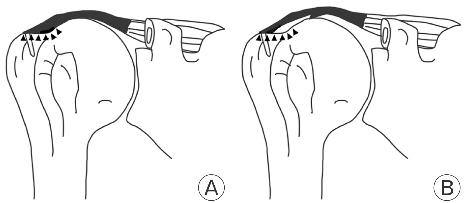
This is a retrospective study with prospectively collected data from July 2013 to March 2017 and approved by the Institutional Review Board of Myongji Hospital (No. 2019-10-002). The require-ment for informed consent was waived due to the nature of retrospective study design. A total of 351 cases of arthroscopic rotator cuff repair during the study period were assessed for eligibility. Inclusion criteria were no history of surgery, fracture, or infection; no inflammatory or degenerative arthritis, available immediate and postoperative MRI and image quality enough to read. The surgical indication was full-thickness or high-grade partial-thickness rotator cuff tears with persistent pain with functional disability after nonoperative treatments of at least 3 months. All surgeries were performed by shoulder joint specialists with 10 years of experience. The study subjects were classified based on the tear size measured by arthroscopic finding. Among those, cases with partial rotator cuff tears, small full-thickness tears (<1 cm), large to massive (>3 cm) rotator cuff tears (n=2) as well as follow-up loss at postoperative 6 months (n=321) were excluded. A total of 30 cases of medium-sized full-thickness rotator cuff tears (1–3 cm) were enrolled in this study. We classified into complete footprint coverage group (CC group, n=19) and incomplete footprint coverage group (IC group, n=11) by arthroscopic and immediate postoperative MRI findings (Figs. 1 and 2).
2. Demographics and variables
Baseline demographic data including age, sex, preoperative pain visual analogue scale (PVAS), and preoperative range of motion (ROM; forward flexion, external rotation at neutral and internal rotation) were collected. Internal rotation was measured by the vertebral spinous process that could be reached with the tip of the patient’s thumb and was converted into continuously numbered groups: T1–12 to 1–12, L1–5 to 13–17, and buttock to 188. The radiographic variables including anteroposterior (AP) tear width (cm), length of retraction (cm), fatty infiltration, and muscle atrophy were measured in MRI5-7.
The footprint coverage, fatty infiltration, and muscle atrophy were evaluated at postoperative 1 day and postoperative 6 months. PVAS and ROM were evaluated at postoperative 6 months.
3. Radiologic evaluation
The radiologic or anatomical evaluation was performed by MRI at preoperative, postoperative 1 day, and postoperative 6 months. Image assessments were subsequently performed using the PACS software (Impax; Agfa, Antwerp, Belgium) with a 3-T Signa HDxt MRI scanner/Discovery MR750w system (General Electric, Milwaukee, WI, USA). T2-weighted coronal oblique, sagittal oblique, and axial images were acquired during follow-up periods. An orthopedic surgeon with 10 years of experience as a shoulder consultant surgeon and orthopedic surgeon who are in the fellowship made an agreement for the findings in MRI. In preoperative MRI, AP tear size, retraction, fatty infiltration, and muscle atrophy were evaluated. In postoperative MRI, the footprint coverage, fatty infiltration, and muscle atrophy were evaluated. And we analyzed interobserver reliability. In inter-observer reliability analysis. Intraclass correlation coefficients showed a good level of reliability for footprint coverage degrees (0.85, p<0.001).
4. Anteroposterior tear size and tendon retraction
AP tear size was measured from T2-weighted sagittal oblique images. AP tear size was defined as the longest straight distance from the anterior tendon edge to the posterior tendon edge. From the T2-weighted coronal oblique images, tendon retraction was measured from the lateral end of greater tuberosity to the medial tendon edge with a straight distance.
5. Fatty infiltration and muscle atrophy
Using the sagittal oblique view of T1-weight MRI, fatty infiltration in all rotator cuff muscles were classified by the modified Goutallier staging system described by Fuchs et al.13. Based on the lateral image on which the scapular spine was in contact with scapular body (Y-shaped view), the patients were classified into five grades: grade 0, normal muscle without fatty streak; grade 1, some fatty streaks in the muscle; grade 2, fatty infiltration is present, but more muscle than fat; grade 3, equal amount of fat and muscle; and grade 4, more fat than muscle. We checked two times at preoperative and postoperative 6 months for comprising before and after operation condition of fatty infiltration.
Measurement of muscle atrophy is based on oblique sagittal plane image medial to coracoid process, according to the classification of Warner et al.14. A line is drawn from the edge of the coracoid to the inferior scapular tip, from the inferior tip of the scapula to the spine, and from the scapular spine to the coracoid process. If the muscle is convex above the line, there is no atrophy. If the muscle contour is even with the line, mild atrophy exists. If the contour of the muscle is concave below the line, moderate atrophy is present. If there is barely any muscle visible, severe atrophy exists.
6. Surgical technique
The arthroscopic surgery was performed by only one experienced surgeon after the patient was sedated with general anesthesia and set in the lateral position. After routine formation of posterior and anterior portals, intraarticular injuries, including long head of the biceps tendon, superior labrum, and subscapularis tendon, were addressed. The arthroscope was inserted into the subacromial space from the posterior portal. Posterolateral viewing and lateral working portals were created, and evaluation of the subacromial space was carried out. In cases of adhesive capsulitis, the capsular release was done in the same setting. Acromioplasty was performed if an acromial spur was defined. After bursectomy and debridement of pathologic cuff tissue, tear size was determined by a 5-mm marked probe. After the tear size was measured with a probe to confirm the size measured from the magnetic resonance arthroprahpy or MRI, the surgeon released any adhesion surrounding the tendon. Mobility of the tendon was evaluated, and the torn rotator cuff tendon was repaired to bone in double-row with suture bridging technique. At the end of the rotator cuff repair, footprint coverage was determined as complete or partial. All patients underwent controlled early passive motion exercise consisting of forward flexion, abduction, and external rotation from 1 day after the operation during the brace-wearing period15. A sling with an abduction pillow (20° of abduction and neutral rotation) was applied for 6 weeks. Progressive active-assisted passive motion exercises were commenced at the 3rd to 4th week for muscle strengthening.
7. Statistical analysis
Medical records and preoperative MRI were reviewed for the evaluation of preoperative variables affecting the amount of footprint coverage. As the data were not normally distributed, we selected the chi-square test to compare proportions of categorical variables between both groups. A Mann-Whitney U-test was used to compare continuous data variables in both groups. Pearson-correlation analysis and Spearman rank-order correlation took a role in determining association between categorical and continuous variables, respectively. These variables, which determined the independent factors affecting healing rate, were analyzed. We created the receiver operating characteristic (ROC) curve of significant predicting factors which correlated with healing of rotator cuff tendons. Intraobserver and interobserver reliabilities were evaluated with intraclass correlation coefficients for categorical and continuous data respectively. Statistical significance was set at a p-value of <0.05. We use the SPSS version 14 (SPSS Inc., Chicago, IL, USA) for all statistical analysis.
Results
Thirty patients (16 male and 14 female, mean age of 60 years [range, 47–76 years]) were included in the analysis. Thirty medium-size rotator cuff tears were defined from MRI and all were confirmed under arthroscopic examination. Overall, 19 and 11 patients underwent complete and partial repair, respectively. In the demographic data, there was no significant difference in age and sex in both groups. On preoperative MRI, AP tear size and retraction were not significantly different between the two groups (Table 1). PVAS and ROM also did not differ significantly between the two groups at preoperative, postoperative 6 months, and change at postoperative 6 months (Table 2). Fatty infiltration of all rotator cuff muscle and muscle atrophy of supraspinatus did not differ significantly between the two groups at preoperative, postoperative 6 months, and change at postoperative 6 months. Out of 19 patients in CC group, nine patients were changed to incomplete repair at postoperative 6 months. Out of 11 patients of IC group, six patients were improved to complete repair at postoperative 6 months. It means that 54.5% of incomplete repair was changed to complete repair during postoperative 6 months (Table 3). The ROC curves were further analyzed on change of footprint coverage at postoperative 6 months to search for each proper cut-off point that gave optimal sensitivity and specificity. The results were as follows; change of footprint coverage at postoperative 6 months the optimal sensitivity and specificity (Table 4). We have found there is no independent factor that significantly affected the healing rate after multivariate logistic regression analysis. The intraobserver reliability ranged from 0.516 to 0.999 with average of 0.868. The interobserver reliability ranged from 0.750 to 0.998 with an average of 0.900.
Discussion
The aim of this study is to verify the preoperative factor that can affect the footprint coverage during arthroscopic rotator cuff repair in full-thickness medium-size cuff tear and the change of footprint coverage at postoperative 6-month MRI. In our study, AP tear size, retraction, PVAS, ROM, fat degeneration, and muscle atrophy do not affect footprint coverage in medium-sized full-thickness rotator cuff tears. In addition, complete footprint coverage may change to incomplete footprint coverage, and incomplete footprint coverage may also change to complete footprint coverage on MRI in postoperative 6 months.
For MRI parameters, there were previous studies that demonstrated the association between tear sizes and partial repair16. Yoo et al.11 reported the sagittal tear size of ≥32 mm and the coronal tear size of ≥31 mm correlated with an inability to achieve satisfactory anatomical rotator cuff repair. Sugihara et al.17 showed that primary rotator cuff repair was usually not feasible when both the length and width of tear are ≥40 mm. According to our study result, AP tear size is not correlated with partial repair group (p=0.642). We found that length of retraction is also not correlated with incomplete coverage group (p=0.681) in this study.
Previously, Burkhart17-19 has described a concept of functional partial repair which reduces pain and improves shoulder function. Furthermore, recent studies showed that partial repair provides inferior functional outcomes, shoulder mobility and strength compared to the anatomical repair20. Our study found that there is no significantly different of functional outcomes between two group; preoperative and postoperative PVAS (p=0.470, p=0.324), preoperative and postoperative forward flexion (p=0.374, p=0.509), preoperative and postoperative external rotation (p=0.710, p=0.835) and preoperative and postoperative internal rotation (p=0.240, p=0.841).
Large-sized and massive rotator cuff tears remain a challenging situation as there is a high re-rupture rate and poor clinical outcomes7,11,21. In our study, even in the medium-sized rotator cuff tears, outcomes are not good to excellent result. Although 19 patients of cases (63.3%) were complete repair at immediate postoperation, nine patients (47.4%) were changed to incomplete repair at postoperative 6 months. Eleven patients (36.7%) of cases were getting in partial repair at immediate postoperative checkup. But, six patients in partial repair cases (54.5%) were changed to complete repair at postoperative 6 months. So, there is a chance for changing of healing process from incomplete to complete. Our study found that there is a positive improvement in healing process in incomplete or partial repair cases. So, we believed that the healing process can be changed not only from complete to incomplete but also from incomplete to complete during healing phase.
Advanced fatty infiltration correlates with an inability to achieve complete rotator cuff repair11,20. Our study demonstrates that there is no correlation between fatty infiltration of rotator cuff muscle and rotator cuff healing in medium-sized rotator cuff tear.
Tendon mobility to cover the footprint could only be demonstrated under the operation. Nonetheless, various options of arthroscopic and open surgery were performed for the partially repaired rotator cuff tears ranging from arthroscopic debridement to reversed total shoulder replacement. The double-row technique is widely used in arthroscopic rotator cuff repairs as it has been shown in several biomechanics studies to have a higher tensile strength than a conventional single-row technique22,23. Although we did not perform the comparative analysis between single, double, and triple row techniques, the previous meta-analysis studies showed that double-row repair had a more complete healing rate, less re-rupture rate, better ROM, and improved clinical outcomes, especially in large-sized and massive rotator cuff tears24-26. Hence, in this study, we intended to arthroscopically repair rotator cuff tendons with double-row technique to completely cover the anatomical footprint but we avoided too much tension at repair site as mentioned above.
Other recent studies have explored the clinical and surgical factors in predicting the reparability of large and massive rotator cuff tears. A study by Holtby and Razmjou4 showed that U-shaped tears, tear size, and tendon quality defined intraoperatively correlated with reparability. On a contrary, there are no preoperative factors that can predict the reparability of large and massive rotator cuff tears4. Dwyer et al.20 studied the association between preoperative MRI and the reparability of large and massive rotator cuff tears. The results were quite similar to previous studies, demonstrating mediolateral tear size, tendon retraction to the glenoid level, muscle atrophy, advanced fatty infiltration, and superior migration of humeral head were associated with partial repairs20. Our study also included clinical, radiographic, and MRI parameters to analyze the correlation with healing rate of rotator cuff tears.
The limitation of this study is a low number of patients. The size of the study subjects was calculated using the G-power program. But this study did not meet the population size due to the study by a single operator at a single center. And no differentiation between traumatic or nontraumatic, no definite interval between MRI and operation time, no measurement of rotator cuff arthropathy and acromiohumoral index in the partial repair group. All such factors were statistically important correlations between healing rate and footprint coverage.
In conclusion, in medium-sized full-thickness rotator cuff tears, AP tear size, retraction, PVAS, ROM, fatty degeneration, and muscle atrophy did not affect foot coverage. And the completeness of the footprint coverage of cuff tear repair is not an absolute indicator of the cuff healing process and is not considered to be directly related.

 XML Download