

 PDF
PDF Citation
 Print
Print


Introduction
Anthracosis is black pigmentation occurring mainly in the bronchi due to inhalation of pollutants, such as dust, particulates, cigarette smoke, and biomass.1) Anthracofibrosis is defined as black anthracotic pigmentation on the bronchial mucosa with bronchial luminal narrowing or obliteration.2) The majority of cases of anthracosis and anthracofibrosis are observed in the lungs, with rare cases of extrapulmonary presentation being reported in the mediastinal and axillary lymph nodes, paratracheal masses and esophagus.3-7) Herein, we report a case of paraesophageal anthracofibrosis with severe adhesion to the recurrent laryngeal nerve (RLN) in a patient with papillary thyroid cancer associated with extensive extrathyroidal extension. Upper paratracheal/paraesophageal black colored anthracofibrosis can be confused with metastatic lymphadenopathy in patients with papillary thyroid cancer. Suspecting anthracofibrosis might prevent unnecessary aggressive surgery with injury to the RLN in patients with papillary thyroid cancer.
Go to : 
Case Report
A 76-year-old woman presented with a several-year history of anterior neck swelling. The patient had a history of hypertension. She was a non-smoker and did not have a history of pulmonary tuberculosis. She did not have a familial history of thyroid disease. Thyroid ultrasonography (US) revealed a 1.8×1.7× 2.2-cm hypoechoic, ill-defined mass with microcalcification and extrathyroidal invasion in the right thyroid gland (Fig. 1A). In addition, the thyroid US showed a 5.7×3.4×5.9-cm mixed-echoic mass on the left side (Fig. 1B). The patient underwent US-guided fine needle aspiration biopsy in the right thyroid mass. Cytologic examination of the right thyroid mass was consistent with papillary carcinoma. Flexible laryngoscopy revealed normal vocal cord movement. Neck computed tomography (CT) with enhancement showed a 2.2×1.8×1.4-cm irregular subtle enhancing low attenuated nodule with calcification and extrathyroidal extension in the right thyroid gland, and a 7×4.5×5-cm heterogeneously enhancing mass on the left side (Fig. 2). Neck CT scan did not reveal any significantly enlarged lymph nodes in both lateral necks. The patient underwent total thyroidectomy with central compartment neck dissection. After thyroidectomy, a 1.5-cm black-colored, right paraeso-phageal mass, suspected as metastatic lymphadenopathy, was identified under the right RLN (Fig. 3). The paraesophageal mass was severely adherent to the RLN. Thus, it was not possible to remove the mass completely without injuring the RLN. To determine the need for shaving the RLN, intraoperative frozen sectional biopsy of the mass was performed, which revealed a negative result for malignancy. The surgery was terminated without attempting complete excision of the mass for functional preservation of the RLN. Permanent histopathological examination of the mass revealed anthracofibrosis with no evidence of neoplasia. The pathologic findings of the mass showed anthracotic pigment deposition and dense fibrotic changes (Fig. 4). To evaluate the cause of the severe adhesion to the surrounding tissue, Masson’s Trichrome (MT) staining was performed. We compared the histopathological findings of four MT-stained samples. This case and another case with a paratracheal anthracotic mass had severe adhesion to adjacent tissues (Fig. 5A). In contrast, two other cases of level IV cervical anthracosis were incidentally detected without adhesion to adjacent tissues during neck dissection (Fig. 5B). A prominent increase in blue collagen fibers was demonstrated in cases with adhesion (Fig. 5A), when compared with that in the cases without adhesion (Fig. 5B). In addition, histopathological examination of the main mass of the right thyroid gland revealed papillary carcinoma with extrathyroidal extension involving the perithyroidal adipose tissue. The patient had an uneventful postoperative recovery with good vocal cord movement.
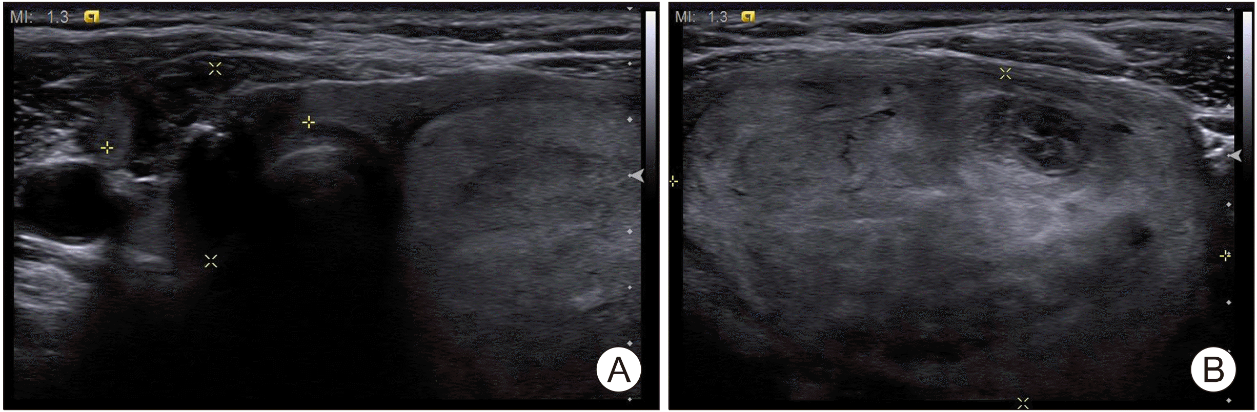 | Fig. 1Thyroid ultrasonography showing a 1.8× 1.7×2.2-cm hypoechoic, ill-defined mass with microcalcification and extrathyroidal invasion in the right thyroid gland (A) and a 5.7× 3.4×5.9-cm mixed-echoic mass in the left thyroid gland (B).
|
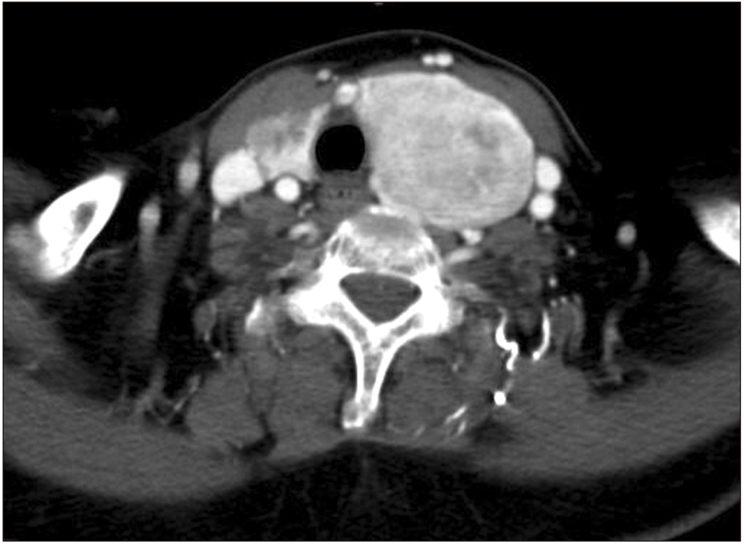 | Fig. 2Neck computed tomography with enhancement showing a 2.2×1.8×1.4-cm irregular subtle enhancing low attenuated nodule with calcification and extrathyroidal extension in the right thyroid gland and a 7×4.5×5-cm heterogeneously enhancing mass in the left thyroid gland.
|
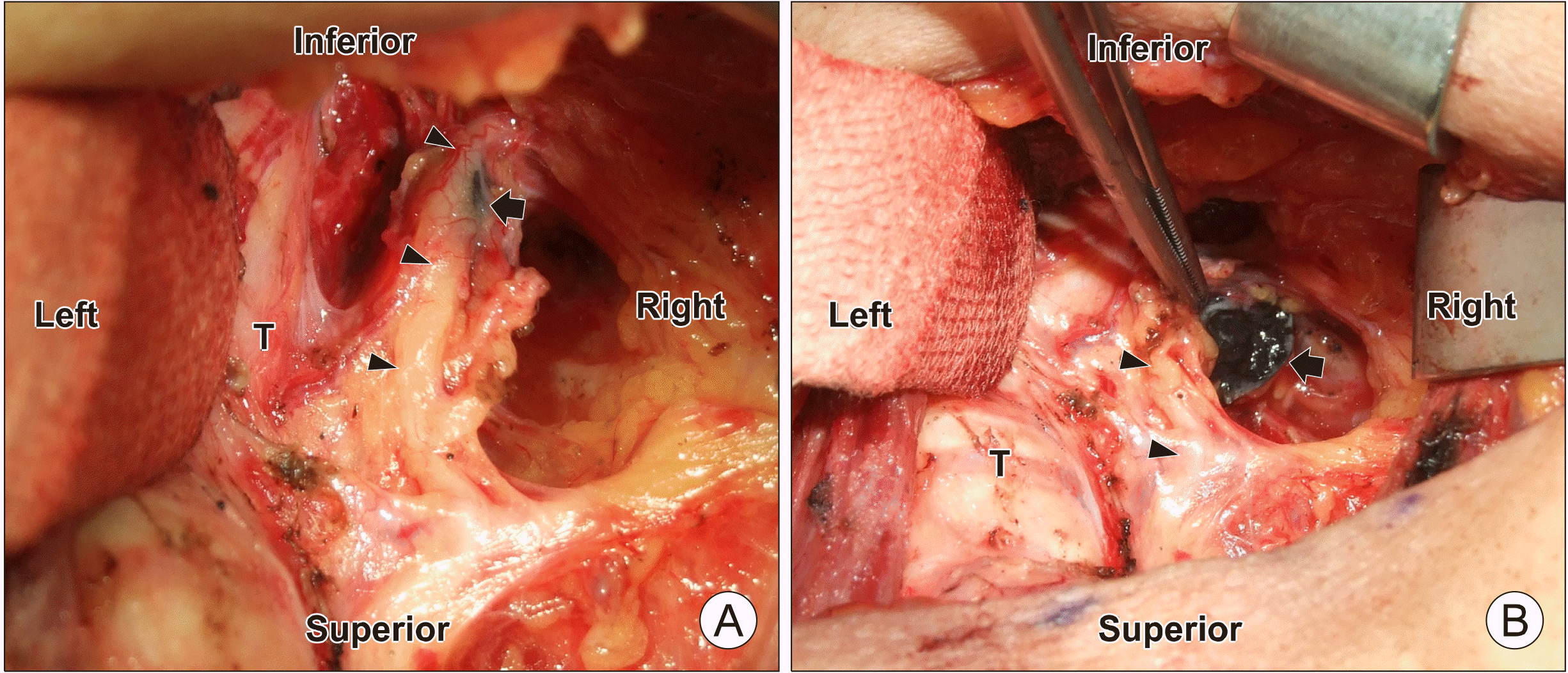 | Fig. 3Operative findings after thyroidectomy shows a 1.5-cm black colored, right paraesophageal mass (black arrow), suspected as metastatic lymphadenopathy, under the right recurrent laryngeal nerve (black arrowheads) (A). The cut surface of the mass (black arrow) obtained for intraoperative frozen sectional biopsy was as black as coal. Black arrowheads indicated the right recurrent laryngeal nerve (B). T: trachea
|
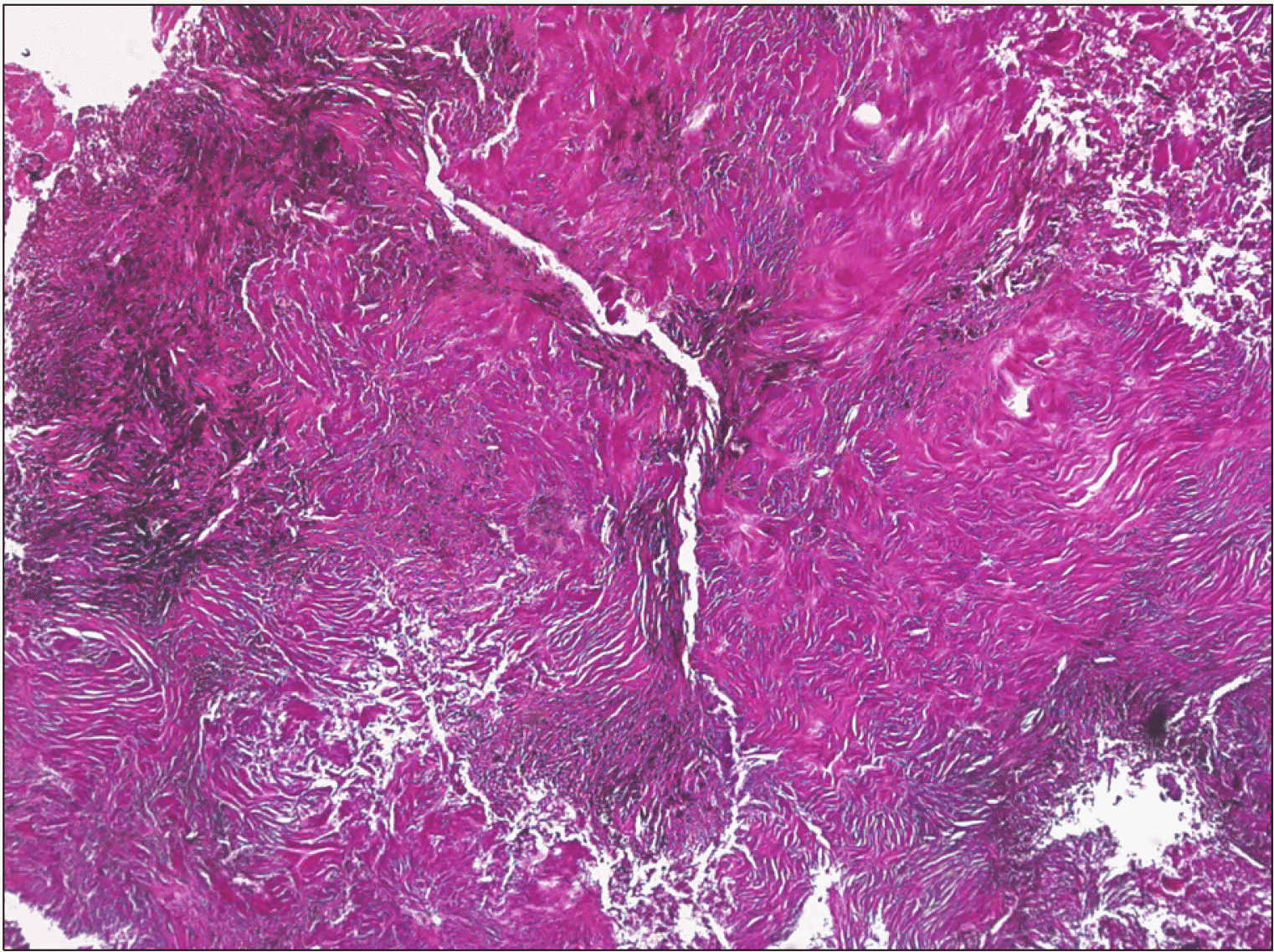 | Fig. 4Histopathological examination of the paraesophageal mass revealed anthracotic pigment deposition and dense fibrotic changes (Hematoxylin and Eosin [H&E] stain, ×40).
|
 | Fig. 5Histopathological findings of Masson’s Trichrome staining (MT, ×200) in 4 cases with anthracosis. (A) This case and another case with a paratracheal mass had severe adhesion to adjacent tissues. A prominent increase in blue collagen fibers were seen on MT staining. (B) Two other cases of cervical anthracosis had no adhesion to adjacent tissues. These cases showed negative on MT staining (H&E stain, ×100).
|
Go to :
Discussion
Bronchial anthracofibrosis has been well documented as a bronchoscopic finding showing dark anthracotic pigmentation associated with bronchial luminal narrowing without a relevant history of pneumoconiosis or heavy smoking.2) Some studies reported, anthracofibrosis could be associated with pulmonary tuberculosis,2,8) but the present patient didn’t have a history of tuberculosis. Anthracofibrosis often causes intrapulmonary lymphadenopathy; however, extrapulmonary anthracofibrosis rarely causes mediastinal or axillary lymphadenopathy.3,4,7) In addition, there are reports of esophageal anthracosis mimicking esophageal submucosal tumors.6) In our case, paraesophageal anthracofibrosis presented as a black-colored hard paraesophageal mass with severe adhesion to the RLN, as found during thyroid surgery, in a patient with advanced thyroid cancer. A paraesophageal mass could suggest metastasis from malignancies of adjacent organs, such as the thyroid, esophagus, lung, thymus, and stomach. Particularly, metastatic lymphadenopathy from thyroid cancer occasionally has a black color and shows adhesion to adjacent tissue with extranodal extension. Thus, the operative finding in this case significantly raised the suspicion of metastatic lymphadenopathy due to thyroid cancer. However, the frozen sectional biopsy helped us exclude metastatic lymphadenopathy and consequently preserve the function of the RLN. The present case highlights that anthracofibrosis should be included in the differential diagnosis of paraesophageal masses, and suspecting anthracofibrosis might prevent unnecessary aggres-sive surgery with injury to the RLN in patients with papillary thyroid cancer. In addition, intraoperative frozen sectional biopsy can help in decision making during surgery.
MT staining is widely used as the collagen fibers are stained blue-green, and it has a greater contrast compared to hematoxylin and eosin staining.9) Therefore, MT staining is useful to differentiate between collagen and smooth muscle in tumors and determine the increase in collagen in cases of liver cirrhosis and kidney disease.9) Anthracosis could be incidentally detected during lower neck dissection, and unlike this case, it usually has no adhesion to the adjacent tissue. To evaluate the cause of the severe adhesion to the surrounding tissue in this case, we performed MT staining in four cases with anthracosis. In our cases, the anthracotic mass with adhesion showed prominent blue collagen fibers when compared to cases without adhesion to the surrounding tissues. These findings could signify that collagen is involved in the adhesion of the anthracotic to the surrounding tissues.
In conclusion, extrapulmonary anthracofibrosis, especially in the paraesophageal location, has rarely been reported, and it could mimic metastatic lymphadenopathy in patients with cancer. This case highlights that anthracofibrosis should be included in the differential diagnosis of paraesophageal masses, and the unnecessary aggressive surgery can be avoided with appropriate suspicion of anthracofibrosis. In addition, frozen sectional biopsy can help in decision making during surgery. Collagen might be involved in the adhesion of the anthracotic mass to the surrounding tissues.
Go to :
 XML Download