Journal List > J Surg Ultrasound > v.9(1) > 1516078681

유방암 재건수술 후 초음파 소견

Abstract

Breast ultrasound is the most important tool for breast cancer surveillance after surgery. In particular, when a breast reconstruction is performed for patients with breast cancer, the sonographer should be provided with surgical information to distinguish between a benign lesion and cancer recurrence. This article describes the sonographic changes after breast reconstruction for breast cancer, which vary, but may be typical. Therefore, if breast surgeons and sonographers are aware of the typical sonographic findings, it would be helpful for the early detection and management of postoperative complications.

서 론

최근 유방암의 수술은 단순히 유방의 부분 또는 전절제술 뿐만 아니라 유방재건술이 함께 시행되는 경우도 많다. 특히 유방의 전절제술 후 재건술에 보험적용이 되면서부터 보형물이나 자가조직을 이용한 유방재건술의 비율이 꾸준히 증가하는 것으로 보고되고 있다.(1,2)
유방암의 추적검사에는 이학적 검사와 유방촬영술, 유방초음파, 유방 자기공명영상(magnetic resonance imaging) 등이 적용될 수 있으며, 수술방법에 따라 가능한 검사방법과 필요한 검사방법이 다소 다를 수 있다.(3,4) 보형물을 이용한 유방재건술 이후에는 전체 유방을 촬영하는 유방촬영술의 적용이 어려워 유두-유륜 복합체 주변만 촬영하거나 반대편 유방만 촬영하는 방법을 선택해야 한다.(4,5) 자가조직으로 유방재건술을 시행한 경우 어떤 수술방법이 적용되었는지 영상검사자가 파악하고 있어야만 이상 소견이 있을 때 유방실질에 발생한 것인지 재건된 피판에 발생한 것인지 구별할 수 있다.
유방암에서 초음파 검사는 암의 재발 및 새로운 병변의 발생을 감시할 뿐만 아니라 재건수술 후 발생하는 합병증도 확인할 수 있다는 장점이 있다. 따라서 각 유방재건술 후의 초음파 소견을 파악하고, 이와 다른 소견이 보이는 경우 치료가 필요한 합병증인지 판단하는 것은 임상에서 매우 중요한 부분이라 할 수 있겠다.

본 론

유방외과의는 대체로 본인이 선호하는 술식이 일정하다. 다양한 수술방법이 존재하지만, 유방암의 크기나 위치에 따라서 수십가지에 달하는 유방재건술식 중 본인이 선호하거나 쉽게 시행가능한 방법을 선택하여 자주 이용하기 때문이다. 따라서 영상검사자는 수술방법을 이해하고 수술 후 변화된 유방에서 보일 수 있는 초음파소견을 미리 알고 있어야 한다. 이와 다른 형태의 소견이 관찰된다면, 합병증이나 다른 문제가 발생한 것일 수 있다.
재건수술의 방법에 따른 유방초음파 소견은 매우 전형적이어서 한두번만 익혀두면 쉽게 구별할 수 있다. 여기에서는 자가조직을 이용한 재건수술방법과 보형물을 이용한 재건수술방법 이후의 유방초음파 소견을 기술하고자 한다.

1. 자가조직을 이용한 부피이동술(volume displacement technique) 후 초음파 소견

유방의 부피이동술은 유방내 절제범위 이외의 유선을 이용하여 결손부위를 채워 넣는 술식이다. 대표적인 부피이동술로는 국소피판술(local flap), 회전피판술(rotation flap), 유방축소술(reduction mammoplasty) 등이 있다.
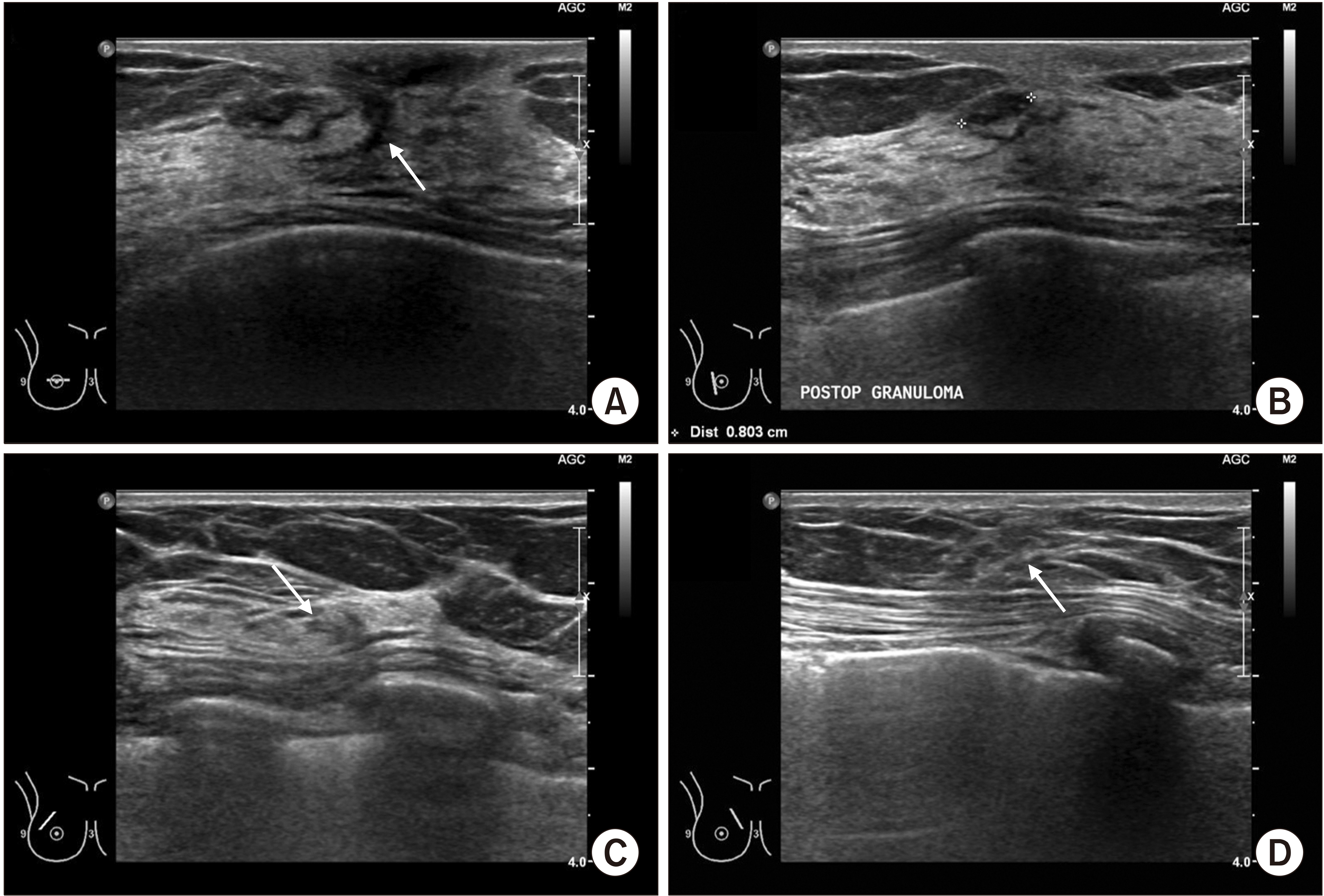
국소피판술은 결손부위에서 인접한 유선을 유선분리법(gland undermining technique)을 통해 흉벽에서 분리한 후 이를 결손부위로 당겨 유선을 봉합하는 방법으로, 절개선은 기존의 절개선에서 크게 변하지 않는다. 유선피판술(glandular flap) 또는 유선재형성술(glandular reshaping)이라고도 하며, 기존의 유방보존술과 초음파 소견이 크게 다르지 않다. 다만, 보이는 절개선보다 더 넓은 부위를 내부에서 박리했을 가능성이 높으므로 절개선에서 다소 떨어진 곳에 유선의 구조왜곡이 관찰될 수 있다는 점을 기억해야 한다 (Fig. 1).
회전피판술은 절개선과 유선의 반흔이 거의 일치하여 보이기 때문에 쉽게 알 수 있다. 절개선을 따라서 탐촉자(probe)를 움직이다 보면 유선의 반흔이 연속적으로 관찰되고, 절개선의 주변에 발생한 결절은 육아종(granuloma)일 가능성이 높으며 유방암의 재발 병변일 가능성은 낮다. 그러나 절개부위 주변이라도 불규칙한 변연부, 저에코성 병변, 고혈류성 병변 등 악성 병변의 특징을 보인다면 조직검사를 시행하여 유방암의 재발여부를 확인하는 것이 필요하다(Fig. 2).
유방축소술은 다른 부피이동술에 비하여 잘렸다가 봉합된 부분이 더 넓고 다양한 위치에서 확인되며, 대체로 유방하수의 정도가 중등도 이상인 환자에서 시행되므로 지방의 분포도 넓은 편이다. 따라서 수술 후 지방괴사성 결절이 비교적 흔히 나타나지만, 시간이 경과하며 결절의 크기가 점차 감소하고 경우에 따라서는 완전히 사라지기도 한다. 유방암의 재발과 구별하기 위해서는 유방축소수술 방법에 대해서 알고 있어야 하며, 절개선이나 반흔의 위치와 일치하는지 확인하는 것도 좋은 방법이다(Fig. 3).

2. 자가조직을 이용한 부피치환술(volume replacement technique) 후 초음파 소견

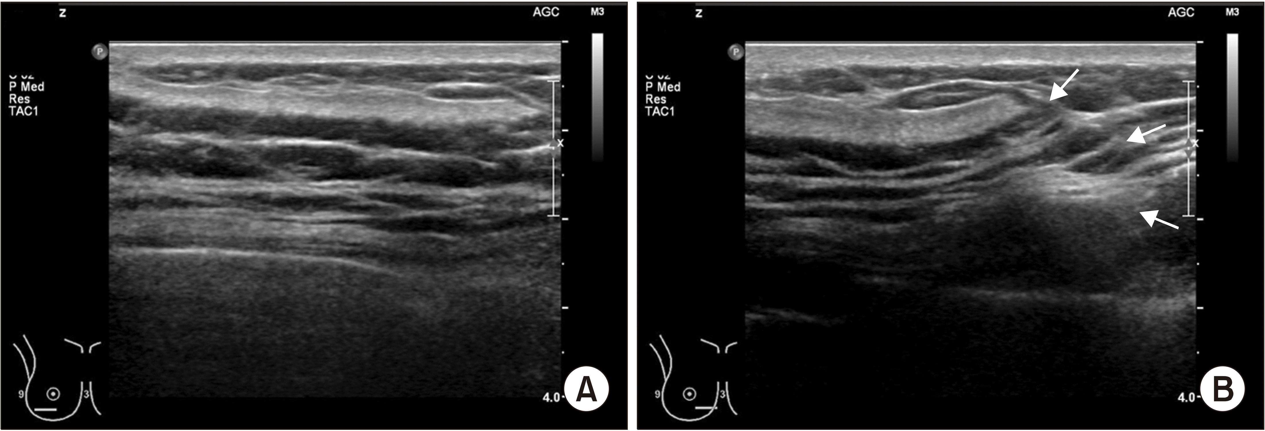
자가조직을 이용한 유방재건술 중 부피치환술은 주로 광배근 피판(Latissimus dorsi muscle flap, LD flap)이나 횡복직근 피판(Transversus rectus abdominis muscle flap, TRAM flap)을 이용한다. 대부분의 외과의사는 유경피판(pedicle flap)의 형태로 피판을 구득하므로 수술방법을 이해한다면 혈관이나 피판의 고정 위치 등을 짐작할 수 있고, 이는 초음파 검사를 하는데 많은 도움이 된다.
피판은 혈류가 풍부해야 잘 유지되므로 근육에 더하여 피부와 피하지방이 함께 포함되고, 따라서 부피치환술 후 초음파 검사에서는 근섬유가 평행선을 이루는 형태가 주로 관찰된다(Fig. 4A). 근육의 주변부에는 피판에 같이 구득된 지방층이 관찰된다. 남은 유방실질과 구득한 피판을 고정한 경계부위 역시 초음파에서 비교적 명확히 구별되는데(Fig. 4B), 이론적으로 남은 유방실질에서 유방암이 재발할 수 있으므로 초음파 추적검사 시 피판의 내부보다는 남은 유선실질을 중점적으로 관찰하는 것이 재발 감시에 중요하다.

3. 보형물을 이용한 유방재건술 후 초음파 소견

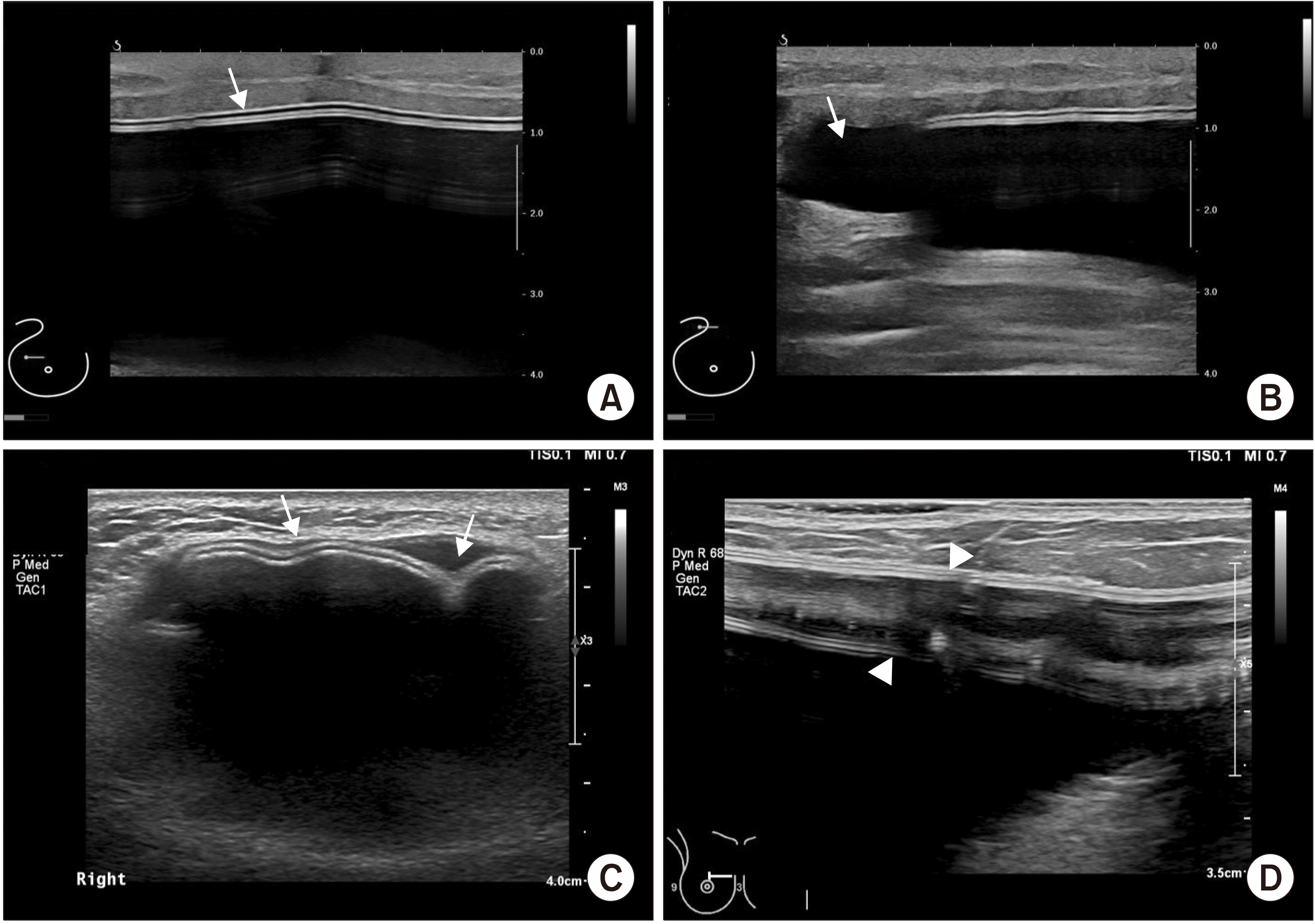
보형물의 초음파 소견은 큰 낭의 형태로 보이며, 보형물의 내부와 외부가 매우 명확하게 구별된다. 보형물은 유방의 전절제술 후 재건술에만 이용되기 때문에 유선의 실질이 없이 피하지방층이나 대흉근 아래에 위치한 보형물만 관찰된다(Fig. 5A). 유선조직과 보형물의 피막(capsule)은 명확하게 구별되므로, 보형물 주변에 발생한 장액종(peri-prosthetic seroma)은 초음파검사에서 보형물과 강(cavity) 사이에 위치한 무에코성(anechoic) 병변으로 관찰된다(Fig. 5B). 장액종은 끝이 뾰족하지 않은 척수바늘(spinal needle)이나 정맥캐뉼라(venous cannula) 등을 이용하여 쉽고 안전하게 흡인할 수 있다.(6)
초음파는 보형물을 이용한 유방재건술 후 발생한 합병증의 진단에도 유용하다.(7) 보형물 강의 피막구축이 발생하면 환자는 주로 유방이 딱딱해지거나 가슴이 조이는 듯한 증상을 호소하는데, 실제로 초음파 검사에서 보형물 강이 좁아지며 보형물에 주름이 형성된 소견을 관찰할 수 있다(Fig. 5C). 드물게 보형물의 파열이 의심되는 경우에는 초음파 탐촉자(probe)로 피막을 연속적으로 따라가며 검사하면 피막의 흐름이 끊기거나 피막이 두 층으로 분리된 것이 보일 수 있는데, 이는 피막의 외부파열 또는 내부파열을 시사하는 소견일 수 있다(Fig. 5D).

결 론

유방암의 수술적 치료는 단순 유방절제술을 넘어 다양한 종양성형술의 기법을 이용한 재건수술이 함께 이루어지고 있다. 대부분의 유방외과 의사는 유방재건 수술방법에 대한 이해도가 뛰어나므로 유방재건술 후 초음파 영상소견을 통해 보다 쉽고 정확하게 임상적 진단을 내릴 수 있다. 유방재건술 시행 후 유방초음파의 전형적인 소견을 숙지하고 있다면 수술 후 합병증의 발생이나 유방암의 재발을 놓치지 않을 수 있겠다.

REFERENCES

1. Korean Breast Cancer Society. 2016. Breast Cancer Facts & Figures 2016. Korean Breast Cancer Society;Seoul:
2. Min SY, Kim Z, Hur MH, Yoon CS, Park EH, Jung KW. 2016; The basic facts of Korean breast cancer in 2013: results of a nationwide survey and breast cancer registry database. J Breast Cancer. 19:1–7. DOI: 10.4048/jbc.2016.19.1.1. PMID: 27066090. PMCID: PMC4822102.
crossref
3. Margolis NE, Morley C, Lotfi P, Shaylor SD, Palestrant S, Moy L, et al. 2014; Update on imaging of the postsurgical breast. Radio-graphics. 34:642–60. DOI: 10.1148/rg.343135059. PMID: 24819786.
crossref
4. Heller SL, Lourenco AP, Niell BL, Ajkay N, Brown A, et al. Expert Panel on Breast Imaging. 2020; ACR Appropriateness Criteria® imaging after mastectomy and breast reconstruction. J Am Coll Radiol. 17(11S):S403–14. DOI: 10.1016/j.jacr.2020.09.009. PMID: 33153553. PMCID: PMC8173332.
crossref
5. Zakhireh J, Fowble B, Esserman LJ. 2010; Application of screening principles to the reconstructed breast. J Clin Oncol. 28:173–80. DOI: 10.1200/JCO.2008.21.7588. PMID: 19884555.
crossref
6. Jeon HJ, Park HY, Jung JH, Kim WW, Chung JH, Moon SH, et al. 2017; Usefulness of ultrasound-guided aspiration using intravenous cannulas for patients with peri-prosthetic seroma. J Surg Ultrasound. 4:18–24.
7. Lee J, Kim SH, Lee JH, Han BK. 2021; Understanding silicone breast implant-associated complications for radiologists. J Korean Soc Radiol. 82:49–65. DOI: 10.3348/jksr.2020.0208.
crossref

Fig. 1
Sonographic findings after breast reconstruction with glandular flap. (A) Since the surgery was done with periareolar incision, the post-operative scar (arrow) is shown as a perpendicular, hypoechoic lesion. (B) A benign looking, oval-shaped hypoe-choic nodule was detected on incision site and it is suspected as post-operative granuloma. (C, D) Although there is only one incision on the breast, minor structural distortions (arrows) were shown around main incision. Based on these ultrasound findings, it can be presumed that the patient had received glandular flap for breast reconstruction.
jsu-9-1-8-f1.tif
Fig. 2
Sonographic findings after breast reconstruction with rotation flap. (A) Structural distortion which is consistent with the axillary incision is shown, and there is a hypoechoic nodule (arrow) which is presumed as postoperative granuloma or fat necrosis. Since the patient had pain with this lesion, the pathologic confirmation was performed with needle biopsy and it was revealed as fat necrosis. (B, C) Continuous glandular scarring is observed along with the incision, indicating that the skin and the parenchyma were rotated simultaneously. (D) Normal glandular structures are found on the lower central part of the breast. There is no specific findings except a small, simple cyst (arrow).
jsu-9-1-8-f2.tif
Fig. 3
Sonographic findings 9 months after breast reconstruction with reduction mammoplasty. The patient had received adjuvant radiotherapy for the right side breast cancer. (A, B) There are multiple, small nodules underneath the fat layer, which are presumed as fat necrosis. Since the multiple nodules which had fairly clear margins were shown simultaneously, they were assumed as fat necrosis rather than local recurrence of breast cancer. About 3-month follow-up is recommended for these lesions, if the pathologic confirmation is not performed. (C) Although the contralateral reduction mammoplasty was also done, superior part of the left breast showed normal breast structures without postoperative scars. (D) However, inferior part of the breast showed postoperative scar which was occurred by reduction mammoplasty.
jsu-9-1-8-f3.tif
Fig. 4
Sonographic findings after breast reconstruction with Latissimus dorsi (LD) muscle flap. (A) On lower outer quadrant of the breast, muscle fibers of LD flap were shown in linear parallel arrangement. (B) The arrows indicate the boundary between the LD muscle and original breast structures, where suture ties were done for holding the LD muscle flap to the breast.
jsu-9-1-8-f4.tif
Fig. 5
Sonographic findings after implant-based breast reconstruction. (A) Two layers of implant capsule(arrow) is located under the subcutaneous fat layer. Based on this finding, it is assumed that pre-pectoral implant insertion technique was done for breast reconstruction. (B) The peri-prosthetic seroma (arrow) is shown as anechoic area outside of implant capsule, and it can be aspirated using blunt needle, such as intravenous cannula or spinal needle. (C) This sonographic finding shows capsular contracture after implant-based breast reconstruction. There are multiple radial folds (arrows), which are occurred due to narrowed implant cavity. (D) This sonographic finding indicates the internal capsular rupture of the implant. The two layers of capsule were separated as two parallel echogenic lines (arrow heads).
jsu-9-1-8-f5.tif
TOOLS
Similar articles