Abstract
Background
Preoperative measurement of the tumor size is essential for deciding on a treatment plan for breast cancer. In particular, in a neoadjuvant setting, the treatment plan is based on preoperative clinical staging. This study evaluated the differences in tumor size measured by ultrasonography and magnetic resonance imaging (MRI) compared to the pathological tumor size.
Methods
This study reviewed the electronic medical records of patients with primary breast cancer who underwent surgery between 2014 and 2021. The clinicopathologic data, including ultrasonographic and MRI findings, were collected. Bland-Altman analysis was used to compare the differences in the tumor size between the imaging modalities and pathological tumor size.
초음파는 유방암의 진단과 함께 유방암 진단 후 수술 전 병기 결정에 중요한 역할을 담당하고 있다.(1,2) 수술 전 종양의 크기를 정확하게 측정함으로써 수술의 범위를 결정하는데 도움을 준다. 더욱이 수술 전 항암화학요법을 시행하는 경우 수술 후 치료는 처음 진단 당시의 영상의학적 검사를 통한 임상병기를 기준으로 진행되므로 정확한 크기를 측정하는 것은 매우 중요하다.(3)
본 연구는 수술 전 병기설정에 영상검사로 이용되고 있는 초음파와 자기공명영상(Magnetic resonance imaging, MRI)의 정확도를 비교해보고자 한다.
2014년 4월부터 2021년 12월까지 조선대학교병원에서 유방암으로 진단받고 수술을 시행한 140명의 환자를 대상으로 의무기록을 이용해 후향적 연구를 시행하였다. 원발성 유방암으로 수술 전 초음파와 MRI에서 단일 병변을 가진 환자를 대상으로 하였다. 선행화학요법을 시행 받은 환자, 다발성 병변, 광범위한 악성 미세석회화를 동반한 환자는 제외하였다. 수술 전 초음파, MRI에서 측정한 종양의 크기와 수술 후 병리검사결과에서 측정한 종양의 크기를 비교하였으며 각 검사별로 종양의 장경(largest diameter)을 기준으로 하였다.
통계 방법으로는 초음파와 MRI에서 측정한 종양의 크기와 병리검사에서 측정한 크기의 상관도를 확인하기 위해 Bland-Altman 분석을 시행하였다. 영상검사에서의 종양 크기와 병리검사에서의 종양 크기와의 평균값의 차이를 계산하였으며 95% 일치한계 값들(limits of agreement, LOA)을 계산했다. 유의수준은 P-value가 0.05 미만일 때를 기준으로 하였으며 통계분석은 SPSS 버전 27.0을 이용하였다(IBM Inc., Armonk, NY, USA).
본 연구는 조선대학교병원의 임상연구심사위원회에서 심의를 받았다(IRB No. 2022-04-036).
전체 140명은 모두 여자 환자였으며 평균 나이는 54.6세(범위 33세-82세)였다. 수술 전 진단은 관내 제자리암종(Ductal carcinoma in situ)이 9명(6.4%), 침윤성 유관암(Invasive ductal carcinoma)이 122명(87.1%), 침윤성 소엽암(Invasive lobular carcinoma)이 9명(6.5%)이었다(Table 1).
초음파, MRI, 병리검사에서 측정한 평균값은 각각 20.6 (±0.9) mm, 23.1 (±0.9) mm, 22.6 (±0.9) mm였다.
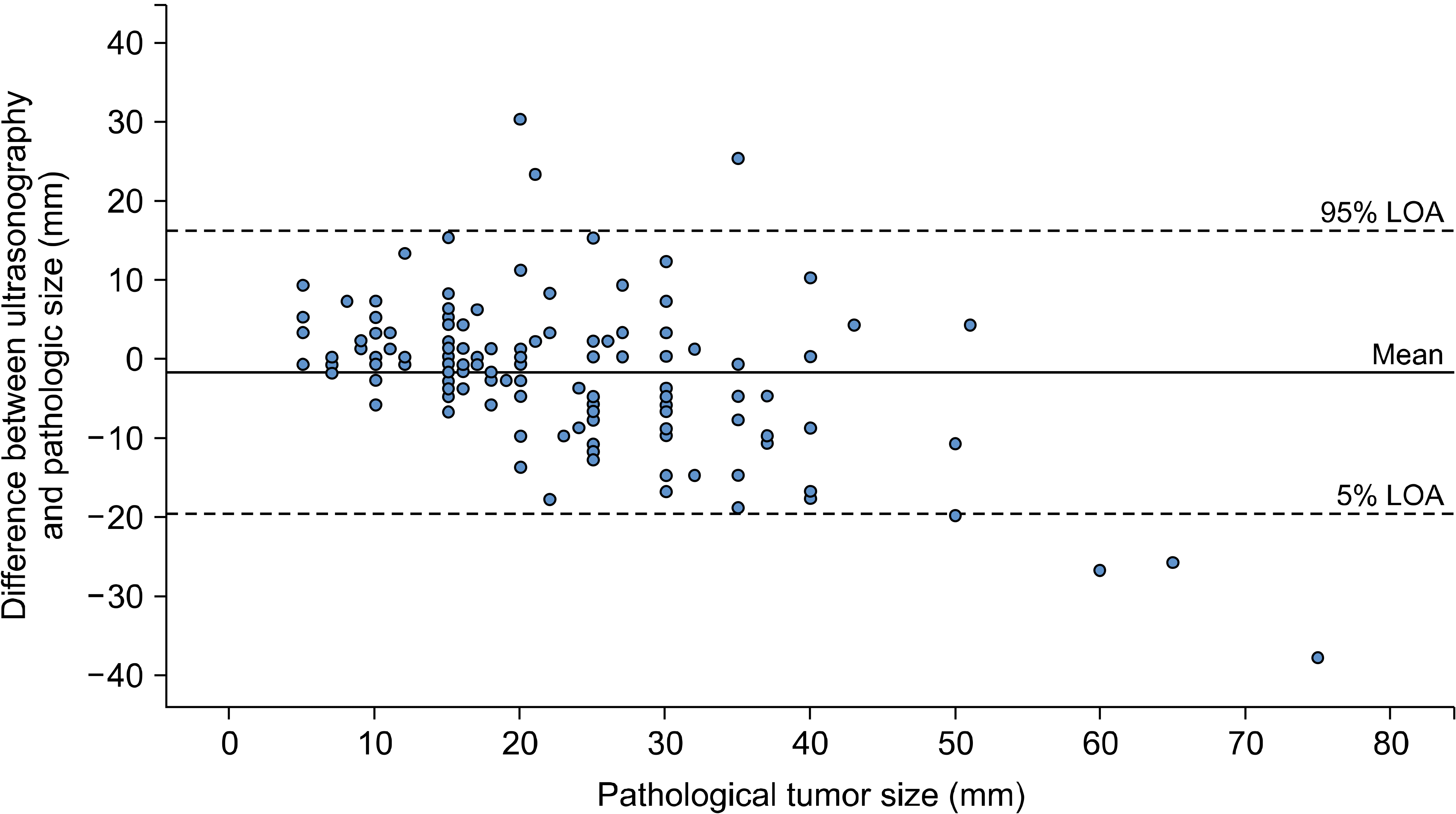
본 연구에서 초음파로 측정한 종양의 크기가 병리결과에 비해 통계적으로 유의하게 2 mm 정도 작게 측정되었음을 확인하였다. 이는 기존의 연구에서도 유사한 결과를 보였다.(4-6) 특히 Bosch 등(4)은 초음파의 탐색자(probe) 크기를 넘어설 정도로 큰 종양일 경우 초음파로 측정한 크기가 작게 측정될 수 있음을 보고했다. 이는 최근 사용되고 있는 파노라마 모드를 이용하여 극복할 수 있을 것으로 보인다.
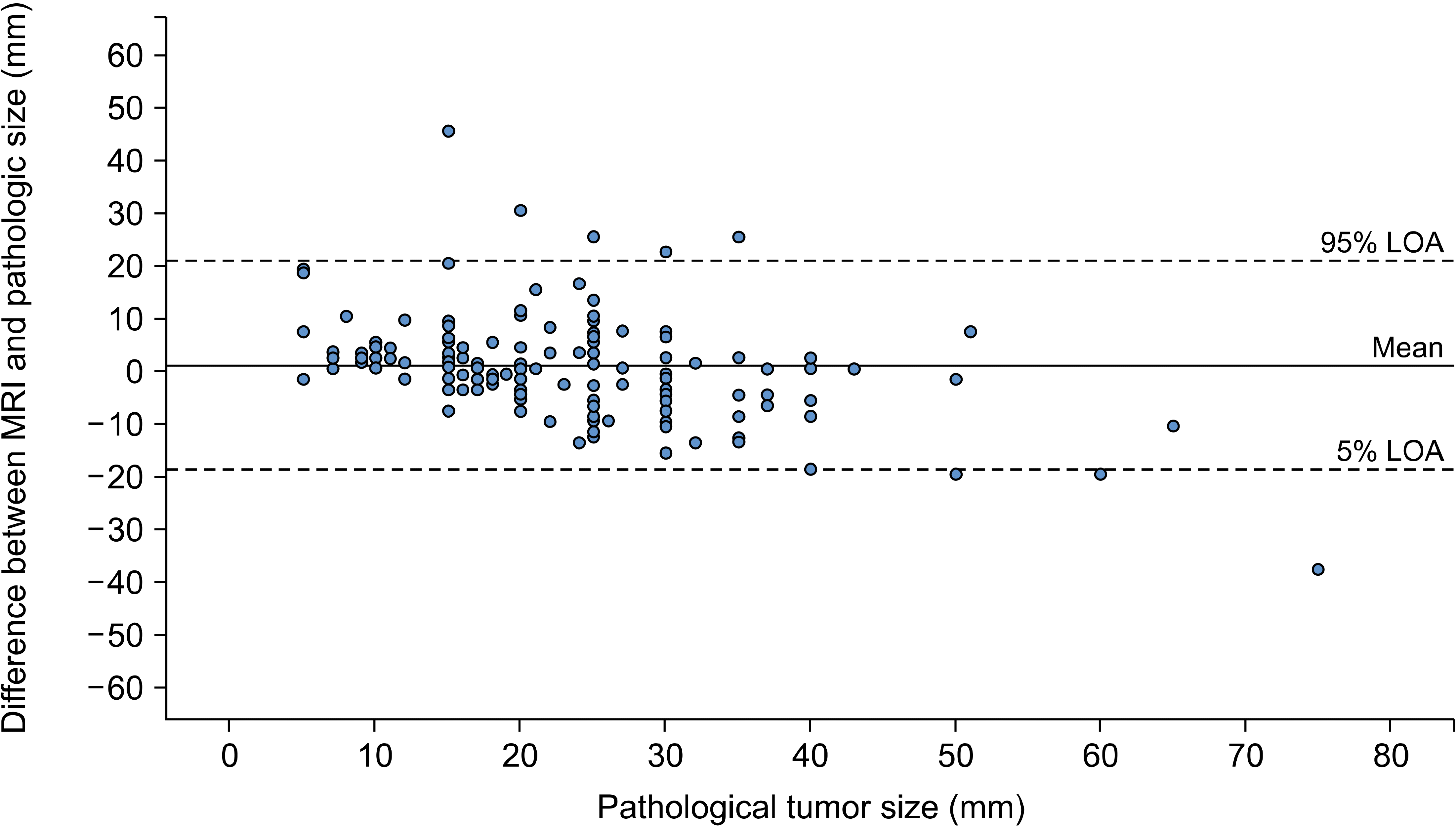
본 연구에서 MRI로 측정한 종양의 크기는 실제 병리검사와 통계적으로 유의한 차이는 보이지 않았다. Onesti 등(7)은 MRI가 실제 크기와 비교해 통계적으로 유의하게 크기를 크게 측정하는 것으로 보고 하였으나 이는 광범위한 미세석회화를 동반한 관내 제자리암종으로 기인하였던 것으로 본 연구에서는 이러한 경우를 제외하여 비교적 정확하게 측정된 것으로 보인다. 또한 Kang 등(8)의 기존 연구에서도 MRI가 실제보다 종양의 크기를 크게 측정하는 것으로 보고하였으나 본 연구에서는 진단 당시 다발성 병변을 보였던 환자는 제외하여 MRI에서 주 병변과 그 외결절(daughter nodule)이 하나의 종괴 형태로 보일 수 있는 가능성을 배제하여 결과의 차이를 보였을 가능성이 있다. 이는 실제 크기와 다양한 영상의학적 검사로 측정한 종양의 크기를 비교한 기존의 연구와 유사한 결과를 보인다. (9-11)
본 연구의 한계로는 단일 기관, 후향적 연구로 표본의 크기가 크지 않아 기존의 연구에서 유방암의 아형별로 영상의학적 검사 방법에 따라 차이가 있는 것을 확인할 수 없었던 한계가 있었다.(12) 과거에 비해 현재 영상의학적 검사의 기술이 발전하고 있고 정확도 또한 높아지고 있어 추후 전향적 다기관 연구를 통한 분석이 필요하다.
REFERENCES
1. Guo R, Lu G, Qin B, Fei B. 2018; Ultrasound imaging technologies for breast cancer detection and management: a review. Ultrasound Med Biol. 44:37–70. DOI: 10.1016/j.ultrasmedbio.2017.09.012. PMID: 29107353. PMCID: PMC6169997.

2. Yang WT. 2011; Staging of breast cancer with ultrasound. Semin Ultrasound CT MR. 32:331–41. DOI: 10.1053/j.sult.2011.02.008. PMID: 21782123.

3. Romeo V, Accardo G, Perillo T, Basso L, Garbino N, Nicolai E, et al. 2021; Assessment and prediction of response to neoadjuvant chemotherapy in breast cancer: a comparison of imaging modalities and future perspectives. Cancers (Basel). 13:3521. DOI: 10.3390/cancers13143521. PMID: 34298733. PMCID: PMC8303777.

4. Bosch AM, Kessels AG, Beets GL, Rupa JD, Koster D, van Engelshoven JM, et al. 2003; Preoperative estimation of the pathological breast tumour size by physical examination, mammography and ultrasound: a prospective study on 105 invasive tumours. Eur J Radiol. 48:285–92. DOI: 10.1016/S0720-048X(03)00081-0. PMID: 14652148.

5. Hieken TJ, Harrison J, Herreros J, Velasco JM. 2001; Correlating sonography, mammography, and pathology in the assessment of breast cancer size. Am J Surg. 182:351–4. DOI: 10.1016/S0002-9610(01)00726-7. PMID: 11720669.

6. Shoma A, Moutamed A, Ameen M, Abdelwahab A. 2006; Ultrasound for accurate measurement of invasive breast cancer tumor size. Breast J. 12:252–6. DOI: 10.1111/j.1075-122X.2006.00249.x. PMID: 16684323.

7. Onesti JK, Mangus BE, Helmer SD, Osland JS. 2008; Breast cancer tumor size: correlation between magnetic resonance imaging and pathology measurements. Am J Surg. 196:844–48. discussion 849–50. DOI: 10.1016/j.amjsurg.2008.07.028. PMID: 19095098.

8. Kang SY, Choi EJ, Youn HJ, Jung SH. 2016; Accuracy of preoperative tumor size assessment on ultrasonography and magnetic resonance imaging in breast cancer. J Surg Ultrasound. 3:24–9.
9. Boetes C, Mus RD, Holland R, Barentsz JO, Strijk SP, Wobbes T, et al. 1995; Breast tumors: comparative accuracy of MR imaging relative to mammography and US for demonstrating extent. Radiology. 197:743–7. DOI: 10.1148/radiology.197.3.7480749. PMID: 7480749.

10. Davis PL, Staiger MJ, Harris KB, Ganott MA, Klementaviciene J, McCarty KS Jr, et al. 1996; Breast cancer measurements with magnetic resonance imaging, ultrasonography, and mammography. Breast Cancer Res Treat. 37:1–9. DOI: 10.1007/BF01806626. PMID: 8750522.

11. Wasif N, Garreau J, Terando A, Kirsch D, Mund DF, Giuliano AE. 2009; MRI versus ultrasonography and mammography for preoperative assessment of breast cancer. Am Surg. 75:970–5. DOI: 10.1177/000313480907501024. PMID: 19886147.

12. Pritt B, Ashikaga T, Oppenheimer RG, Weaver DL. 2004; Influence of breast cancer histology on the relationship between ultrasound and pathology tumor size measurements. Mod Pathol. 17:905–10. DOI: 10.1038/modpathol.3800138. PMID: 15105809.

Fig. 1
Bland Altman Plots illus-trating the size difference between ultrasonography and pathology com-pared to the pathological tumor size.

Fig. 2
Bland Altman Plots illus-tra-ting the size difference between MRI and pathology compared to the pathological tumor size.

Table 1
Clinicopathologic Features of Enrolled Patients (N = 140)
Table 2
Comparison of the Imaging Size and the Pathological Tumor Size
| Difference between | ||||||
|---|---|---|---|---|---|---|
|
|
||||||
| Ultrasonography and pathology | MRI and pathology | |||||
|
|
|
|||||
| Mean (mm) | LOA (mm) | r | Mean (mm) | LOA (mm) | r | |
| -2.0* | -19.8 to 15.8 | 0.133 | 0.5 | -19.1 to 20.2 | 0.902 | |


 PDF
PDF Citation
 Print
Print


 XML Download