

 PDF
PDF Citation
 Print
Print


INTRODUCTION
Melanoma is a malignant neoplasm arising from melanocytes and is primarily of a cutaneous origin. On the other hand, only 15% of all melanomas are encountered in various extracutaneous sites, including the respiratory, gastrointestinal, and urogenital tracts.1 Primary melanoma in the gastrointestinal tract is rare. Falling under the term mucosal melanoma, it constitutes approximately 1.4% of melanoma cases. Primary mucosal melanoma can arise in any site of the gastrointestinal mucosa, but it is most common in the anorectal (31.4% in the anal canal and 22.2% in the rectum), followed by the oropharyngeal area (32.8%). The esophagus (5.9%), stomach (2.7%), small bowel (2.3%), gallbladder (1.4%), and large bowel (0.9%) are extremely rare sites of origin.2
Primary esophageal melanoma (PEM) is a rare disease characterized by aggressive behavior, early metastasis, and abysmal prognosis. There is no established treatment strategy for PEM because of the rarity and aggressiveness of the disease. Currently, a radical surgical resection is the primary component of management. Chemotherapy and radiotherapy (RT) play only minor roles in the treatment of PEM.1,3
Literature searches were undertaken using two databases, Medline and KoreaMed, using the following keywords: primary, esophagus, and melanoma with no date restrictions. To date, 18 cases of PEM have been documented in the Korean medical literature. This paper reports four additional cases of PEM with a review of the relevant literature.
Go to : 
CASE REPORT
1. Case 1
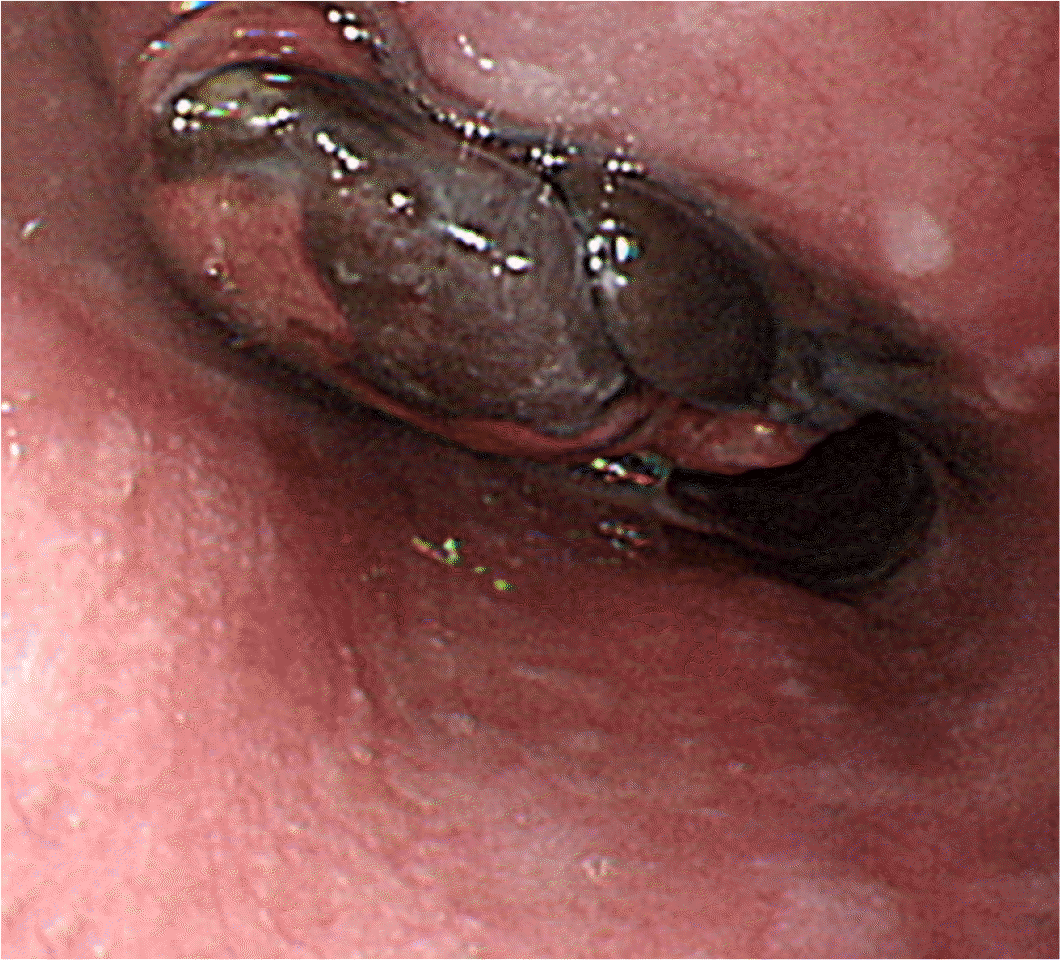
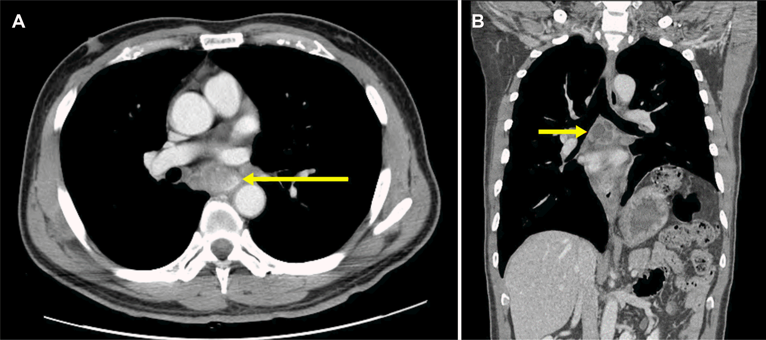
A 38-year-old woman was admitted to the Chonnam National University Hwasun Hospital with a 3-month history of dysphagia and odynophagia. On admission, the patient’s vital signs were normal, and she denied any previous relevant medical history, including gastrointestinal disease, abdominal surgery, or significant medical illness. A physical examination revealed no cutaneous pigmented lesions, lymphadenopathy, or hepatosplenomegaly. Laboratory investigations showed normal hemoglobin and hematocrit levels, white blood cell count, and hepatic and renal function. The CEA level was 0.845 ng/mL, and CA 19-9 was 5 U/mL, which was all within the normal ranges. The LDH level was increased to 722 U/L. Esophagogastroduodenoscopy (EGD) revealed a 4.0 cm sized black pigmented polypoid mass in the mid-esophagus (Fig. 1), and a biopsy was performed from the lesion. Chest CT revealed eccentric irregular wall thickening with enhancement in the mid-esophagus and multiple necrotizing lymphadenopathies at the carina (Fig. 2). Abdominal CT showed no definite abnormalities. The routine histology using H&E revealed brown-colored atypical melanocytes (Fig. 3A).
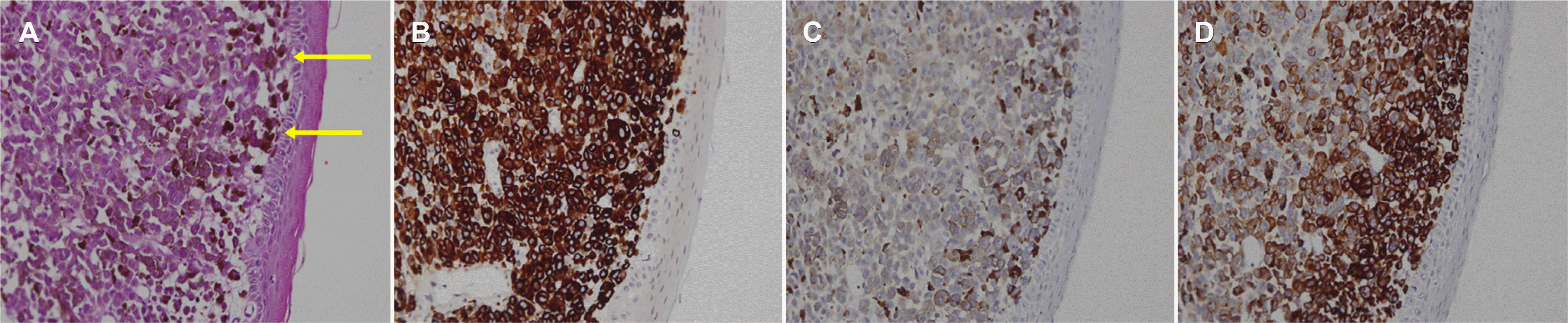
Immunohistochemical staining was performed to clarify the nature of the cells. The tumor cells exhibited strong reactivity for Melan A, S-100, and HMB45 proteins (Fig. 3B-D). The biopsy specimens were interpreted to be malignant melanomas. Torso PET-CT showed esophageal cancer without evidence of a distant metastasis. The patient was diagnosed with stage III disease (T3N1M0). Chemotherapy was performed. Unfortunately, the follow-up endoscopy after 3 months showed disease progression. The patients expired 10 months after the diagnosis of PEM.
2. Case 2
A 59-year-old man was admitted to the Chonnam National University Hwasun Hospital with a 2-month history of dysphagia. The CEA level was 2.06 ng/mL, and the CA 19-9 level was 2.37 U/mL. EGD revealed a 2.5 cm sized protruding mass without black pigmentation in the mid-esophagus (Fig. 4A), and a biopsy was performed on the lesion. Chest CT revealed a 1.1 cm-sized heterogeneously enhanced lesion in the mid-esophagus. PET-CT showed no distant metastasis. The patient was diagnosed with stage IA disease (T1N0M0). Esophagectomy and intrathoracic esophagogastrostomy were performed. The specimen showed a 2.5×1.1 cm sized fungating mass 9.5 cm from the esophagogastric junction. In microscopy, tumor cells showed muscularis mucosa invasion without lymph node involvement. In the immunohistochemical stains, tumor cells showed strong reactivity for HMB45 proteins. The patient was diagnosed with malignant melanoma with a muscularis mucosa invasion. Thirteen months after surgery, the follow-up examinations revealed multiple lung, pleura, thyroid, bone, peritoneum, and lymph node metastases. The patient declined further treatment and was transferred to a hospice facility. The patients expired 14 months after diagnosis.

3. Case 3
A 66-year-old man visited the outpatient clinic for health screening. The CEA level was 1.58 ng/mL, and the CA 19-9 level was 2.0 U/mL. Chest CT showed focal enhancing wall thickening in the distal esophagus. PET-CT showed no distant metastasis. EGD revealed multiple flat and black pigmented lesions in the lower esophagus with a maximum size of 2.0 cm (Fig. 4B). H&E and immunohistochemical staining of the biopsy specimens confirmed malignant melanoma. The patient was diagnosed with stage IA disease (T1N0M0). Accordingly, esophagectomy and intrathoracic esophagogastrostomy were performed. The specimen showed a 5.5×5.0 cm sized irregular lesion 5 cm from the esophagogastric junction. The tumor cells exhibited strong reactivity for Melan A and HMB45 proteins. No lymph node metastases were noted, and the resection margins showed no tumor cell involvement. The patient was diagnosed with malignant melanoma in situ. After surgery, he was alive 36 months after surgery without evidence of recurrence in the follow-up CT and EGD.
4. Case 4
A 61-year-old man was admitted to the Chonnam National University Hwasun Hospital with a 2-month history of epigastric pain and dysphagia. The CEA level was 2.53 ng/mL. EGD revealed a 2.0 cm-sized, black-pigmented polypoid mass in the mid-esophagus. H&E and immunohistochemical staining of the biopsy specimens confirmed malignant melanoma. Chest CT revealed eccentric irregular wall thickening with enhancement in the mid-esophagus, multiple pulmonary metastases, and necrotizing lymphadenopathies at the carina. The patient was diagnosed with stage IV disease (T3N1M1), but he declined further treatment. The patient expired 13 months after diagnosis. Informed consent was obtained from all four patients for the purpose of publication.
Go to :
DISCUSSION
PEM is a rare and aggressive disease, accounting for only 0.1-0.5% of malignant tumors of the esophagus,1,3 and has a poor prognosis. To date, 22 cases of PEM (including those reported in the present article) have been reported in Korea. Table 1 lists the clinical features, management, and prognosis of 22 patients with PEM reported in Korea.4-19
Table 1
Summary of the Reported Cases of Primary Esophageal Melanoma in Korea
| Patient No | Study | Age (years)/Sex | Symptoms (duration) | Location | Endoscopic finding | Stage (TNM) | Metastasis | Treatment | Follow up period (months) | Outcome |
|---|---|---|---|---|---|---|---|---|---|---|
| 1 | Kim et al. (1991)4 | 28/Female | Dysphagia, odynophagia (6 months) | Lower | Protruding mass with yellowish discoloration | IV (NA) | LN | RT | NA | NA |
| 2 | Park et al. (1997)5 | 47/Male | Dysphagia (3 months) | Lower | Multiple black pigmented polypoid mass | I (T1N0M0) | Absent | Surgery | NA | NA |
| 3 | Jung et al. (1997)6 | 58/Male | Dysphagia, substernal pain (3 months) | Mid | Protruding mass with friable mucosa and easy touch bleeding | NA | NA | Surgery | 8 | Alive |
| 4 | Lee et al. (1998)7 | 60/Male | Dysphagia, epigastric discomfort (3 months) | Lower | Protruding mass with central ulceration | IIB (T2N1M0) | LN | Surgery | NA | NA |
| 5 | Park et al. (1998)8 | 36/Female | Dysphagia, odynophagia (2 months) | Mid | Protruding mass | IIB (T1N1M0) | LN | Surgery | 11 | Recurrence (stomach) |
| 6 | Lee et al. (1998)9 | 47/Male | Dysphagia (3 months) | Lower | Black pigmented polypoid mass | I (T1N0M0) | Absent | Surgery | 4 | Death (liver metastasis) |
| 7 | Lee et al. (1998)9 | 39/Female | Dysphagia (6 months) | Lower | Protruding mass with central ulceration | I (T1N0M0) | Absent | Surgery | 4 | Alive |
| 8 | Lee et al. (2001)10 | 67/Male | Epigastric discomfort, dyspepsia (1 month) | Lower | Black pigmented polypoid mass | I (T1N0M0) | Absent | Refuse | 8 | Alive |
| 9 | Lee et al. (2007)11 | 58/Male | Dysphagia, substernal discomfort (4 months) | Lower | Protruding mass | IIA (T2N0M0) | Absent | Surgery | 9 | Recurrence (brain, liver, lung metastases) |
| 10 | Lee et al. (2004)12 | 46/Male | Dysphagia (3 months) | Lower | Black pigmented polypoid mass with friable mucosa and easy touch bleeding | IV (NA) | Liver, LN, brain | Chemothe rapy + RT | 1 | Death |
| 11 | Park et al. (2006)13 | 55/Female | Dysphagia, weight loss (1 month) | Lower | Black pigmented polypoid mass | IV (NA) | LN | Surgery | 10 | NA |
| 12 | Lee et al. (2007)14 | 60/Male | Dysphagia, upper abdominal discomfort (3 months) | Lower | Black pigmented polypoid mass | IVA (T3N0M1) | LN at the lesser curvature of the stomach | Surgery + chemotherapy | 35 | Alive |
| 13 | Lee et al. (2007)14 | 63/Male | Dysphagia, odynophagia (2 months) | Lower | Protruding mass with friable mucosa and easy touch bleeding | IV (NA) | LN, lung | Chemothe rapy + RT | 4 | Progression (skin, perirenal, retroperitoneal, inguinal metastases) |
| 14 | Kim et al. (2009)15 | 56/Male | Absent | Lower | Black pigmented and flat lesion | I (T1N0M0) | Absent | Surgery | 4 | Alive |
| 15 | Kang et al. (2009)16 | 56/Male | Epigastric pain (10 days) | Lower | Multiple black pigmented and flat lesions | I (T1N0M0) | Absent | NA | NA | NA |
| 16 | Kim et al. (2011)17 | 61/Male | Dysphagia (1 month) | Mid | Black pigmented polypoid mass | III (T3N0M0) | Absent | Surgery + RT | 4 | Progression (LN, bone metastases) |
| 17 | Lee et al. (2015)18 | 51/Female | Epigastric discomfort, belching | Mid | Multiple black pigmented and flat lesions | IA (T1N0M0) | Absent | Surgery | 15 | Alive |
| 18 | Choi et al. (2018)19 | 65/Male | Absent | Lower | Multiple black pigmented and flat lesions | IA (T1N0M0) | Absent | Surgery | NA | NA |
| 19 | Present case 1 | 38/Male | Dysphagia, odynophagia (3 months) | Mid | Black pigmented polypoid mass | III (T3N1M0) | LN | Chemothe rapy | 7 | Alive |
| 20 | Present case 2 | 59/Male | Dysphagia (2 months) | Mid | Protruding mass | IA (T1N0M0) | Absent | Surgery | 13 | Progression (lung, pleura, thyroid, bone, peritoneum, LN) |
| 21 | Present case 3 | 66/Male | Absent | Lower | Multiple black pigmented and flat lesions | IA (T1N0M0) | Absent | Surgery | 33 | Alive |
| 22 | Present case 4 | 61/Male | Epigastric pain, dysphagia (2 months) | Mid | Black pigmented polypoid mass | IV (T3N1M1) | LN, lung | NA | NA | NA |
![]()
The patients with PEM ranged in age from 28 to 67 years (mean age, 53.6 years), of whom 17 (77.2%) were male and five (22.8%) were female. The most common symptom was dysphagia (72.7% [16/22]), followed by epigastric pain and discomfort, odynophagia, and weight loss. These clinical findings are non-specific manifestations and are similar to those of other esophageal malignancies.1,3
Regarding the endoscopic features, the most frequently involved site was the lower esophagus (68.2% [15/22]), followed by the mid-esophagus (31.8% [7/22]). The frequency of observation was in the order of a black-pigmented polypoid mass (40.9% [9/22]), protruding mass without black pigmentation (36.4% [8/22]), and black pigmented and flat lesions (22.7% [5/22]).
Routine H&E of the endoscopic biopsy PEM specimens typically displays brown-colored atypical melanocytes. Histopathological diagnosis of PEM can be made using immunohistochemical staining of Melan A, S-100, and HMB45 proteins. In the present study, most cases were confirmed as malignant melanoma by a combination of H&E and immunohistochemical staining of endoscopic biopsy specimens, which was consistent with previous reports.1,3
The staging workup was available for all cases reviewed, except for one. According to the TNM staging system (from the American Joint Committee on Cancer),20 10 (47.6%) were stage I, three (14.3%) were stage II, two (9.5%) were stage III, and six (28.6%) were stage IV. Regarding lymph node metastases, none were found in 12 (57.1%) cases, but lymph node involvement was found in nine (42.9%) cases. No distant metastasis was found in 19 patients (85.7%), and distant metastasis was found in three (14.3%). Distant metastatic sites included the lung, liver, and brain.
To date, the treatment guidelines for PEM have not been established. Surgery, chemotherapy, RT, or combination therapy has been applied in the treatment of PEM. A radical surgical resection is still recommended as the primary treatment for PEM, but its therapeutic efficacy is unclear because of its rarity.1,3 In the present study, 13 patients underwent surgery, one chemotherapy, and one RT. Two patients underwent combination chemotherapy plus RT. One patient underwent combination surgery plus chemotherapy, and another received RT.
In the present study, data regarding follow-up period or clinical outcome were available for 16 and 15 cases, respectively, of all the cases reviewed. Seven patients experienced recurrence, metastasis, and death after treatment. The most common metastatic sites included the lymph nodes, followed by the lung, liver, and bone. The mean follow-up duration of patients with available data was 10.6 months (range, 1-33 months). Follow-up data for 46.7% of patients indicated recurrence, distant metastasis, and death during follow-up. Owing to the short follow-up period and the small number of patients in the present study, it was difficult to evaluate the prognosis accurately. In previous reports, the five-year survival rate ranged from 4% to 37%.1,3
In Korea, PEMs are prevalent in males in their 6th decade of life. Regarding the endoscopic features, the most frequently involved site was the lower esophagus, and black pigmented polypoid mass and flat lesions were observed predominantly. PEMs were treated in various ways, including surgery, chemotherapy, RT, or combination therapy. The prognosis is abysmal owing to the high recurrence rate and distant metastases. In summary, PEM in Korea is a rare disease characterized by aggressive behavior, early metastasis, and poor prognosis.
Go to :
 XML Download