Journal List > Korean J Orthod > v.52(3) > 1516078644

Yassir, Nabbat, McIntyre, and Bearn: Which anchorage device is the best during retraction of anterior teeth? An overview of systematic reviews

Abstract

Objective

To evaluate the available evidence regarding the clinical effectiveness of different types of anchorage devices.

Methods

A comprehensive literature search of different electronic databases was conducted for systematic reviews investigating different anchorage methods published up to April 15, 2021. Any ongoing systematic reviews were searched using PROSPERO, and a grey literature search was undertaken using Google Scholar and OpenGrey. No language restriction was applied. Screening, quality assessment, and data extraction were performed independently by two authors. Information was categorized and narratively synthesized for the key findings from moderate- and high-quality reviews.

Results

Fourteen systematic reviews were included (11 were of moderate/high quality). Skeletal anchorage with miniscrews was associated with less anchorage loss (and sometimes with anchorage gain). Similarly, skeletal anchorage was more effective in retracting anterior teeth and intruding incisors and molars, resulting in minor vertical skeletal changes and improvements in the soft tissue profile. However, insufficient evidence was obtained for the preference of any anchorage method in terms of the duration of treatment, number of appointments, quality of treatment, patient perception, or adverse effects. The effectiveness of skeletal anchorage can be enhanced when directly loaded, used in the mandible rather than the maxilla, used buccally rather than palatally, using dual rather than single miniscrews, used for en-masse retraction, and in adults.

Conclusions

The level of evidence regarding anchorage effectiveness is moderate. Nevertheless, compared to conventional anchorage, skeletal anchorage can be used with more anchorage preservation. Further high-quality randomized clinical trials are required to confirm these findings.

INTRODUCTION

Orthodontic treatment of malocclusion such as a full Class II relationship, dentoalveolar protrusion, or severe crowding often requires premolar extraction. The treatment outcomes in these cases depend on the closure of the extraction spaces while adequately controlling the anchorage teeth.1 Orthodontic anchorage is defined as the resistance to unwanted tooth movement.2 Anchorage control is of great importance in extraction and during the space closure stage.3,4 Conventionally, different methods and devices have been used for anchorage control, e.g., bonding of second molars, the use of a transpalatal arch (TPA) with or without a Nance button, lingual arches, headgear, or intermaxillary elastics.1 Each method can be used according to the clinical situation and has its advantages and disadvantages. For instance, extraoral appliances require greater patient compliance5 and may be associated with facial trauma.6,7 In contrast, intraoral appliances (such as TPAs) have not been shown to be effective despite being widely used.8 Recently, orthodontic temporary anchorage devices (TADs) were introduced as skeletal anchorage devices that can provide maximum to absolute anchorage9,10 and are compliance-free. TADs can take the form of implants, plates, screws, or screw-retained devices, which are inserted into the bone to provide resistance to unwanted tooth movement (indirect anchorage) or serve as a point from which orthodontic traction can be applied (direct anchorage).11,12
Studies have suggested that anchorage could be better preserved when using two-step retraction by initially retracting the canine followed by retraction of the incisors than when using en-masse retraction of the six anterior teeth. However, this is still a controversial subject clinically, especially when considering the variety of anchorage designs.13,14
Although TPAs have been found to have very limited effectiveness in terms of anchorage control, especially the mesial movement of the anchored molars, by using finite element analysis,15,16 some clinical trials have suggested using them as secondary anchorage devices when maximum anchorage is not required.9,17-19
Several systematic reviews8,20-32 have compared the clinical effectiveness of different types of anchorage devices in terms of the amount of anchorage loss; sagittal and vertical dental, skeletal, and soft tissue changes; duration of treatment; quality of treatment; patient perception; and adverse effects. However, to date, researchers have not provided a robust and clear answer to the following question: which anchorage method is the best during retraction of anterior teeth according to these different treatment outcomes?
This overview was designed to evaluate the available evidence (on a systematic review level) regarding the clinical effectiveness of different types of anchorage devices during the retraction of anterior teeth. The reason for conducting an overview of systematic reviews was that all the available systematic reviews varied in terms of the types of anchorage devices, method and time of assessment, types of tooth movement, and treatment outcomes. Therefore, this overview aimed to gather all the available evidence for each intervention, evaluate the level of evidence, and then categorize the outcomes to translate the evidence into practice. This overview should be able to identify what is known in this topic area, what remains unknown, and where investigators should focus their efforts in future research projects.

MATERIALS AND METHODS

Ethical approval was not required for this study as there was no individual participation, intervention, or personal data collection. This overview was prepared in line with the Preferred Reporting Items for Systematic Review and Meta-Analyses (PRISMA) statement,33 and according to the methodological guidelines for conducting overviews of systematic reviews of health care intervention, as provided by Smith et al.34

Protocol and registration

The protocol for the present review was registered with PROSPERO (registration number: CRD42020218197).

Eligibility criteria

The eligibility criteria were determined according to the Population, Intervention, Comparison, Outcome, and Study design (PICOS) scheme:
Population: patients of any age undergoing orthodontic treatment with fixed appliances and requiring retraction of anterior teeth with anchorage control after premolar extraction.
Intervention: orthodontic treatment with fixed appliances and any type of anchorage reinforcement device.
Comparison: orthodontic treatment with fixed appliances and any other type of anchorage reinforcement device.
Outcome: comparison of the effectiveness of anchorage devices.
Study design: systematic reviews with or without meta-analyses. In case of Cochrane reviews, the most recent publication was included, and all previous versions were excluded. Studies with any other design or without orthodontic treatment were also excluded, as were systematic reviews of in vitro or animal studies.

Information sources, search strategy, and study selection

A comprehensive literature search was conducted for systematic reviews published up to April 15, 2021 by using the following key terms: “anchorage”, “conventional”, “transpalatal”, “screw”, “implant”, “retraction”, “systematic review”, and “meta-analysis”. The search was accomplished using the following electronic databases: MEDLINE via OVID (1946 to April 15, 2021), EMBASE (1974 to April 15, 2021), AMED (Allied and Complementary Medicine Database, 1985 to April 15, 2021), PubMed (inception to April 15, 2021), and Web of Science (1900 to 2021). Any ongoing systematic reviews were searched using PROSPERO, and a grey literature search was undertaken using Google Scholar and OpenGrey (www.opengrey.eu/). No restrictions were applied in terms of language, date, and status of publication, or the age of treated patients. All relevant articles were identified, retrieved, and assessed for eligibility of inclusion by two authors (Y.A.Y. and S.A.N.). Any disagreement was resolved by discussion to reach consensus or, alternatively, by consulting a third author (D.R.B.).

Data items and collection

After screening the eligible systematic reviews, the following data were extracted independently and in duplicate by two authors (Y.A.Y. and S.A.N.): (1) authors; (2) year of publication; (3) study design; (4) number of studies included; (5) type of studies; (6) number of participants; (7) period of search; (8) name of journal; and (9) objectives of the study (Table 1).

Quality assessment in individual studies

Two authors (Y.A.Y. and S.A.N.) assessed the included reviews independently by using the AMSTAR 2 quality assessment tool (A Measurement Tool to Assess Systematic Reviews)35 (Table 2). Any disagreement was initially resolved by discussion or in conjunction with a third author (D.R.B.), if necessary. The level of evidence according to the AMSTAR 2 is presented in Table 3.

Summary measures and approach to synthesis

Data pooling was planned to qualitatively assess the effectiveness of the critically appraised topic as systematic reviews per se do not have primary data.

RESULTS

Study selection and characteristics

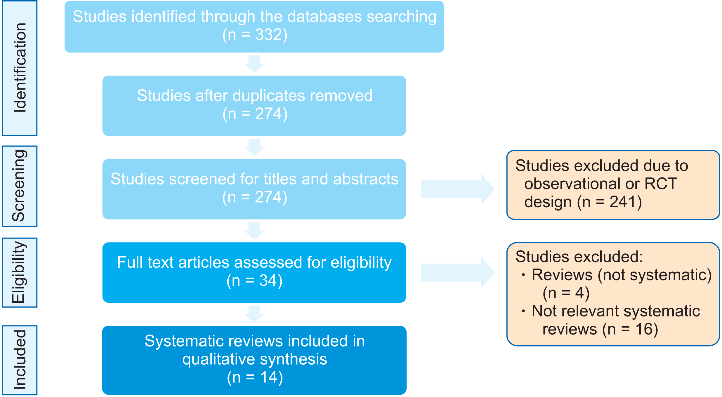
A total of 332 potentially eligible studies were identified. After excluding duplicates, 274 studies were left. Thereafter, initial screening of the titles and abstracts reduced the number of studies to 34. After full-text assessment, 20 reviews were excluded (4 reviews were not systematically designed or were older versions of Cochrane reviews and 16 were not relevant to the aim of this overview), and the remining 14 systematic reviews were included (Figure 1).
A summary of the characteristics of the included systematic reviews is presented in Table 1. The included studies were published between 2006 and 2021. Meta-analyses were carried out in 85.7% of the systematic reviews (12/14 systematic reviews).

Data synthesis

Owing to the lack of primary data, differences in types of anchorage devices used, method, and time of anchorage loss measurement, further meta-analysis was not possible. The data were, therefore, synthesized qualitatively by using thematic synthesis by identifying the most prominent and important themes with the findings summarized accordingly.

Quality of evidence

According to the AMSTAR 2 checklist, the quality of the included reviews was variable: 2 (14.3%) had critically low quality, 1 (7.1%) had low quality, 9 (64.3%) had moderate quality, and 2 (14.3%) had high quality (Table 1). Most of the AMSTAR 2 items were covered to a varying degree. Only one review22 reported the source of funding of the included studies (Table 2). Complete consensus was obtained between the reviewers regarding quality assessment.
In this overview, the main findings from the moderate- and high-quality systematic reviews were considered in the thematic synthesis. Therefore, the results of Feldmann and Bondemark8 (critically low quality), Li et al.20 (critically low quality), and Jayaratne et al.25 (low quality) were not considered further.

Method and time of measuring anchorage loss

Anchorage loss was measured using different methods, e.g., study model analysis (including three-dimensional methods), cephalometric analyses, or using reference points clinically. The time of measurement was also variable, i.e., from the start of treatment until achieving a Class I canine relationship or to the end of space closure, from the start to the end of the anchorage phase, during space closure, and at the start and end of active orthodontic treatment.22,30

Amount of anchorage loss

The mean anchorage loss, as represented by the mesial movement of the maxillary first molar, was significantly greater with conventional anchorage devices (TPAs, headgear, Nance appliances, banding of second molars, and differential anchorage methods) than with miniscrews.
The results of the included systematic reviews in terms of the amount of anchorage loss and the influence of anchorage method on vertical change of the maxillary first molar, anterior teeth retraction, vertical change of maxillary incisors, skeletal changes, soft tissue changes, duration of space closure, overall duration of treatment, number of appointments, quality of treatment, patient perception, and adverse effects are described in detail in Table 4.

DISCUSSION

Since the effectiveness of anchorage devices had been investigated by various heterogenous systematic reviews, the logical next step was to perform an overview of these reviews. This would allow the findings of these separate reviews to be appraised, compared, and contrasted in order to highlight and summarize the best available evidence from more than one systematic review in a single document. This consequently aids in evidence-based clinical decision-making.34,36

Amount of anchorage loss

All the included systematic reviews showed a consensus in terms of greater anchorage preservation with skeletal anchorage than with conventional anchorage. The amount of anchorage loss between the two methods was roughly 2 mm, and this can be considered of clinical importance since it represents about 25% of the extraction space. This consistent finding can be attributed to the fact that miniscrews are solely anchored by bone, and so they usually provide maximum anchorage with zero effect on the first molars. Alternatively, they either fail or migrate; however, the included systematic reviews provided no evidence regarding the side effects of migration. Becker et al.27 and Khlef et al.28 reported that skeletal anchorage not only preserved the anchorage but also induced distalization of the molars. This might be explained by the friction between the archwire and molar tube during the sliding of the wire during incisor retraction. This friction is the result of the direction of retraction (distal and intrusive) that could cause binding of the archwire in the molar tube, and hence, the force will be transmitted through the archwire to the dentition.27 The greater distal tipping of the molars with miniscrews, albeit non-statistically significant, could be the result of the lack of a reactive force on the molars during the retraction of the anterior teeth.23 Additionally, this distal tipping could be attributed to the friction between the archwire and molar tube when the distal force is transmitted through the archwire and results in distal tipping of the molars.
The finding of less anchorage loss with dual miniscrews than with a single miniscrew21,22 is logical and related to the greater stability and correspondingly lower failure with dual miniscrews. On the other hand, the reduced anchorage loss in the mandible than in the maxilla whether between miniscrews and conventional anchorage or between miniscrews themselves21 may be due to the higher density and less resilience of the mandibular bone, which provide more stability for skeletal anchorage, as miniscrews rely on mechanical retention and not osseointegration. The finding that anchorage loss was greater for younger patients could also be attributed to the quality of bone, which was denser with higher cortical thickness at specific sites in the maxilla and mandible of older patients than of younger patients, and hence provided greater stability for mesial molar movement.21,31 Nevertheless, further investigations are needed to confirm this finding.
Anchorage loss was greater when the miniscrew/implant was placed palatally rather than buccally.21,22 This can be correlated with another finding where greater preservation was observed with direct anchorage than with indirect anchorage.21,27 In general, with indirect anchorage, the force of retraction is applied to the tooth that is ligated by the miniscrew. Therefore, any resilience/deformation of the connecting wire, or loose ligation, can lead to anchorage loss. The fact that miniscrews that are placed palatally are mostly used for indirect anchorage can explain these findings.
According to the results of Papadopoulos et al.,21 one should be aware that any pretreatment mesial drifting of the molars can be a risk factor for anchorage loss. Hence, the implementation of skeletal anchorage may be necessary during treatment.
A TPA alone does not prevent molar mesial movement and is comparable to “no anchorage”. Using headgear can enhance anchorage when compared to that obtained using TPAs. This is due to the use of the extraoral skeletal component of the headgear. However, the headgear is only used part-time, and generally, it is less acceptable than are miniscrews. Furthermore, both TPAs and headgears showed greater anchorage loss than did miniscrews.24
When en-masse retraction with miniscrews was compared with two-step or en-masse retraction with conventional anchorage, the result was in line with the above findings and led to the conclusion that even with increasing numbers of retracted teeth, the skeletal anchorage did not have any adverse effect on the anchored teeth.28,29 Miniscrews were also more effective in preserving the anchorage than were conventional anchorage methods in the two-step retraction technique.32

Anchorage method and vertical change of the maxillary first molar

The vertical force vector associated with retraction using miniscrews explains the finding of intrusion of the molars,27-29,31 as the incisors will be subject to distal and intrusive force vectors due to the position of the miniscrew, and hence, the intrusive force will be transferred to the molars via the archwire. Therefore, the intrusive force on the molars while retracting the incisors with miniscrews could be beneficial for patients with clockwise rotation of the mandible, anterior openbite, and Class II malocclusion.28,31 Even minor intrusion of the posterior teeth of approximately 1 mm has been reported to produce a significant upward and forward movement of the chin of approximately 3–4 mm.37

Anchorage method and anterior teeth retraction

Although incisor retraction was greater with miniscrews and was statistically significant,23,26,28,29,31 this was of less clinical significance (0.46–1.5 mm) when compared to the amount of anchorage loss. The greater incisor retraction may be related to the greater stability of the bone than that of the tooth as an anchorage unit as well as to the greater space available for incisor retraction with miniscrews (due to less anchorage loss).23,28,31 The same is true for canine retraction.32
Incisor inclination and tipping depend on factors such as the size of the archwire, point of force application, and presence or absence of third-order bends, which do not have a direct relationship with the anchorage method.23 Khlef et al.28 reported that when using temporary skeletal anchorage devices, the incisors would be retracted via controlled tipping and bodily movement, whereas with conventional anchorage, the incisors would be retracted via controlled and uncontrolled tipping. Therefore, optimal incisor inclination can be achieved with skeletal anchorage devices.

Anchorage method and vertical change of the maxillary incisors

As with molar vertical changes, the incisors will be subject to an intrusive force when retracted with miniscrews.28,29,31 This is the effect of the line of force application that is higher with miniscrews than with conventional anchorage methods where the point of force application is on the molars, which may result in a downward vector and extrusion. The length of the power arm can also play a role in the amount of vertical change, as decreasing the height of the power arm results in increasing the intrusive force on the incisors.28

Anchorage method and skeletal changes

Since the effect of incisor retraction, whether with miniscrews or with conventional anchorage methods, is mainly dental, its reflection on skeletal components is mainly limited to the skeletal regions that are affected indirectly by the dentition. Consequently, the difference in incisor retraction between the different anchorage methods will rarely influence sagittal jaw position, and hence, it is insufficient to be shown as a difference in the sella-nasion-point A (SNA), sella-nasion-point B (SNB), and point A-nasion-point B (ANB) angles. However, intruding the molars with miniscrews and extruding them with conventional anchorage can influence the vertical skeletal measurements as shown with the Sella-Nasion to Mandibular plane angle (SN-MP) angle.26,29 Nevertheless, this difference can be masked if the amount of vertical molar change is minimal between the two anchorage methods, as detailed in the systematic review by Khlef et al.,28 where the molar vertical change was only 0.61 mm, and hence, no significant difference in the SN-MP angle was revealed. The meta-analyses by Xu and Xie26 and Khlef et al.29 found similar differences in the SN-MP angle between skeletal and conventional anchorage (1.12˚), which in general was not of clinical significance.

Anchorage method and soft tissue changes

The greater amount of incisor retraction with miniscrews can explain the increase in the nasolabial angle, greater reduction of the upper lip to the E-line, and the tendency of decreasing the facial convexity.26,28,29 However, it is difficult to explain that when the mean differences in incisor retraction between the two anchorage methods were 1.5 mm26 and 0.46 mm,28 the mean differences in the nasolabial angle increased by 3.52˚ and 4.73˚, respectively, unless a growth factor is considered to play a role in this process.

Anchorage method and duration of space closure/treatment

According to the decision algorithm by Jadad et al.,38 the evidence about the non-significantly shorter duration of treatment with miniscrews than with conventional anchorage22,28-30 outweighs that of the significantly shorter duration with miniscrews.23 This together with the non-significant difference between skeletal anchorage and conventional anchorage in the duration of extraction space closure22,30 could be explained by the same reason. If miniscrew failures occur, greater time will be required for bone healing before reinsertion and treatment resumption. Moreover, despite the more effective retraction with miniscrews as anchorage, the greater anchorage loss with conventional anchorage devices results in a smaller extraction space; hence, the time required to close that space with conventional anchorage will be shortened.30
The difference in the duration of space closure between single and dual miniscrews (2.19 months in favor of a single miniscrew) was not statistically significant, and was derived from the findings of only one study, and hence, no meta-analysis was performed.22 Therefore, this finding may be due to chance.

Anchorage method and number of appointments

The number of appointments to complete treatment correlated with the technique used for space closure. Since skeletal anchorage is usually used with en-masse retraction and conventional anchorage with two-step retraction, this may indicate the need for less appointments with skeletal anchorage than with conventional methods. However, the number of appointments was reported with contradicting results in two reviews,22,30 each of which took its information from one study, and did not consider factors such as the miniscrew failure rate and surgical healing of the mid-palatal implant.

Anchorage method and quality of treatment

Not enough evidence was obtained to determine which anchorage methods were associated with better outcomes, because the available information was derived from only one meta-analysis,30 which in turn obtained data from one study.

Anchorage method and patient perception

Both miniscrews and conventional anchorage methods resulted in a comparable level of patient discomfort as each had disadvantages. Miniscrews are associated with pain during insertion and removal, while conventional methods are characterized by their bulkiness, which is unpleasant for patients. However, the general feedback with miniscrews is positive, especially if they are the pre-drilling type. Interestingly, some of the discomfort reported by Jambi et al.22 was due to a surgical procedure that was not relevant to most of the skeletal anchorage.

Anchorage method and adverse effects

One meta-analysis22 found that the failure rate of the anchorage method with conventional anchorage methods was greater than that with miniscrews. However, miniscrew success depends on biological factors, while that of conventional methods is mainly dependent on the durability of the cementing material. Reusing conventional methods seems easier and quicker than using miniscrews, but evidence on this aspect is insufficient. Miniscrew failure rate was approximately 10–12%,21,27 and it did not differ between early and delayed loading.22

CONCLUSION

  1. The level of evidence regarding the effectiveness of anchorage methods is moderate. High-quality randomized clinical trials are hence warranted. According to the available evidence, skeletal anchorage with miniscrews is clinically more effective than are conventional anchorage methods, especially in preventing horizontal mesial molar movement.

  2. During space closure, skeletal anchorage is more effective than conventional anchorage for the following:

    • Retraction of anterior teeth.

    • Intrusion of the incisors and molars.

    • Minor decrease in the vertical skeletal relationship.

    • Improving the soft tissue profile.

  3. There was insufficient evidence to determine the benefit of specific anchorage methods for the following:

    • Duration of space closure and overall duration of treatment.

    • Number of appointments.

    • Quality of treatment.

    • Patient perception and adverse effects.

  4. The effectiveness of skeletal anchorage can be enhanced as follows:

    • When directly loaded.

    • When placed in the mandible rather than in the maxilla.

    • When placed buccally rather than palatally.

    • When using dual miniscrews rather than a single miniscrew.

    • When used for en-masse retraction.

    • When used in adult patients.

Notes

CONFLICTS OF INTEREST

No potential conflict of interest relevant to this article was reported.

REFERENCES

1. Wahl N. 2008; Orthodontics in 3 millennia. Chapter 15: skeletal anchorage. Am J Orthod Dentofacial Orthop. 134:707–10. DOI: 10.1016/j.ajodo.2008.04.015. PMID: 18984405.
crossref
2. Proffit WR, Fields HW, Sarver DM. 2006. Contemporary orthodontics. Elsevier Health Sciences;Louis: DOI: 10.1016/j.ajodo.2008.04.015.
3. Melsen B, Bosch C. 1997; Different approaches to anchorage: a survey and an evaluation. Angle Orthod. 67:23–30. DOI: 10.1043/0003-3219(1997)067<0023:DATAAS>2.3.CO;2. PMID: 9046396.
4. Lai EH, Yao CC, Chang JZ, Chen I, Chen YJ. 2008; Three-dimensional dental model analysis of treatment outcomes for protrusive maxillary dentition: comparison of headgear, miniscrew, and miniplate skeletal anchorage. Am J Orthod Dentofacial Orthop. 134:636–45. DOI: 10.1016/j.ajodo.2007.05.017. PMID: 18984395.
crossref
5. Cole WA. 2002; Accuracy of patient reporting as an indication of headgear compliance. Am J Orthod Dentofacial Orthop. 121:419–23. DOI: 10.1067/mod.2002.122369. PMID: 11997767.
crossref
6. Samuels RH, Willner F, Knox J, Jones ML. 1996; A national survey of orthodontic facebow injuries in the UK and Eire. Br J Orthod. 23:11–20. DOI: 10.1179/bjo.23.1.11. PMID: 8652493.
crossref
7. Blum-Hareuveni T, Rehany U, Rumelt S. 2004; Blinding endophthalmitis from orthodontic headgear. N Engl J Med. 351:2774–5. DOI: 10.1056/NEJM200412233512623. PMID: 15616219.
crossref
8. Feldmann I, Bondemark L. 2006; Orthodontic anchorage: a systematic review. Angle Orthod. 76:493–501.
9. Kuroda S, Yamada K, Deguchi T, Kyung HM, Takano-Yamamoto T. 2009; Class II malocclusion treated with miniscrew anchorage: comparison with traditional orthodontic mechanics outcomes. Am J Orthod Dentofacial Orthop. 135:302–9. DOI: 10.1016/j.ajodo.2007.03.038. PMID: 19268827.
crossref
10. Park HS, Kwon TG. 2004; Sliding mechanics with microscrew implant anchorage. Angle Orthod. 74:703–10. DOI: 10.1043/0003-3219(2004)074<0703:SMWMIA>2.0.CO;2. PMID: 15529508.
11. Odman J, Lekholm U, Jemt T, Thilander B. 1994; Osseointegrated implants as orthodontic anchorage in the treatment of partially edentulous adult patients. Eur J Orthod. 16:187–201. DOI: 10.1093/ejo/16.3.187. PMID: 8062859.
crossref
12. Ren Y. 2009; Mini-implants for direct or indirect orthodontic anchorage. Evid Based Dent. 10:113. DOI: 10.1038/sj.ebd.6400687. PMID: 20023620.
crossref
13. Heo W, Nahm DS, Baek SH. 2007; En masse retraction and two-step retraction of maxillary anterior teeth in adult Class I women. A comparison of anchorage loss. Angle Orthod. 77:973–8. DOI: 10.2319/111706-464.1. PMID: 18004930.
14. Xu TM, Zhang X, Oh HS, Boyd RL, Korn EL, Baumrind S. 2010; Randomized clinical trial comparing control of maxillary anchorage with 2 retraction techniques. Am J Orthod Dentofacial Orthop. 138:544.e1–9. discussion 544–5. DOI: 10.1016/j.ajodo.2009.12.027. PMID: 21055588.
crossref
15. Bobak V, Christiansen RL, Hollister SJ, Kohn DH. 1997; Stress-related molar responses to the transpalatal arch: a finite element analysis. Am J Orthod Dentofacial Orthop. 112:512–8. DOI: 10.1016/S0889-5406(97)90100-1. PMID: 9387838.
crossref
16. Kojima Y, Fukui H. 2008; Effects of transpalatal arch on molar movement produced by mesial force: a finite element simulation. Am J Orthod Dentofacial Orthop. 134:335.e1–7. discussion 335–6. DOI: 10.1016/j.ajodo.2008.03.011. PMID: 18774078.
crossref
17. Liu YH, Ding WH, Liu J, Li Q. 2009; Comparison of the differences in cephalometric parameters after active orthodontic treatment applying mini-screw implants or transpalatal arches in adult patients with bialveolar dental protrusion. J Oral Rehabil. 36:687–95. DOI: 10.1111/j.1365-2842.2009.01976.x. PMID: 19602104.
crossref
18. Sharma M, Sharma V, Khanna B. 2012; Mini-screw implant or transpalatal arch-mediated anchorage reinforcement during canine retraction: a randomized clinical trial. J Orthod. 39:102–10. DOI: 10.1179/14653121226878. PMID: 22773673.
crossref
19. Al-Sibaie S, Hajeer MY. 2014; Assessment of changes following en-masse retraction with mini-implants anchorage compared to two-step retraction with conventional anchorage in patients with class II division 1 malocclusion: a randomized controlled trial. Eur J Orthod. 36:275–83. DOI: 10.1093/ejo/cjt046. PMID: 23787192.
crossref
20. Li F, Hu HK, Chen JW, Liu ZP, Li GF, He SS, et al. 2011; Comparison of anchorage capacity between implant and headgear during anterior segment retraction. Angle Orthod. 81:915–22. DOI: 10.2319/101410-603.1. PMID: 21299412. PMCID: PMC8916170.
crossref
21. Papadopoulos MA, Papageorgiou SN, Zogakis IP. 2011; Clinical effectiveness of orthodontic miniscrew implants: a meta-analysis. J Dent Res. 90:969–76. DOI: 10.1177/0022034511409236. PMID: 21593250.
crossref
22. Jambi S, Walsh T, Sandler J, Benson PE, Skeggs RM, O'Brien KD. 2014; Reinforcement of anchorage during orthodontic brace treatment with implants or other surgical methods. Cochrane Database Syst Rev. 2014:CD005098. DOI: 10.1002/14651858.CD005098.pub3. PMID: 25135678. PMCID: PMC6464832.
crossref
23. Antoszewska-Smith J, Sarul M, Łyczek J, Konopka T, Kawala B. 2017; Effectiveness of orthodontic miniscrew implants in anchorage reinforcement during en-masse retraction: a systematic review and meta-analysis. Am J Orthod Dentofacial Orthop. 151:440–55. DOI: 10.1016/j.ajodo.2016.08.029. PMID: 28257728.
crossref
24. Diar-Bakirly S, Feres MF, Saltaji H, Flores-Mir C, El-Bialy T. 2017; Effectiveness of the transpalatal arch in controlling orthodontic anchorage in maxillary premolar extraction cases: a systematic review and meta-analysis. Angle Orthod. 87:147–58. DOI: 10.2319/021216-120.1. PMID: 27504820. PMCID: PMC8388582.
crossref
25. Jayaratne YSN, Uribe F, Janakiraman N. 2017; Maxillary incisors changes during space closure with conventional and skeletal anchorage methods: a systematic review. J Istanb Univ Fac Dent. 51(3 Suppl 1):S90–101. DOI: 10.17096/jiufd.52884. PMID: 29354313. PMCID: PMC5750832.
26. Xu Y, Xie J. 2017; Comparison of the effects of mini-implant and traditional anchorage on patients with maxillary dentoalveolar protrusion. Angle Orthod. 87:320–7. DOI: 10.2319/051016-375.1. PMID: 27684189. PMCID: PMC8384353.
crossref
27. Becker K, Pliska A, Busch C, Wilmes B, Wolf M, Drescher D. 2018; Efficacy of orthodontic mini implants for en masse retraction in the maxilla: a systematic review and meta-analysis. Int J Implant Dent. 4:35. DOI: 10.1186/s40729-018-0144-4. PMID: 30357551. PMCID: PMC6200826.
crossref
28. Khlef HN, Hajeer MY, Ajaj MA, Heshmeh O. 2018; Evaluation of treatment outcomes of En masse retraction with temporary skeletal anchorage devices in comparison with two-step retraction with conventional anchorage in patients with dentoalveolar protrusion: a systematic review and meta-analysis. Contemp Clin Dent. 9:513–23. DOI: 10.4103/ccd.ccd_661_18. PMID: 31772456. PMCID: PMC6868609.
crossref
29. Khlef HN, Hajeer MY, Ajaj MA, Heshmeh O. 2019; En-masse retraction of upper anterior teeth in adult patients with maxillary or bimaxillary dentoalveolar protrusion: a systematic review and meta-analysis. J Contemp Dent Pract. 20:113–27. DOI: 10.5005/jp-journals-10024-2485. PMID: 31058623.
crossref
30. Alharbi F, Almuzian M, Bearn D. 2019; Anchorage effectiveness of orthodontic miniscrews compared to headgear and transpalatal arches: a systematic review and meta-analysis. Acta Odontol Scand. 77:88–98. DOI: 10.1080/00016357.2018.1508742. PMID: 30350741.
crossref
31. Liu Y, Yang ZJ, Zhou J, Xiong P, Wang Q, Yang Y, et al. 2020; Comparison of anchorage efficiency of orthodontic mini-implant and conventional anchorage reinforcement in patients requiring maximum orthodontic anchorage: a systematic review and meta-analysis. J Evid Based Dent Pract. 20:101401. DOI: 10.1016/j.jebdp.2020.101401. PMID: 32473793.
crossref
32. Tian H, Xie C, Lin M, Yang H, Ren A. 2020; Effectiveness of orthodontic temporary anchorage devices in canine retraction and anchorage preservation during the two-step technique: a systematic review and meta-analysis. BMC Oral Health. 20:278. DOI: 10.1186/s12903-020-01271-8. PMID: 33036593. PMCID: PMC7547464. PMID: 3253d012366242d28e413266a7f1c4da.
crossref
33. Moher D, Liberati A, Tetzlaff J, Altman DG. PRISMA Group. 2010; Preferred reporting items for systematic reviews and meta-analyses: the PRISMA statement. Int J Surg. 8:336–41. DOI: 10.1016/j.ijsu.2010.02.007. PMID: 20171303.
crossref
34. Smith V, Devane D, Begley CM, Clarke M. 2011; Methodology in conducting a systematic review of systematic reviews of healthcare interventions. BMC Med Res Methodol. 11:15. DOI: 10.1186/1471-2288-11-15. PMID: 21291558. PMCID: PMC3039637.
crossref
35. Shea BJ, Reeves BC, Wells G, Thuku M, Hamel C, Moran J, et al. 2017; AMSTAR 2: a critical appraisal tool for systematic reviews that include randomised or non-randomised studies of healthcare interventions, or both. BMJ. 358:j4008. DOI: 10.1136/bmj.j4008. PMID: 28935701. PMCID: PMC5833365.
crossref
36. Becker L, Oxman AD. Higgins JP, Green S, editors. 2009. Overviews of reviews. Cochrane handbook for systematic reviews of interventions. The Cochrane Collaboration;Oxford: DOI: 10.1002/9780470712184.ch22.
crossref
37. Upadhyay M, Yadav S, Nagaraj K, Patil S. 2008; Treatment effects of mini-implants for en-masse retraction of anterior teeth in bialveolar dental protrusion patients: a randomized controlled trial. Am J Orthod Dentofacial Orthop. 134:18–29.e1. DOI: 10.1016/j.ajodo.2007.03.025. PMID: 18617099.
crossref
38. Jadad AR, Cook DJ, Browman GP. 1997; A guide to interpreting discordant systematic reviews. CMAJ. 156:1411–6. PMID: 9164400. PMCID: PMC1227410.

Figure 1
PRISMA (Preferred Reporting Items for Systematic Review and Meta-Analyses) flow diagram of the literature selection process.
RCT, randomized controlled trial.
kjod-52-3-220-f1.tif
Table 1
Systematic reviews that assessed anchorage reinforcement devices during orthodontic treatment
Author Year Study design No. of studies No. of participants Intervention anchorage Comparison anchorage Type of studies Period of search Journal Main outcomes Quality of evidence
Feldmann and Bondemark8 2006 Systematic review 14 (7 of these are related to the aim of the current study) 388 Space closure with different techniques Different anchorage devices/methods 2 RCTs January, 1966 to December, 2004 Angle Orthodontist Due to contradictory results and the vast heterogeneity in study methods, the scientific evidence was too weak to evaluate anchorage efficiency during space closure Critically low
3 Retrospective studies Score: 4
2 Prospective studies
Li et al.20 2011 Systematic review and meta-analysis 8 392 Midpalatal implant, mini-implant, and onplant Headgear 4 RCTs Not reported Angle Orthodontist The skeletal anchorage of the midpalatal implant, mini-implant, and onplant offer better alternatives to headgear, with less anchorage loss and more anterior teeth retraction Critically low
1 Prospective cohort study Score: 7
3 Retrospective studies
Papadopoulos et al.21 2011 Meta-analysis 8 206 Mini-implants Different types (TPA, headgear, banding the second molar and application of differential moments). 3 RCTs Up to June, 2010 Journal of Dental Research The use of mini-implants significantly decreased or negated loss of anchorage Moderate
Mini-implants were also compared according to location, number, and etc. 5 CCTs Score: 10
Jambi et al.22 2014 Systematic review and meta-analysis (Cochrane Review) 15 (11 for meta-analysis) 561 Mid-palatal implants, onplants, mini-screw implants, spider screws, titanium plates and zygomatic wires Conventional methods (headgear, chin caps, face masks, transpalatal arches, Nance buttons, lingual arches and interarch elastics). RCTs Up to October 28, 2013 Cochrane Database of Systematic Reviews Reinforcement of anchorage is more effective with surgical anchorage than conventional anchorage methods High
Studies with two methods of surgically assisted anchorage were also included. Score: 15
Antoszewska-Smith et al.23 2017 Systematic review and meta-analysis 14 616 Miniscrew and miniplate TPA and headgear 7 RCTs 1990 to March, 2016 American Journal of Orthodontics and Dentofacial Orthopedics Skeletal anchorage devices are more effective for en-masse retraction than conventional methods of anchorage reinforcement Moderate
7 CCTs Score: 10
Diar-Bakirly et al.24 2017 Systematic review and meta-analysis 14 (13 for meta-analysis) 579 TPA Other types of anchorage including skeletal anchorage (miniscrews, onplants), and headgear 9 RCTs Up to April 2015 Angle Orthodontist Transpalatal arch alone should not be recommended to provide maximum anchorage during retraction of anterior teeth in extraction cases Moderate
5 Non-RCTs Score:11
Jayaratne et al.25 2017 Systematic review 6 327 Mini-implants Different types (TPA, headgear, banding the second molar and application of differential moments) RCTs Up to May, 2015 Journal of Istanbul University Faculty of Dentistry The amount of incisor retraction and intrusion was greater with buccally placed mini-implants when compared to conventional anchorage techniques Low
Score: 8
Xu and Xie26 2017 Systematic review and meta-analysis 14 450 Mini-implants Conventional anchorage 8 RCTs December, 1966 to March, 2016 Angle Orthodontist Mini-implant anchorage was more effective in retracting the anterior teeth, produced less anchorage loss, and had a greater effect on SN-MP for the high-angle patients than did conventional anchorage Moderate
6 CCTs Score: 9
Alharbi et al.30 2019 Systematic review and meta-analysis 7 (6 for meta-analysis) 271 Miniscrews Different types (TPA, headgear, banding the second molar and application of differential moments) RCTs Up to March 16, 2018 Acta Odontologica Scandinavica Miniscrews are more effective in preserving orthodontic anchorage than conventional appliances Moderate
Score: 14
Becker et al.27 2018 Systematic review and meta-analysis 12 (7 for meta-analysis) 393 Mini-implants Different types (TPA, headgear, Nance button, lingual arch, mushroom loops, intrusion arch, banding the second molar, application of different moments) 9 RCTs 1992 to December 31, 2017 International Journal of Implant Dentistry Maximum anchorage en-masse retraction can be achieved by orthodontic mini-implants and direct anchorage Moderate
1 CCT Moderate
Score: 12
1 Cohort study
Khlef et al.28 2018 Systematic review and meta-analysis 4 150 En-masse retraction of the upper anterior teeth associated skeletal anchorage (mini-implant, miniplates, and C-tube) Two-step retraction of the upper anterior associated with conventional anchorage 2 RCTs January, 1990 to April, 2018 Contemporary Clinical Dentistry There is a very weak-to-moderate evidence that using skeletal anchorage devices with en-masse retraction would cause better posterior anchorage and incisors inclination, and greater anterior teeth retraction than using conventional anchorage with two-step retraction Moderate
2 CCTs Score: 14
Khlef et al.29 2019 Systematic review and meta-analysis 8 (5 for meta-analysis) 255 En-masse retraction of maxillary anterior teeth with skeletal anchorage En-masse retraction of maxillary anterior teeth with conventional or no anchorage system and without any approach for tooth movement acceleration 6 RCTs January, 1990 to April, 2018 The Journal of Contemporary Dental Practice There is weak to moderate evidence that using skeletal anchorage devices would lead to better posterior anchorage than using conventional anchorage Moderate
2 CCTs Score: 13
Liu et al.31 2020 Systematic review and meta-analysis 12 Not reported Mini-implants Different types (TPA, headgear, Nance button, lingual arch) 4 RCTs Up to July, 2018 The Journal of Evidence-Based Dental Practice Mini-implants seem to be more effective than the conventional anchorage devices in terms of minimizing unintended mesial movement of molars with maximum retraction of anterior teeth High
3 Prospective controlled trials Score: 13
5 Retrospective studies
Tian et al.32 2020 Systematic review and meta-analysis 8 146 Miniscrew (during the first phase of the two-step retraction technique) Different types (TPA, lingual arch, and dental anchorage) 3 RCTs Up to June 30, 2019 BMC Oral Health Anchorage with miniscrew is more efficient than conventional anchorage during canine retraction Moderate
5 CCTs Score: 12

Score of each review represents the number of “YES” answers in the A Measurement Tool to Assess Systematic Reviews (AMSTAR 2) checklist. However, this may not always reflect the quality as items do not have the same weight.

RCT, randomized controlled trial; CCT, controlled clinical trial; TPA, transpalatal arch; SN-MP, sella-nasion to mandibular plane angle.

Table 2
A Measurement Tool to Assess Systematic Reviews (AMSTAR 2) items
AMSTAR 2 items Meeting the criteria
Yes Partial yes No
1. Did the research questions and inclusion criteria for the review include the components of PICO? 13 1
2. Did the report of the review contain an explicit statement that the review methods were established prior to the conduct of the review and did the report justify any significant deviations from the protocol? 8 6
3. Did the review authors explain their selection of the study designs for inclusion in the review? 14
4. Did the review authors use a comprehensive literature search strategy? 6 7 1
5. Did the review authors perform study selection in duplicate? 14
6. Did the review authors perform data extraction in duplicate? 14
7. Did the review authors provide a list of excluded studies and justify the exclusions? 7 4 3
8. Did the review authors describe the included studies in adequate detail? 5 7 2
9. Did the review authors use a satisfactory technique for assessing the risk of bias in individual studies that were included in the review? 12 1 1
10. Did the review authors report on the sources of funding for the studies included in the review? 1 13
11. If meta-analysis was performed did the review authors use appropriate methods for statistical combination of results? 12
12. If meta-analysis was performed, did the review authors assess the potential impact of risk of bias in individual studies on the results of the meta-analysis or other evidence synthesis? 11 1
13. Did the review authors account for risk of bias in individual studies when interpreting/ discussing the results of the review? 11 3
14. Did the review authors provide a satisfactory explanation for, and discussion of, any heterogeneity observed in the results of the review? 12 2
15. If they performed quantitative synthesis did the review authors carry out an adequate investigation of publication bias (small study bias) and discuss its likely impact on the results of the review? 5 7
16. Did the review authors report any potential sources of conflict of interest, including any funding they received for conducting the review? 8 6

PICO, Population, Intervention, Comparison, and Outcome.

Table 3
Level of evidence according to the AMSTAR 2 assessment tool
Level Description
High No or one non-critical weakness: the systematic review provides an accurate and comprehensive summary of the results of the available studies that address the question of interest.
Moderate More than one non-critical weakness*: the systematic review has more than one weakness but no critical flaws. It may provide an accurate summary of the results of the available studies that were included in the review.
Low One critical flaw with or without non-critical weaknesses: the review has a critical flaw and may not provide an accurate and comprehensive summary of the available studies that address the question of interest.
Critically low More than one critical flaw with or without non-critical weaknesses: the review has more than one critical flaw and should not be relied on to provide an accurate and comprehensive summary of the available studies.

AMSTAR 2, A Measurement Tool to Assess Systematic Reviews.

*Multiple non-critical weaknesses may diminish the confidence in the review, and it may be appropriate to move the overall appraisal down from moderate to low confidence.

Table 4
The results of the included systematic reviews in terms of anchorage methods and their influence on different factors
Amount of anchorage loss
Buccal miniscrews vs. conventional anchorage
  • The mean difference of anchorage loss between miniscrews and conventional anchorage was −2.4 mm (95% CI: −2.9 to −1.8 mm),21 −1.68 mm (95% CI: −2.27 to −1.09 mm),22 −1.87 mm (95% CI: −2.21 to −1.53 mm),23 −2.01 mm (95% CI: −2.45 to −1.58 mm),26 −1.94 mm (95% CI: −3.46 to −0.42 mm)30

  • Miniscrews were more effective for anchorage reinforcement than conventional anchorage methods in the mandible (mean difference −3.1 mm) than in the maxilla (mean difference −2.2 mm) and in adults than in young patients.21

  • There was greater but non-statistically significant distal molar tipping with miniscrews when compared to conventional anchorage methods.23 On the other hand, another meta-analysis32 reported (from one study) that mesial tipping of maxillary and mandibular molars was significantly greater with the conventional anchorage methods than with miniscrews by 2.15˚ and 2.5˚, respectively.

  • Anchorage loss was significantly lower in the miniscrew group when compared to TPA alone (mean difference −2.09 mm, 95% CI: −2.38 to −1.8 mm), TPA and headgear (mean difference −1.71 mm, 95% CI: −2.6 to −0.81 mm), and TPA and utility arch (mean difference −0.63 mm, 95% CI: −1.15 to −0.12 mm).24

  • Miniscrews are either associated with no anchorage loss or with “anchorage gain” in contrast to the conventional anchorage methods (mainly TPA) and the significant mean difference was −2.79 mm (95% CI: −3.56 to −2.03 mm).27

  • Miniscrews achieved maximum anchorage with significantly less mesial movement of first molar of −1.48 mm (95% CI: −2.25 to −0.72 mm) than conventional anchorage. This difference between the two methods was greater for patients aged less than 18 years (−2.36 mm, 95% CI: −4.18 to −0.53 mm) than those older than 18 years (−1.2 mm, 95% CI: −2.01 to −0.39 mm).31

Mid-palatal implant vs. conventional anchorage
  • Anchorage loss was greater with conventional anchorage compared to mid-palatal implants (mean difference −1.02 mm, 95% CI: −2.31 to 0.26 mm) and alveolar miniscrews (mean difference −2.17 mm, 95% CI: −2.58 to −1.77 mm).22

Different applications of miniscrews
  • Anchorage loss with miniscrews was significantly lower in the following situations: when the miniscrews were placed in the mandible than in the maxilla (−0.6 mm vs. 0.2 mm), when the miniscrews were placed between the second premolar and first molar than palatally (−0.2 mm vs. 1.3 mm), when two miniscrews were placed rather than one (−0.2 mm vs. 1.3 mm), when miniscrews were loaded directly rather than indirectly (−0.2 mm vs. 0.8 mm), and when there was absence of pre-treatment space loss rather than existing loss (−0.4 mm vs. 0.9 mm).21

  • Anchorage loss was in favor of dual miniscrews than single miniscrews (mean difference −1.62 mm, 95% CI: −2.26 to −0.98 mm).22

  • Indirect anchorage with miniscrews was associated with greater anchorage loss than that of direct anchorage with miniscrews but still lower than that of the conventional anchorage methods.27

En-masse vs. two-step retraction
  • En-masse retraction with miniscrews andtwo-step retractionwith conventional anchorage methods: the maxillary first molar was moved distally with miniscrews and moved mesially with conventional anchorage with a significant mean difference of −3.03 mm (95% CI: −3.65 to −2.42 mm).28

  • En-masse retraction with miniscrews and conventional anchorage: There was significantly greater anchorage loss with conventional anchorage (mean difference −1.17 mm, 95% CI: −1.81 to −0.53 mm).29

  • Two-step retraction technique: anchorage preservation was greater with miniscrews than with the conventional anchorage methods both in the maxilla (mean difference −1.56 mm, 95% CI: −1.98 to −1.14 mm) and the mandible (mean difference −1.62 mm, 95% CI: −2.01 to −1.24 mm) with an overall greater effect of the direct anchorage method.32

Anchorage method and vertical change of the maxillary first molar
  • Miniscrews were associated with less vertical anchorage loss (extrusion) compared to the conventional anchorage (mean difference −1.76 mm, 95% CI: −2.56 to −0.97 mm27; −0.61 mm, 95% CI: −1.08 to −0.15 mm28; and −1.26 mm, 95% CI: −1.86 to −0.67 mm29) and in the majority of the studies, molar intrusion was associated with miniscrews. These differences were statistically significant. Similar results were also found but this was not statistically significantly different.31

Anchorage method and anterior teeth retraction
  • There was statistically significantly greater incisor retraction in favour of miniscrews when compared to conventional anchorage methods. The difference was 1.37 mm (95% CI: 0.83 to 1.91 mm),23 1.5 mm (95% CI: 1.17 to 1.84 mm),26 0.46 mm (95% CI: 0.04 to 0.87 mm) with better incisor inclinations (mean difference 0.74˚, 95% CI: 0.25˚ to 1.23˚),28 and 0.47 mm (95% CI: 0.07 to 0.87 mm) (however, this significance was only for patients older than 18 years).31

  • Incisor tipping was slightly greater but the difference was not statistically significant with miniscrews,23 while the reverse was found with another review26 but again with no significant difference.

  • When both anchorage methods were used with en-masse retraction, no significant differences in incisor retraction and incisor inclination were reported.29

  • There was significantly greater canine retraction in the two-step retraction technique with the use of miniscrews than with conventional anchorage methods both in the maxilla (mean difference 0.43 mm, 95% CI: 0.16 to 0.69) and the mandible (mean difference 0.26 mm, 95% CI: 0.02 to 0.49). Distal tipping of the canines was also greater in the miniscrew group than in the conventional anchorage group in both arches by about 3˚, however this difference was not statistically significant.32

Anchorage method and vertical change of the maxillary incisors
  • Incisors were intruded with miniscrews and extruded with conventional anchorage methods with a significant mean difference of 2.48 mm (95% CI: 1.77 to 3.19 mm)28 and 1.87 mm (95% CI: 0.09 to 3.65 mm).29 Similar results were found with another review but with no significant difference.31

Anchorage method and skeletal changes
  • Miniscrews as compared to conventional anchorage methods did not show a significant difference in SNA angle, but there was a significant reduction in SN-MP angle with miniscrews by 1.12˚ (95% CI: 0.03˚ to 2.21˚).26

  • En-masse retraction with miniscrews and two-step retraction with conventional anchorage: no significant differences in terms of SNA, SNB, ANB, and SN-MP angles.28

  • En-masse retraction with miniscrews and conventional anchorage methods: no significant differences of SNA, SNB, and ANB, while, SN-MP was significantly increased with conventional anchorage (mean difference 1.12˚).

Anchorage method and soft tissue changes
  • The nasolabial angle increased with miniscrews significantly by 3.52˚ (95% CI: 1.17˚ to 5.87˚)26 and 4.73˚ (95% CI: 1.30˚ to 8.17˚).28

  • Two reviews found a significant reduction of upper lip with miniscrews.26,29 The reduction of upper lip to E-line (0.73 mm, 95% CI: 0.28 to 1.17 mm) with miniscrews rather than conventional anchorage methods.26 Another review28 found that there was a greater but not statistically significant reduction of upper lip to E-line in the miniscrew group.

  • There was a significantly greater lower lip to E-line reduction with miniscrews compared to conventional anchorage methods (0.95 mm, 95% CI: 0.21 to 1.69 mm).28 While, no significant difference in lower lip was reported in a different review.29

  • There was a tendency to a decrease in the facial convexity angle with skeletal anchorage methods than with conventional anchorage methods.26,28,29

Anchorage method and duration of space closure
  • The duration of space closure was not significantly shorter with surgical anchorage than with conventional anchorage methods (the difference was only 12 days).22

  • No significant difference in the duration of space closure between miniscrews and TPA groups.30

  • Although the duration of space closure was not significantly different between single and dual miniscrews, the difference was 2.19 months (95% CI: –1.97 to 6.35 months) in favour of single miniscrews.22

Anchorage method and overall duration of treatment
  • A non-significant reduction in the overall duration of treatment was found with surgical anchorage (miniscrews and mid-palatal implants) by 0.15 years (95% CI: –0.07 to 0.37 years) than that with conventional anchorage methods.22 Using miniscrews also did not show a significant difference in the duration of treatment in comparison to TPA, Nance appliances, or headgear. Overall duration of treatment was shorter by 1.1 months (95% CI: –1.79 to 3.98 months) in favor of those treated using miniscrews for anchorage.30

  • A similar finding was identified of no significant shorter duration of treatment with miniscrews than conventional anchorage when both were used with en-masse retraction (mean difference 1.15 months).28,29

  • One meta-analysis found significant shorter treatment duration when using miniscrews by 4 months (95% CI: 2.21 to 5.79 months) than when using conventional anchorage.23

Anchorage method and number of appointments
  • A Cochrane review found (from one study) that the mean number of appointments to complete the treatment was shorter by seven appointments for conventional anchorage.22

  • A meta-analysis found (from one study) that number of appointments was shorter in the miniscrews group compared to headgear and Nance appliance groups by one and three appointments, respectively.30

Anchorage method and quality of treatment
  • Using Peer Assessment Rating index (PAR index), the quality of treatment was better when using miniscrews as compared to headgear (statistically significant) and Nance appliance (not significantly different). But again, this was from one study and no meta-analysis was conducted.30

Anchorage method and patient perception
  • Pain was reported to last slightly longer with the conventional anchorage than with the surgical anchorage. While, discomfort was highest on the evening after onplant surgery. Placement and removal of implants was also associated with pain perception. Pain perception was reported to be lower with pre-drilling than with self-drilling miniscrews.22

  • Although a mild level of discomfort was reported during the insertion and removal of miniscrews and Nance appliances, the positive feedback, comfort, and compliance were greater with miniscrews than that with headgear and Nance appliance.30

Anchorage method and adverse effects
  • Although no pooled data is available, a higher failure rate was found with conventional anchorage than with surgically-placed anchorage.22

  • Albeit few and with minimal complications, the failure rate was greater with miniscrews than with conventional anchorage methods.30

  • It was reported that the failure rate of miniscrews was about 10% which sometimes can be replaced immediately or it may lead to peri-implant inflammation that may need discontinuation of treatment until improvement of oral hygiene.27

  • The failure rate of miniscrews was reported to be about 12%.21

  • Early and delayed loading of surgical anchorage have similar success rates.22

CI, confidence interval; TPA, transpalatal arch; SNA, sella-nasion-point A angle; SN-MP, sella-nasion to mandibular plane angle; SNB, sella-nasion-point B angle; ANB, point A-nasion-point B angle.

TOOLS
Similar articles