

 PDF
PDF Citation
 Print
Print


INTRODUCTION
Successful orthodontic treatment is largely dependent on the control of anchorage, and nowadays, it is dependent on the success of skeletal anchorages. Among various measurements used to assess stability,1-6 the torque is a clinically effective and important parameter for verifying the integration of the bone to the implant.2,3,7
Generally, bone damage inevitably occurs during the placement of microimplants, and this is followed by active remodeling of the bone. A previous study on rabbits reported that new bone formation at the bone-to-implant interface in the self-tapping (pre-drilling) group was greater after 5 weeks than after 3 weeks.8 Similar findings have also been obtained in human studies using the resonance frequency test which reported an increase in the implant stability quotient values on stable miniscrews after 4 weeks.4,9 Moreover, a previous human study evaluating torque changes over time showed a decrease in torques from 8 to 4 N cm after treatment.7
These studies have helped establish a deep understanding of microimplant stability during treatment. However, knowledge on the long-term stability of microimplants during the retention period remains insufficient, despite the increased use of microimplants as retention devices to prevent relapse or to correct minor relapse.10 Moreover, their prolonged use might raise concerns over their post-treatment long-term stability and possibility of fracture during removal because of excessive osseointegration, which might affect their fracture resistance.
Therefore, the aim of this study was to compare the removal torques of microimplants with loading during the treatment period to those of microimplants with little to no loading during the retention period and to determine the potential factors that influence the removal torque. The null hypothesis was that there will be no difference between post-use removal and post-retention removal of the microimplants.
MATERIALS AND METHODS
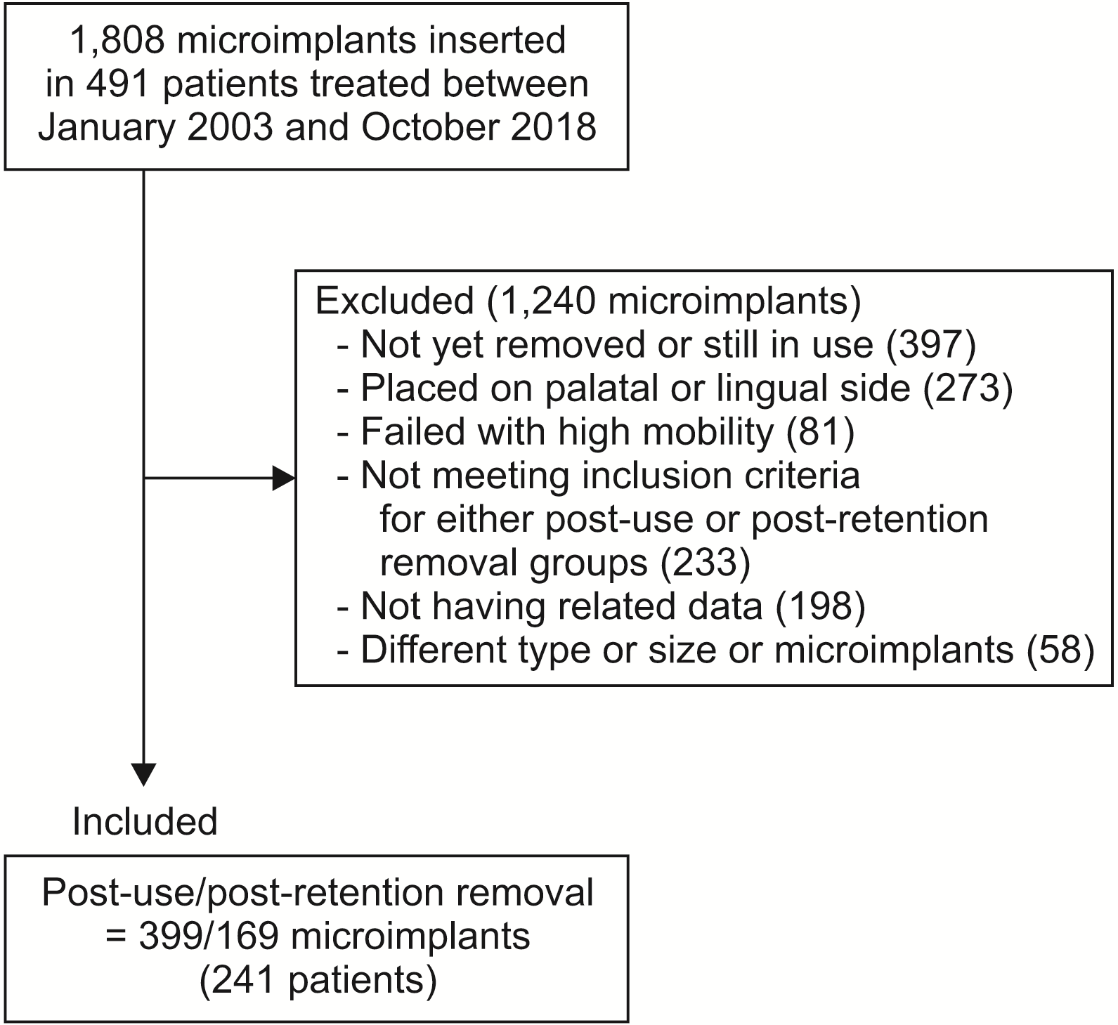
This study was approved by the institutional review board at Kyungpook National University Dental Hospital (KNUDH-2021-03-02-00). The sample group included 241 patients (83 male and 158 female; age, 30.25 ± 12.2 years; age range, 11.9–71 years) with 568 microimplants. Every individual in the sample group underwent orthodontic treatment at the Department of Orthodontics at Kyungpook National University Dental Hospital, Daegu, Korea, performed by one clinician (H.S.P.) between January 2003 and October 2018 (Figure 1, Table 1). All patients provided informed consent prior to the placement of the microimplants.
This study focused on microimplants placed on the buccal side because of the difficulty in measuring torques on the palatal or lingual side. The inclusion criteria for patients with microimplants were as follows: the microimplants were properly used without failure; they were retained for at least 6 months; and data on their removal torques were readily available. On the other hand, the microimplants that failed to apply force owing to high mobility or those without the necessary removal torque data were excluded from the sample. The microimplants used in this study were 1.3, 1.4, or 1.5 mm in diameter and 6, 7, or 8 mm in length (Absoanchor; Dentos, Daegu, Korea; Figure 2). Under local anesthesia and without flap surgery, patients in the sample group underwent microimplant insertion mostly at an angle of 30° to 40° to the long axis of the tooth, and either a no-drilling or pre-drilling method was used in accordance with the bone density at the implantation site.
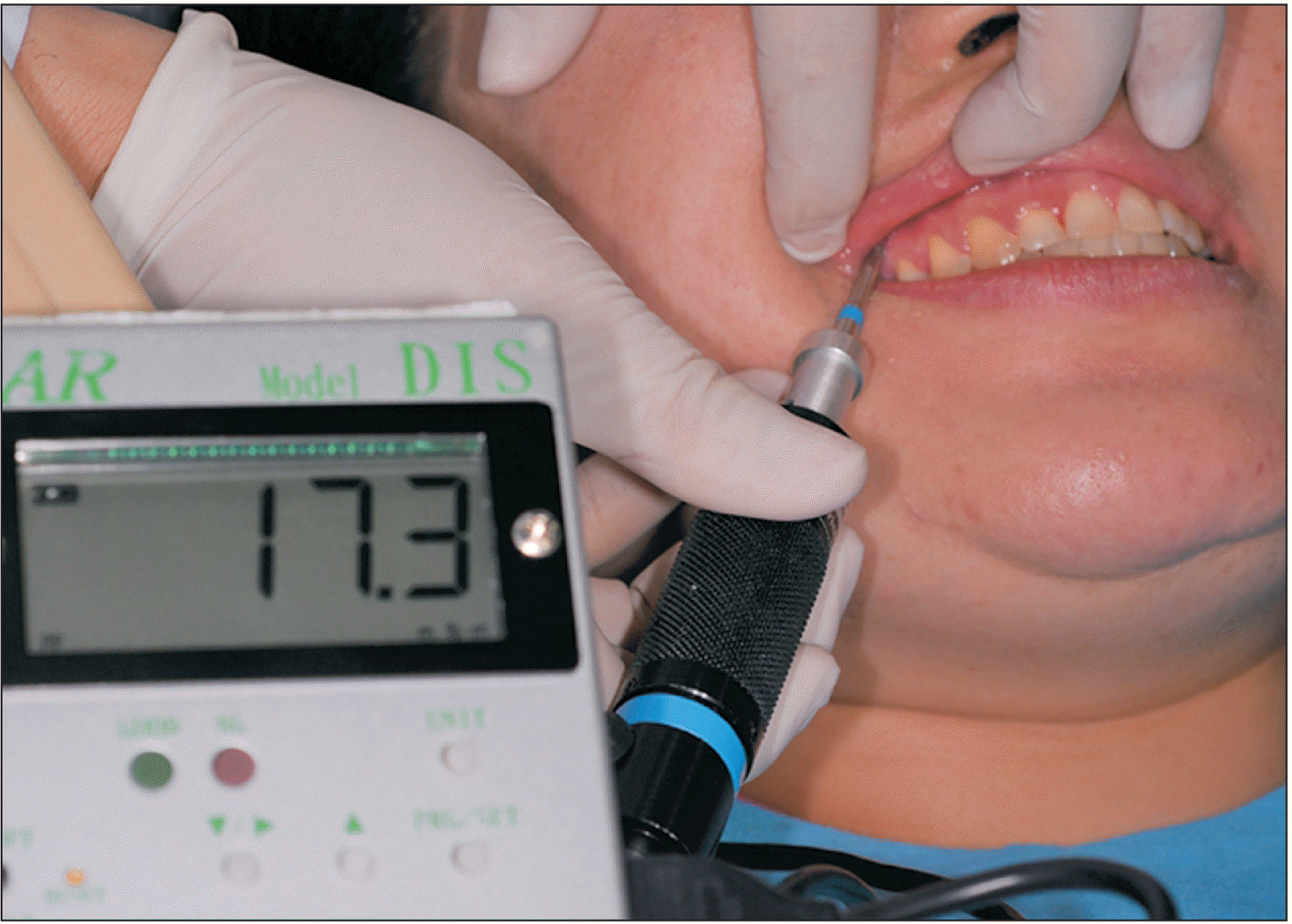
In the event of pre-drilling at the alveolar bone, 0.9-, 1.0-, or 1.1-mm-diameter drills were used. All the procedures related to the microimplants, including the placement, removal, and measurements, were conducted by one investigator (H.S.P.). During treatment, immediate loading was performed with a light force (approximately 50 g), and 3–4 weeks later, the loading force was increased up to 200 g. The peak placement torque was measured during placement, and the peak removal torque was measured at the first turn during the removal procedure by using a digital gauge (DIS-RL05; Sugisaki Meter Co., LTD., Ibaraki, Japan; Figure 3).
The sample group was divided into two main groups—the post-use and post-retention removal groups—depending on the time of removal. The post-use removal group comprised microimplants removed immediately after their intended use or the completion of treatment. The post-retention removal group comprised microimplants removed during the retention period and which required more than 6 months of maintenance after the end of treatment. For further evaluation of both groups, each group was subdivided according to sex, age, placement site, and placement method.
Statistics
As all the groups showed normal distribution when assessed using the Kolmogorov–Smirnov test, an independent t-test was carried out to compare the differences in the placement/removal torques and in-bone stay period of the microimplants between the post-use and post-retention removal groups and/or between the placement sites or the placement methods.
The Pearson correlation coefficient was calculated to determine the relationship between the removal torques and other variables, such as age, placement torques, microimplant length and diameter, and the total/unloading in-bone stay period. Thereafter, multiple linear regression analysis with stepwise selection was performed to demonstrate the correlation between the removal torques and associated variables. All statistical analyses were performed using IBM SPSS Statistics for Windows/Macintosh, Version 22.0 (IBM Corp., Armonk, NY, USA), and the significance level was set at p < 0.05.
RESULTS
In-bone stay period of microimplants (Table 2)
The mean total in-bone stay period of the microimplants in the post-retention removal group was 1,237 days, which was significantly longer than that in the post-use removal group (656.28 days; p < 0.001). However, no significant difference was observed in the in-bone stay period during treatment between the groups. In the post-retention removal group, the unloading in-bone stay period of the microimplants was 507.43 days.
Comparison of the removal torques between the post-use and post-retention removal groups and between sexes or age groups (Table 3)
The removal torques in the post-retention removal group were significantly higher than those in the post-use removal group in the total sample (p < 0.001). Moreover, the post-retention removal group showed significantly higher removal torques than did the post-use removal group in all subgroups of sex and age (p < 0.01), except for the age group ≥ 30 years. However, when comparing the removal torques between sexes or between age groups, no significant difference was found in either the post-use or post-retention removal groups.
Comparison of torques between the post-use and post-retention removal groups and between the placement sites or placement methods (Table 4, Figure 4)
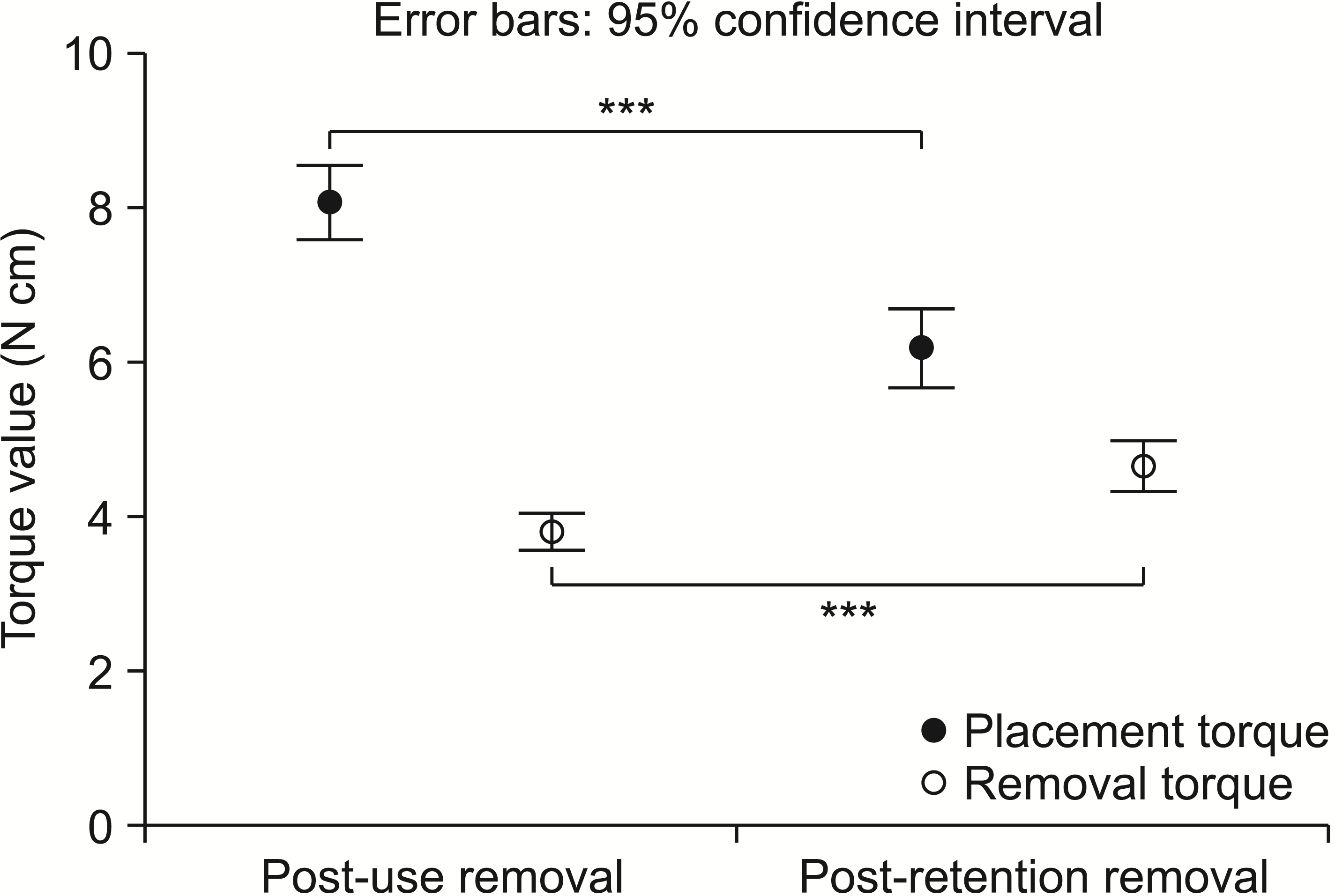
The mean removal torque was higher in the post-retention removal group (4.66 N cm) than in the post-use removal group (3.80 N cm; p < 0.001). However, the mean placement torque was higher in the post-use removal group.
The removal torques in both the maxilla and mandible were significantly higher in the post-retention removal group (maxilla, 4.41 N cm; mandible, 5.51 N cm) than in the post-use removal group (maxilla, 3.46 N cm, p < 0.001; mandible, 4.19 N cm, p < 0.05). As for the placement sites of the microimplant, in the maxilla, the mean placement torque in the post-use removal group (7.34 N cm) was significantly higher than that in the post-retention removal group (5.61 N cm; p < 0.001). In the mandible, however, no significant difference was observed in the placement torques between the groups.
When the microimplants were inserted using the no-drilling method, the post-retention removal group showed significantly higher removal torques than did the post-use removal group (post-retention removal, 4.42 N cm; post-use removal, 3.42 N cm; p < 0.001). In contrast, when using the pre-drilling method, the difference in the removal torques between the two groups was not statistically significant (p = 0.102). Regardless of the placement method of the microimplant, the placement torques were significantly higher in the post-use removal group (with no-drilling, 7.08 N cm, p < 0.001; with pre-drilling, 10.00 N cm, p < 0.05) than in the post-retention removal group (with no-drilling, 5.50 N cm; with pre-drilling, 8.06 N cm).
Regarding the placement sites and placement methods, the mandible and pre-drilling method groups showed significantly higher placement and removal torques than did the maxilla and no-drilling method groups.
Pearson correlation coefficients between the removal torques and potential variables (Table 5)
In the post-use removal group, the removal torque exhibited a significant positive correlation with the placement torque (r = 0.225, p < 0.001) and microimplant length (r = 0.145, p < 0.05) in the no-drilling method. In contrast, no significant correlation was found in the pre-drilling method group.
As for the microimplants in the post-retention removal group, the removal torques showed a significant positive correlation with the unloading in-bone stay period (r = 0.203, p < 0.05) and microimplant diameter (r = 0.376, p < 0.05) in the no-drilling and pre-drilling methods, respectively.
Multiple linear regression analysis with stepwise selection (Table 6)
A stepwise multiple linear regression analysis was performed in both the post-use and post-retention removal groups. To further examine the determinant factors that influenced the removal torque, independent variables such as age, placement torque, microimplant length and diameter, and the total or unloading in-bone stay period were included. We determined that the removal torque with the use of the no-drilling method could be predicted by the placement torque and microimplant length in the post-use removal group. The following equation represented the linear regression for the removal torque with the use of the no-drilling method in the post-use removal group:
Y1 = –0.479 + 0.101 × X11 + 0.464 × X12
(Y1, removal torque, N cm; X11, placement torque, N cm; X12, microimplant length, mm).
Regarding the microimplants placed using the no-drilling and pre-drilling methods in the post-retention removal group, the unloading in-bone stay period and microimplant diameter could explain their removal torques, respectively. The following equations illustrated the linear regression for the removal torque when using the no-drilling and pre-drilling methods in the post-retention removal group, respectively:
Y21 = 3.923 + 0.001 × X21 (Y21, removal torque, N cm; X21, unloading in-bone stay period, days);
Y22 = –9.987 + 12.075 × X22 (Y22, removal torque, N cm; X22, microimplant diameter, mm).
DISCUSSION
The removal torques in the post-retention removal group were significantly higher than those in the post-use removal group. This means that the removal torque tends to increase with time. In the pre-drilling method, no statistically significant difference was observed in the removal torques between the groups, even though the post-retention removal group had higher removal torques. This might indicate less microdamage on the surrounding cortical bone during placement, followed by less bone remodeling and little increase in the removal torque. Moreover, the no-drilling method could result in the formation of more bone microcracks11 and higher subsequent remodeling activity, thereby resulting in a gradual increase in stability over time. This remodeling could be completed in 6 months.12 Another possible explanation is the development of macro-bone fractures on the surface cortical bone during microimplant placement and following bone remodeling. The microimplants in this study were placed at angles to the bone. When the microimplants were placed into the bone at an angle by using the no-drilling method, extensive bone macro-destruction occurred.13 This surface bone fracture produces extensive bone remodeling, and bone repair or consolidation of the fractured bone may occur over time; this may positively influence the increase in the removal torques in the post-retention removal group.
We also obtained some interesting results when studying the variables influencing the removal torques in the no-drilling samples. We found that the removal torque positively correlated with the placement torque in the post-use removal group and with the unloading in-bone stay period in the post-retention removal group. Hence, microimplant stability seems closely associated with the placement torque during treatment and it may be increasingly enhanced over time during retention. There are several possible explanations for these observations. The placement torque, within the proper range, directly relates to the primary stability and may even have long-term effects on the subsequent secondary stability during clinical use. Similarly, previous studies have often stressed the importance of the primary stability on the secondary stability.2,3,9
However, it is important to understand the implications of this result as the microimplants inserted with excessive placement torque may have been excluded from the current sample because of their failure (Figure 1), thus yielding a more significant relationship between the placement and removal torques. This is an essential consideration when placing the microimplants because too high a placement torque may result in a higher chance of failure because of severe microcracks on the surrounding bone or root contacts. Accordingly, a pre-drilling method is commonly recommended in the cortical bone with high density, such as that in the molar or retromolar area of the mandible.14
Considering the changes seen in the removal torque after treatment, the unloading in-bone stay period showed a significant positive correlation, indicating that residual bone remodeling had been taking place gradually over time. The little to no orthodontic loading during retention facilitated the increase in stability. Moreover, the inflammation caused by elastic threads disappeared after treatment, and this may have had a positive effect on subsequent bone remodeling, as shown in an earlier study that reported higher success rates to be highly associated with no inflammation.15 According to the mechanostat theory of Frost,16 bone adaption occurs differently within the four microstrain zones, namely, the acute disuse window zone, adapted window zone, mild overload zone, and pathologic overload zone. As previously stated by Degidi et al.,17 repetitive orthodontic loading to the microimplant is likely to be in the mild overload zone, which shows a higher bone remodeling rate and increased woven-bone formation.18 Therefore, during treatment, less mineralized and organized bone may be present at the bone-to-implant interface. In contrast, during the retention period, the microimplant with little to no orthodontic loading could be categorized as being in the adapted window zone, indicating a reduced remodeling rate. Hence, it might be primarily relevant to well-organized and mineralized lamellar bone at the interface.
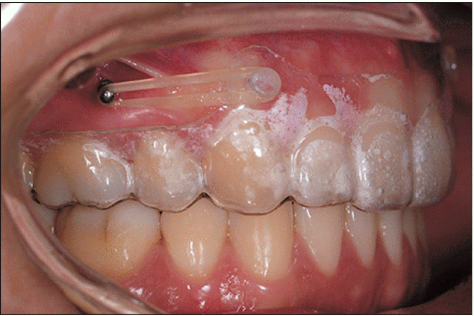
The placement torques in the post-use removal group were higher than those in the post-retention removal group. This can be explained as follows. In this study, the removal of the used microimplants was purposefully delayed after the completion of active treatment in patients who were at a high risk for relapse, particularly those with hyperdivergent skeletons or open bites. Post-treatment relapse can be prevented or corrected by applying an elastic force between the retention appliances or clear retainers and the unremoved microimplants (Figure 5).10 Accordingly, it could be assumed that the inclusion of high-angle patients, showing a relatively thin cortical bone,19,20 in the post-retention removal group likely affected the lower placement torques seen in that group rather than in the post-use removal group (mandibular plane angle to Frankfort horizontal plane: 30.53 ± 6.77° in the post-retention removal group [n = 169]; 28.33 ± 6.73° in the post-use removal group [n = 399]; p < 0.001, independent t-test between the two groups).2 On the other hand, the removal torques in the post-retention removal group were higher than those in the post-use removal group. Therefore, the removal torque seems irrelevant to the placement torque over time. Once the bone is remodeled, the removal torque would be rather related to the period of in-bone stay and local factors such as bone thickness and density and the integrity and surface area of the bone-to-implant interface. Meanwhile, pre-drilling was performed at the site with a high bone density to avoid excessive bone microdamage during microimplant placement, and this probably caused the difference in the torques based on the placement methods.
Excessively high osseointegration might be associated with miniscrew fracture during removal. This study showed an appropriate removal torque of approximately 4–5 N cm without a single fracture even in the post-retention removal group. The torques seen in this study are clinically acceptable not only for the safe removal of microimplants, but also for their continued use during retention. Furthermore, the regression coefficient corresponding to the period of retention was not large enough to induce a fracture during removal.
On the basis of these results, the null hypothesis was rejected because significant differences were observed between the post-use and post-retention removal groups.
Nevertheless, this study has some limitations. Although this study successfully evaluated the removal torque of the microimplant with prolonged retention and the associated variables influencing the removal torque, it was limited by the duration of force loading during treatment. In fact, employing an exact duration of force loading for this analysis was not feasible; thus, the whole-treatment duration was used to evaluate the relevant stability in this study.
Another limitation of this study was that some of the microimplants in the post-retention removal group were loaded while most of them were not loaded. Only seven microimplants, however, were loaded for anchorage because of slight relapse during the retention period. A light force with a 3-oz elastic to a clear retainer was applied to the microimplants placed in patients with an open bite and/or Class II or Class III malocclusion to prevent relapse movements, such as extrusion of the intruded posterior teeth or mesial movement of distalized teeth. Therefore, considering that loaded microimplants were present in a small proportion of the post-retention removal group (7 of 169) and a light elastic force (50–60 g) was applied to them, the influence of the load on this group might be insignificant. In fact, we performed statistical analysis after excluding these seven patients and found it had no significant influence on the results.
Finally, the angulation of microimplant placement was not precisely evaluated in this study. Therefore, the relationship between stability and placement angulation warrants further examination in future studies.
CONCLUSION
By assessing the removal torques, we showed it was possible to demonstrate the stability of the microimplants with prolonged maintenance.
The post-retention removal group showed significantly higher removal torques than did the post-use removal group, except for microimplants placed using the pre-drilling method. The removal torques in the post-retention removal group, approximately 4–5 N cm, were clinically acceptable not only for safe removal but also for continued use.
The placement torque and microimplant length showed significant positive correlations with the removal torques in the no-drilling post-use removal group, and the unloading in-bone stay period and microimplant diameter showed positive correlations with the removal torques in the post-retention removal group in the no-drilling and pre-drilling methods, respectively.

 XML Download