

 PDF
PDF Citation
 Print
Print


INTRODUCTION
MATERIALS AND METHODS
Eligibility criteria
Search strategy
Data collection
Skeletal expansion: zygomatic width (ZMW), nasal width (NW), jugular width (J-J), and the midpalatal suture at the posterior nasal spine (PNS) and the anterior nasal spine (ANS)
Alveolar expansion: alveolar molar width (AMW)
Dental expansion: inter-canine width (ICW), inter-premolar width (IPMW), and inter-molar width (IMW).
Meta-analysis
Heterogeneity test (Forest plots, Cochrane Q-test, and I2 index)
Publication bias (Funnel plot and Egger’s test)
Sensitivity analysis
Subgroup analysis
Risk of bias assessment in individual studies
RESULTS
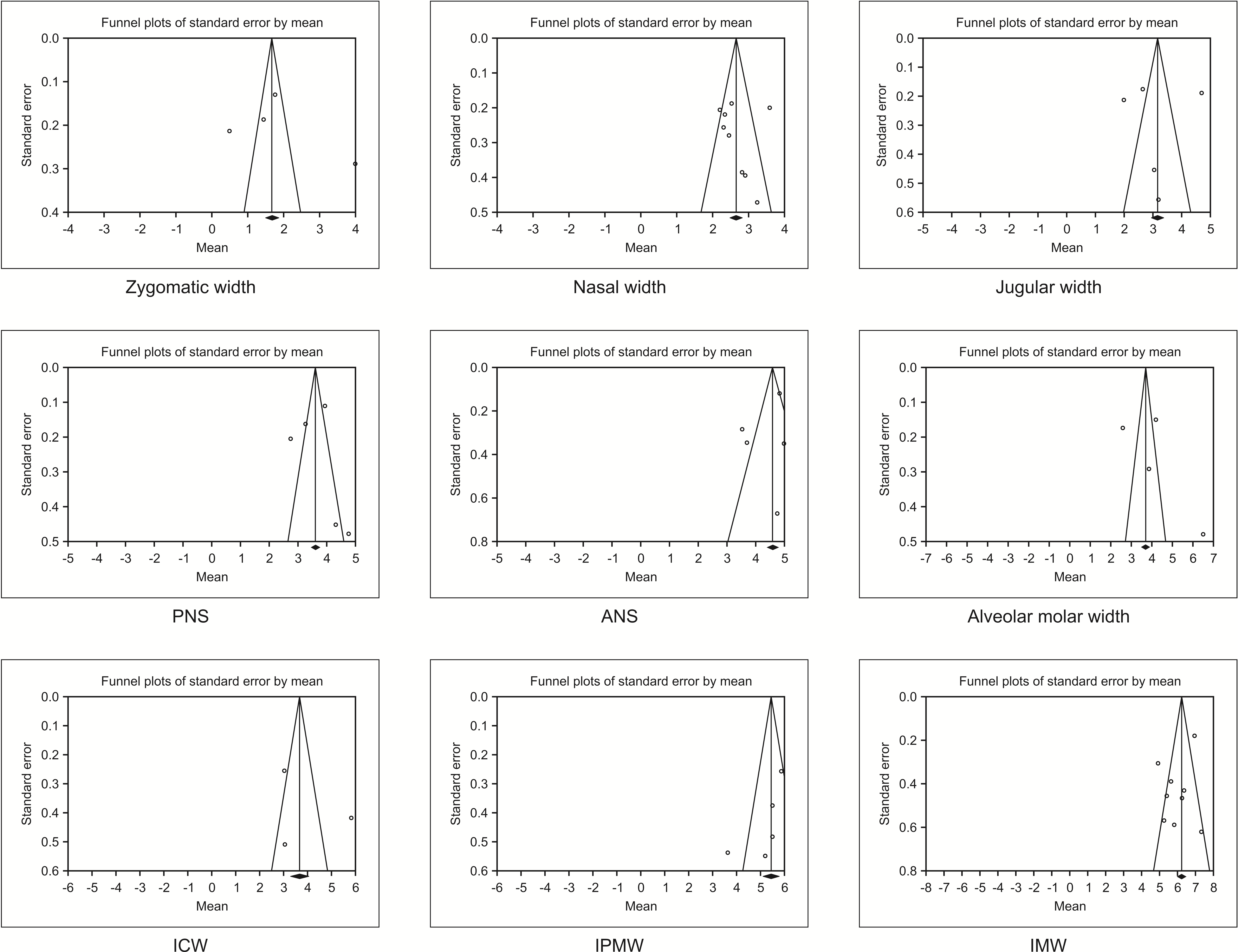
Search strategy
Study characteristics
Table 2
| Author | N | Study design | Setting | Race | Compare | Appliance | Appliance design |
Diameter length |
Mean age (SD) | Activation |
Activation period |
Success rate |
|---|---|---|---|---|---|---|---|---|---|---|---|---|
|
Calil et al.13 (2021) |
16 | Retrospective cohort |
University Dentistry Institute |
Brazil | Self ligate vs. MARPE | PecLab appliance (Belo Horizonte, Brazil) | Molar without extension | 4 titanium mini-implants of 1.8 mm diameter and 8 mm length |
24.92 (7.6) |
2/4-turn a day | Until the palatal cusps of maxillary first molars touch the buccal cusps of the mandibular first molars | ND |
|
Cantarella et al.16 (2017) |
15 | Retrospective | University | USA | No | BioMaterials Korea (Hanam, Korea) | Molar without extension | ND |
17.2 (4.2) |
2 turns per day, then 1 activation per day | Until interincisal diastema appear, then complete when maxillary width was equal to mandibular width | 100%* |
|
Clement et al.20 (2017) |
10 | Prospective | University | India | No | BioMaterials Korea | Molar without extension | 4 titanium mini-implants of 1.8 mm diameter and 11 mm length |
21.5 (ND) |
2 turns per day | Until the required expansion was achieved | 100%* |
|
Elkenawy et al.25 (2020) |
31 | Retrospective | University | USA | No | BioMaterials Korea | Molar without extension | 4 titanium mini-implants of 1.5 mm diameter and 11 mm length |
20.4 (3.2) |
2 turns per day, then 1 activation per day | Until interincisal diastema appear, then complete when maxillary width was equal to mandibular width | ND |
|
Jesus et al.22 (2021) |
12 | Retrospective cohort | ND | Brazil | SARPE with/without cinch | PecLab appliance | Molar without extension | 4 titanium mini-implants, ND |
Range 15–39 |
2 turns a day | For 14 to 18 days, until full correction | ND |
|
Li et al.26 (2020) |
22 | Retrospective | University | China | No | BioMaterials Korea | Molar without extension | 4 titanium mini-implants of 1.5 mm diameter and 11 mm length |
22.6 (4.5) |
2 turns every other day | Maxillary skeletal width was no longer less than mandible (mean 38 days) | 100%* |
|
Li et al.11 (2020) |
48 | Retrospective | University | China | No | BioMaterials Korea | Molar without extension | 4 titanium mini-implants of 1.5 mm diameter and 11 mm length |
19.4 (3.3) |
One-sixth of turn (0.13 mm) each day | Maxillary skeletal width was no longer less than mandible | ND |
|
Lim et al.15 (2017) |
24 | Retrospective | Hospital | Korea | No | Hyrax II (Dentaurum, Ispringen, Germany) | Molar with anterior arm extension | 4 titanium mini-implants diameter, 1.8 mm; length, 7 mm; Orlus |
21.6 (3.1) |
Once a day (0.2 mm) | Until the required expansion was achieved (5 week) | 86.84% |
|
Moon et al.27 (2020) |
24 | Retrospective | University | Korea | C-expander | BioMaterials Korea | Molar without extension | 4 titanium mini-implants of 1.5 mm diameter and 11 mm length |
19.2 (5.9) |
Once a day (0.2 mm) | Until the required expansion was achieved (5 week) | ND |
|
Ngan et al.18 (2018) |
8 | Retrospective | University | USA | No | BioMaterials Korea | Molar without extension | 4 titanium mini-implants of 1.8 mm diameterand 11 mm length |
21.9 (1.5) |
Varied with the severity of transverse discrepancy | Until the occlusal aspect of lingual cusp of the maxillary first molars contacted occlusal aspect of the buccal cusp of the mandibular first molars. The 2–3 mm of overexpansion | 100%* |
|
Nguyen et al.28 (2021) |
20 | Retrospective | University | Korea | No | BioMaterials KoreaMSE type II | Molar without extension | 4 titanium mini-implants of 1.8 mm diameterand 11 mm length |
22.4 (17.6–27.1) |
2 turns per day (0.13 mm/turn) | Until diastema appeared, after which the rate was reduced to one turn per day, stop when required amount was achieved 20% overexpansion | 100%* |
|
de Oliveira et al.24 (2021) |
17/15 | Retrospectivecohort | University | Brazil | MARPE vs. SARPE | PecLab appliance9 mm expander | Molar without extension | 4 miniscrew unknown diameter and length |
MARPE 26 (11) SARPE 28.5 (10.5) |
2/4 immediately after mini implant placement and 2/4 turns daily (14–18 days) | Until full correction | 86.96% |
|
Park et al.29 (2017) |
14 | Retrospective | University | Korea | No | Hyrax II(Dentaurum, Ispringen, Germany) | Molar with anterior arm extension | Diameter, 1.8 mm; length, 7 mm; Orlus |
20.1 (2.4) |
Once a day (0.2 mm) | Until the required expansion was achieved | 84.21% |
|
Tang et al.7 (2021) |
31 | Retrospective | University | China | No | BioMaterials Korea | Molar without extension | 4 titanium mini-implants of 1.5 mm diameterand 11 mm length |
22.14 (4.76) |
Once a day (0.13 mm) | Ranging from 40 to 60 turns | 92% |
![]()
Meta-analysis
Table 3
| Variable | Author | N | Mean | SD | Combined measurements |
|---|---|---|---|---|---|
| ZMW | Elkenawy et al.25 (2020) | 31 | 3.99 | 1.60 | |
| Li et al.26 (2020) | 22 | 0.50 | 1.00 | ||
| Li et al.11 (2020) | 48 | 1.77 | 0.90 | 3 groups* | |
| Tang et al.7 (2021) | 31 | 1.45 | 1.04 | ||
| NW | Calil et al.13 (2021) | 16 | 2.82 | 1.54 | |
| Jesus et al.22 (2021) | 12 | 3.46 | 1.95 | ant./post.* | |
| Li et al.26 (2020) | 22 | 2.30 | 1.20 | ||
| Li et al.11 (2020) | 48 | 3.58 | 1.39 | 3 groups* | |
| Lim et al.15 (2017) | 24 | 2.20 | 1.01 | ||
| Moon et al.27 (2020) | 24 | 2.45 | 1.37 | ||
| Ngan et al.18 (2018) | 8 | 2.53 | 0.53 | ||
| de Oliveira et al.24 (2021) | 17 | 2.91 | 1.62 | ant./post.* | |
| Tang et al.7 (2021) | 31 | 2.33 | 1.22 | ||
| J-J | Calil et al.13 (2021) | 16 | 3.06 | 1.81 | |
| Jesus et al.22 (2021) | 12 | 3.20 | 1.92 | ||
| Li et al.26 (2020) | 22 | 2.00 | 1.00 | ||
| Li et al.11 (2020) | 48 | 4.69 | 1.31 | 3 groups* | |
| Tang et al.7 (2021) | 31 | 2.65 | 0.98 | ||
| PNS | Cantarella et al.16 (2017) | 15 | 4.33 | 1.74 | |
| Elkenawy et al.25 (2020) | 31 | 4.77 | 2.65 | ||
| Ngan et al.18 (2018) | 8 | 3.27 | 0.46 | ||
| Nguyen et al.28 (2021) | 20 | 3.95 | 0.50 | ||
| de Oliveira et al.24 (2021) | 17 | 2.75 | 0.85 | ||
| ANS | Cantarella et al.16 (2017) | 15 | 4.75 | 2.59 | |
| Elkenawy et al.25 (2020) | 31 | 4.98 | 1.94 | ||
| Ngan et al.18 (2018) | 8 | 3.53 | 0.80 | ||
| Nguyen et al.28 (2021) | 20 | 4.83 | 0.53 | ||
| de Oliveira et al.24 (2021) | 17 | 3.69 | 1.42 |
N, sample size; SD, standard deviation; ZMW, zygomatic width; NW, nasal width; J-J, jugular width; PNS, midpalatal suture at the posterior nasal spine; ANS, midpalatal suture at the anterior nasal spine.
*Combined mean and SD from multiple groups using an online calculator https://www.statstodo.com/CombineMeansSDs.php
![]()
Table 4
| Variable | Author | N | Mean | SD |
Combined measurements |
|---|---|---|---|---|---|
| AMW | Clement et al.20 (2017) | 10 | 6.50 | 1.51 | |
| Lim et al.15 (2017) | 24 | 2.60 | 0.85 | ||
| Nguyen et al.28 (2021) | 20 | 4.19 | 0.67 | ||
| de Oliveira et al.24 (2021) | 17 | 3.86 | 1.20 | ||
| ICW | Calil et al.13 (2021) | 16 | 3.04 | 2.03 | |
| Clement et al.20 (2017) | 10 | 5.83 | 1.32 | ||
| Lim et al.15 (2017) | 24 | 3.02 | 1.25 | ||
| IPMW | Calil et al.13 (2021) | 32 | 3.63 | 2.14 | 1st/2nd PM* |
| Clement et al.20 (2017) | 20 | 5.50 | 1.52 | 1st/2nd PM* | |
| Lim et al.15 (2017) | 48 | 5.87 | 1.26 | 1st/2nd PM* | |
| de Oliveira et al.29 (2021) | 17 | 5.21 | 2.25 | ||
| Park et al.29 (2017) | 14 | 5.50 | 1.40 | ||
| IMW | Calil et al.13 (2021) | 16 | 6.37 | 1.72 | |
| Clement et al.20 (2017) | 10 | 7.33 | 1.96 | ||
| Jesus et al.22 (2021) | 12 | 5.82 | 2.03 | ||
| Li et al.11 (2020) | 48 | 6.95 | 1.25 | 3 groups* | |
| Lim et al.15 (2017) | 24 | 5.63 | 1.90 | ||
| Moon et al.27 (2020) | 24 | 4.91 | 1.50 | ||
| Ngan et al.18 (2018) | 8 | 6.26 | 1.31 | ||
| de Oliveira et al.24 (2021) | 17 | 5.25 | 2.34 | ||
| Park et al.29 (2017) | 14 | 5.40 | 1.70 |
N, sample size; SD, standard deviation; AMW, alveolar molar width; ICW, inter-canine width; IPMW, inter-premolar width; IMW, inter-molar width; PM, premolar.
*Combined mean and SD from multiple groups using an online calculator https://www.statstodo.com/CombineMeansSDs.php
![]()
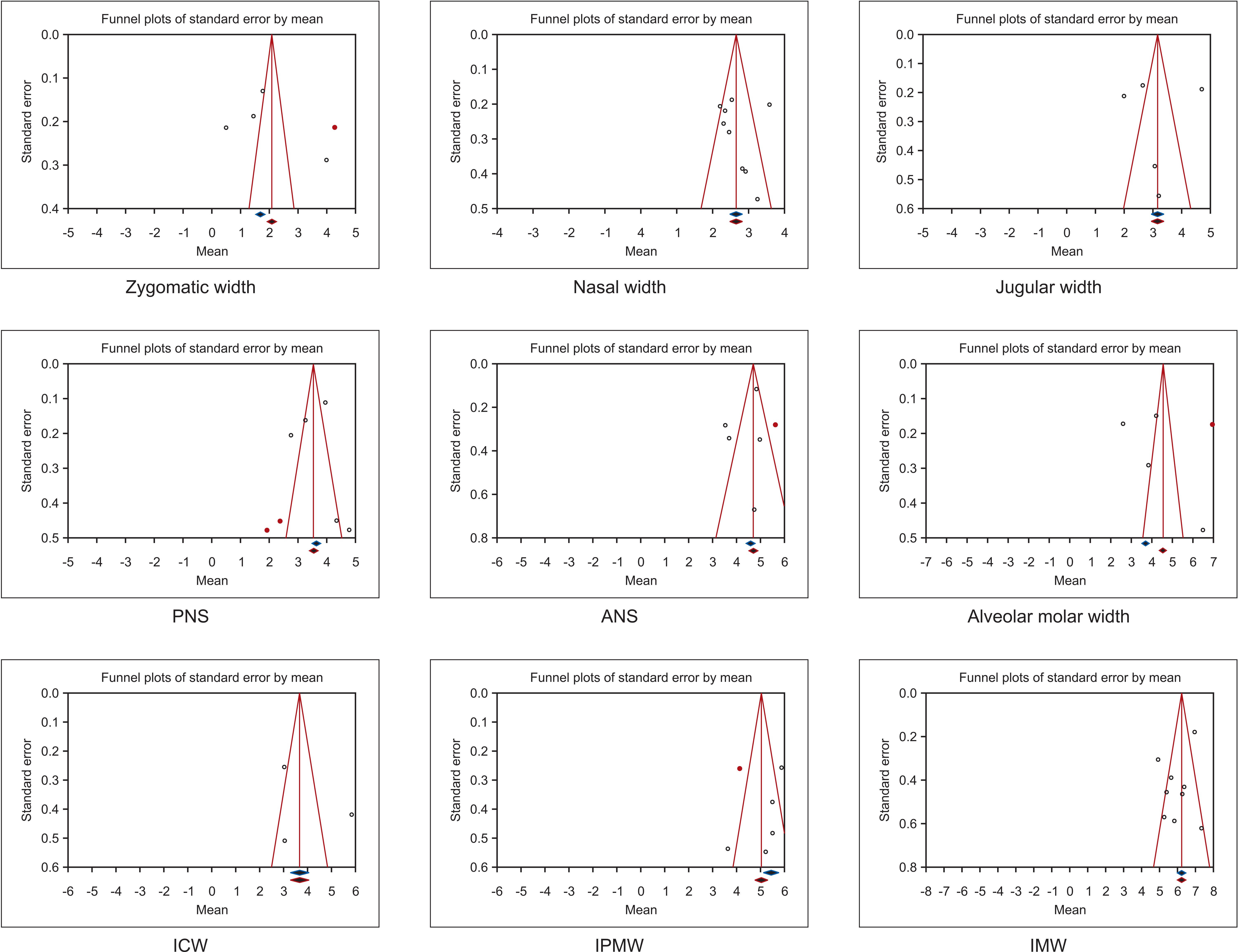
Heterogeneity test (Forest plots, Cochrane Q-test, and I2 index)

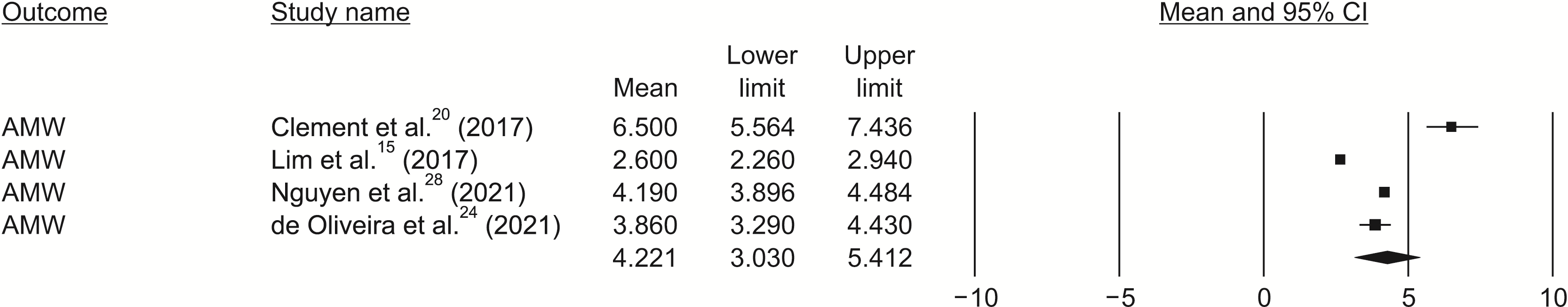
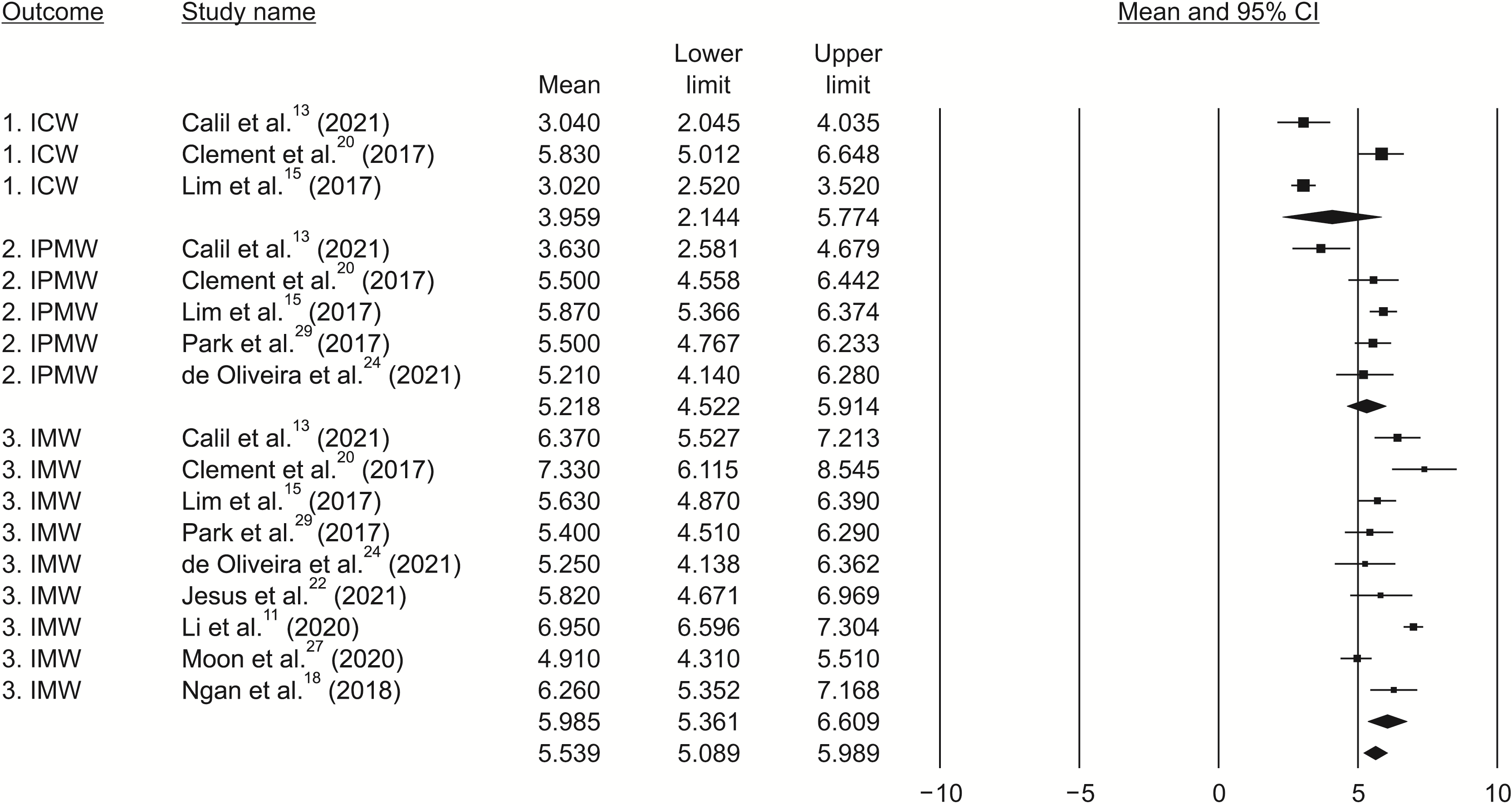
Table 5
![]()
Estimation of the pooled means with 95% CI
Table 6
| Group | Parameter | n | N | Mean | SE | 95% CI |
|---|---|---|---|---|---|---|
| Skeletal expansion | ZMW | 4 | 132 | 1.910 (2.385)** | 0.548 | 0.835, 2.985 (1.120, 3.649)** |
| NW | 9 | 202 | 2.675 | 0.179 | 2.325, 3.026 | |
| J-J | 5 | 129 | 3.120 | 0.576 | 1.990, 4.250 | |
| PNS | 5 | 91 | 3.715 (3.337)** | 0.303 | 3.121, 4.310 (2.754, 3.919)** | |
| ANS | 5 | 91 | 4.335 (4.562)** | 0.338 | 3.674, 4.997 (3.938, 5.187)** | |
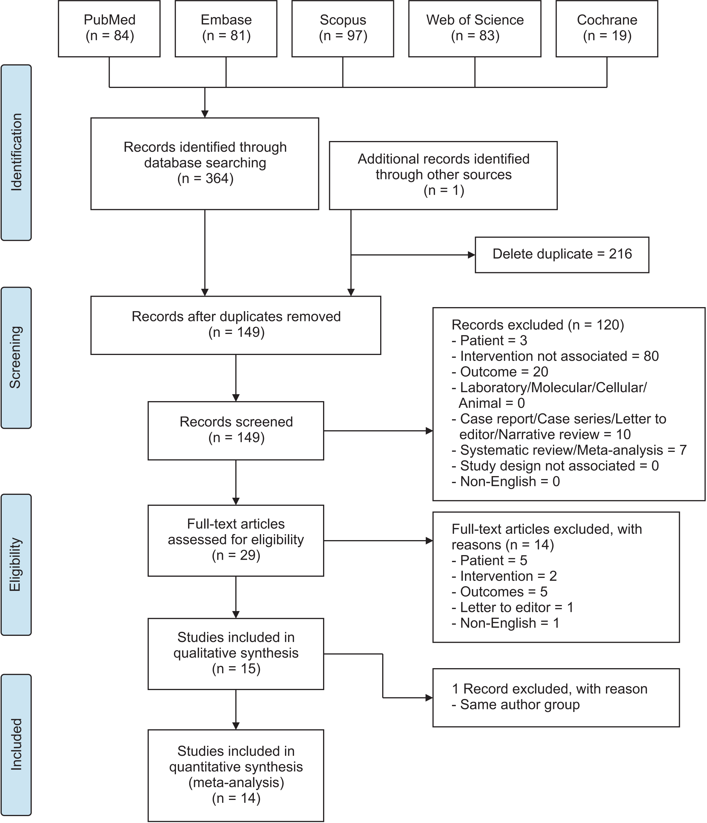
| Alveolar expansion | AMW | 4 | 71 | 4.221 (4.799)** | 0.608 | 3.030, 5.412 (3.112, 6.486)** |
| Dental expansion | ICW | 3 | 50 | 3.959 | 0.926 | 2.144, 5.774 |
| IPMW | 5 | 131 | 5.218 (4.992)** | 0.355 | 4.522, 5.914 (4.229, 5.755)** | |
| IMW | 9 | 173 | 5.985 | 0.318 | 5.361, 6.609 |
n, number of articles; N, number of subjects; SE, standard error; CI, confidence interval; ZMW, zygomatic width; NW, nasal width; J-J, jugular width; PNS, midpalatal suture at the posterior nasal spine; ANS, midpalatal suture at the anterior nasal spine; ICW, inter-canine width; IPMW, inter-premolar width; IMW, inter-molar width.
![]()
Publication bias
Sensitivity analysis
Figure 6
![]()
Risk of bias within studies
Table 7
| Study | Study design | Sample size | Selection description | Valid measurement method | Method error analysis | Blinding | Statistics | Confounding factors | Total score | Judged quality standard | |||||||
|---|---|---|---|---|---|---|---|---|---|---|---|---|---|---|---|---|---|
| Calil et al.13 (2021) | 0 | 1 | 1 | 1 | 1 | 0 | 1 | 0 | 5 | Low | |||||||
| Cantarella et al.16 (2017) | 0 | 1 | 1 | 1 | 1 | 0 | 1 | 0 | 5 | Low | |||||||
| Clement et al.20 (2017) | 1 | 0 | 1 | 1 | 0 | 0 | 1 | 0 | 4 | Low | |||||||
| Elkenawy et al.25 (2020) | 0 | 1 | 1 | 1 | 1 | 0 | 1 | 0 | 5 | Low | |||||||
| Jesus et al.22 (2021) | 0 | 0 | 1 | 1 | 1 | 0 | 1 | 1 | 5 | Low | |||||||
| Li et al.26 (2020) | 0 | 1 | 1 | 1 | 1 | 0 | 1 | 0 | 5 | Low | |||||||
| Li et al.11 (2020) | 0 | 1 | 1 | 1 | 1 | 0 | 1 | 0 | 5 | Low | |||||||
| Lim et al.15 (2017) | 0 | 0 | 1 | 1 | 1 | 0 | 1 | 0 | 4 | Low | |||||||
| Moon et al.27 (2020) | 0 | 0 | 1 | 1 | 1 | 0 | 1 | 0 | 4 | Low | |||||||
| Ngan et al.18 (2018) | 0 | 0 | 1 | 1 | 1 | 0 | 1 | 0 | 4 | Low | |||||||
| Nguyen et al.28 (2021) | 0 | 0 | 1 | 1 | 1 | 0 | 1 | 0 | 4 | Low | |||||||
| de Oliveira et al.24 (2021) | 0 | 0 | 1 | 1 | 1 | 0 | 1 | 1 | 5 | Low | |||||||
| Park et al.29 (2017) | 0 | 0 | 1 | 1 | 1 | 0 | 1 | 0 | 4 | Low | |||||||
| Tang et al.7 (2021) | 0 | 0 | 1 | 1 | 1 | 0 | 1 | 0 | 4 | Low | |||||||
| Overall estimate | 4.5 | Low | |||||||||||||||
Study design: Randomized controlled trial = 3, Prospective = 1, Retrospective = 0, Case series/Case report = 0; Sample size: Adequate (number of samples at least 25 patients) = 1, Inadequate = 0; Selection description: Adequate = 1, Need recalculation = 0; Measurement method: Valid = 1, Invalid = 0; Method error analysis: Yes = 1, No = 0; Blinding in measurement: Yes = 1, No = 0; Statistics: Adequate = 1, Inadequate = 0; Confounding factor stated: Yes = 1, No = 0.
Criteria was modified from the method by Bondemark and Feldmann (Angle Orthod 2006;76:493-501).23
![]()
DISCUSSION
Figure 7
![]()
Skeletal expansion
Midpalatal suture separation
Dental effect
Percentage expansion
Indirect comparison with SARPE
Limitations
CONCLUSION
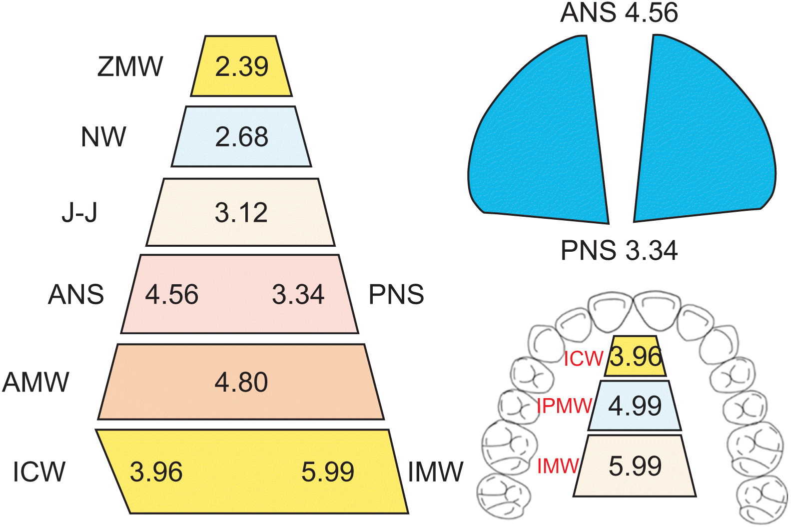
In the coronal view, MARPE resulted in skeletal and dental expansion following a pyramidal pattern.
The pooled mean effects of skeletal expansion were as follows: ZMW, 2.39 mm; NW, 2.68 mm; J-J, 3.12 mm; PNS, 3.34 mm; and ANS, 4.56 mm.
Midpalatal suture split demonstrated a V-shape pattern with greater expansion at the ANS than PNS.
Posterior-anterior ratio (PNS/ANS) of midpalatal suture separation was 73.24%.
The pooled mean effect of AMW was 4.80 mm.
The pooled mean effects of dental expansion were as follows: ICW, 3.96 mm; IPMW, 4.99 mm; and IMW, 5.99 mm.
The percentage of effects of the skeletal (PNS), AMW, and dental (IMW) expansion were 55.76%, 19.87%, and 24.37%, respectively.
MARPE could expand the constricted maxilla in late adolescents to adult patients.
 XML Download