Journal List > Korean J Orthod > v.52(3) > 1516078638

Kwon, Park, Jung, Hwang, Choi, Chung, and Kim: Comparison of the bite force and occlusal contact area of the deviated and non-deviated sides after intraoral vertical ramus osteotomy in skeletal Class III patients with mandibular asymmetry: Two-year follow-up

Abstract

Objective

The objectives of this study were to compare the time-dependent changes in occlusal contact area (OCA) and bite force (BF) of the deviated and non-deviated sides in mandibular prognathic patients with mandibular asymmetry before and after orthognathic surgery and investigate the factors associated with the changes in OCA and BF on each side.

Methods

The sample consisted of 67 patients (33 men and 34 women; age range 15–36 years) with facial asymmetry who underwent 2-jaw orthognathic surgery. OCA and BF were taken before presurgical orthodontic treatment, within 1 month before surgery, and 1 month, 3 months, 6 months, 1 year, and 2 years after surgery. OCA and BF were measured using the Dental Prescale System.

Results

The OCA and BF decreased gradually before surgery and increased after surgery on both sides. The OCA and BF were significantly greater on the deviated side than on the non-deviated side before surgery, and there was no difference after surgery. According to the linear mixed-effect model, only the changes in the mandibular plane angle had a significant effect on BF (p < 0.05).

Conclusions

There was a difference in the amount of the OCA and BF between the deviated and non-deviated sides before surgery. The change in mandibular plane angle affects the change, especially on the non-deviated side, during the observation period.

INTRODUCTION

Facial asymmetry can cause esthetic and functional problems in patients. Therefore, patients with facial asymmetry seek orthognathic surgery.1,2 Bailey et al.1 reported that more than one-third of the patients who visited the Dentofacial Clinic of the University of North Carolina had facial asymmetry. Anistoroaei et al.3 investigated the prevalence of facial asymmetry in orthodontic clinics before treatment and found that 13.8% of the patients had facial asymmetry. Facial asymmetry occurs more in the chin area (74–75%), followed by the midface (36%) and the upper face (5%).1,2 Growth modification, orthodontic treatment, and orthognathic surgery are the alternative treatments for facial asymmetry. The treatment method in this study was selected based on the patient’s age and the severity of the deformity.
The objectives of orthognathic surgery are to improve jaw function and enhance facial appearance. It is difficult to evaluate the jaw function quantitatively. Currently, a simple method using a thin pressure-sensitive sheet is widely used to measure the occlusal contact area (OCA) and bite force (BF). Since the launch of the device, several studies have been conducted to observe the changes in OCA and BF as an evaluation index of masticatory efficiency before and after orthognathic surgery in patients with mandibular prognathism.4-18 The typical results of several previous studies are as follows: (1) the OCA and BF improved after surgery; (2) the absolute values of the OCA and BF after surgery were still lower than those of the control group.7,8,10,11,13,17,18 The above studies involved patients with facial asymmetry, and few studies have observed the difference between the deviated and non-deviated sides of patients with asymmetry. Goto et al.15 carried out a study of patients with skeletal asymmetry to investigate masticatory function differences between the deviated and non-deviated sides. They also compared the parameters of the asymmetry group with those of the symmetric control group. They found that OCA and BF were greater on the deviated side than on the non-deviated side. Both the OCA and BF were lower for the patient group than for the control group. Moroi et al.19 compared the postoperative changes between the asymmetric and symmetric groups. Both groups showed postoperative improvement in the OCA and BF. These studies did not compare the differences between the deviated and non-deviated sides.
The objectives of this study were as follows: (1) to observe the time-dependent changes in OCA and BF on the deviated and non-deviated sides in mandibular prognathic patients with mandibular asymmetry and (2) investigate the factors associated with the changes in OCA and BF on each side. The first hypothesis was that there would be no difference in the OCA and BF between the deviated and non-deviated sides for all observation periods. The second hypothesis was that no skeletal variables affect the time-dependent changes in OCA and BF on the deviated and non-deviated sides.

MATERIALS AND METHODS

Subjects

The study sample was selected from 173 patients diagnosed with skeletal Class III malocclusion who visited Gangnam Severance Dental Hospital for orthognathic surgery between 2011 and 2015. A posteroanterior cephalogram was used to divide the study samples. Patients whose chin deviated by more than 3 mm from the midline were deemed to have facial asymmetry. The completion of mandibular growth was confirmed by a hand-wrist X-ray film. Forty-eight patients who did not meet this condition were excluded. The additional inclusion criteria were the absence of previous orthodontic treatment, missing teeth except for the third molars, temporomandibular disorder, systemic diseases, cleft lip and palate, and craniofacial deformity. Due to the incomplete follow-up data, 58 patients whose OCA and BF were not obtained more than 2 time points were exluded. Finally, the sample consisted of 67 patients (33 men and 34 women; age range 15–36 years) with facial asymmetry. This prospective study was approved by the Gangnam Severance Hospital Institutional Review Board (No. 3-2015-0196), and all participants signed an informed consent form.
The pre- and post-surgical orthodontic treatments were performed with fixed appliances by one orthodontist in the same hospital. Orthognathic surgery was performed by a single surgeon. The patients underwent intraoral vertical ramus osteotomy with Le Fort I osteotomy. The duration of maxillomandibular fixation was approximately 2 weeks. The duration of the pre- and post-surgical orthodontics were 11.4 months (range 9.2–15.5 months) and 7.6 months (range 5.3–12.8 months), respectively.

Measurements

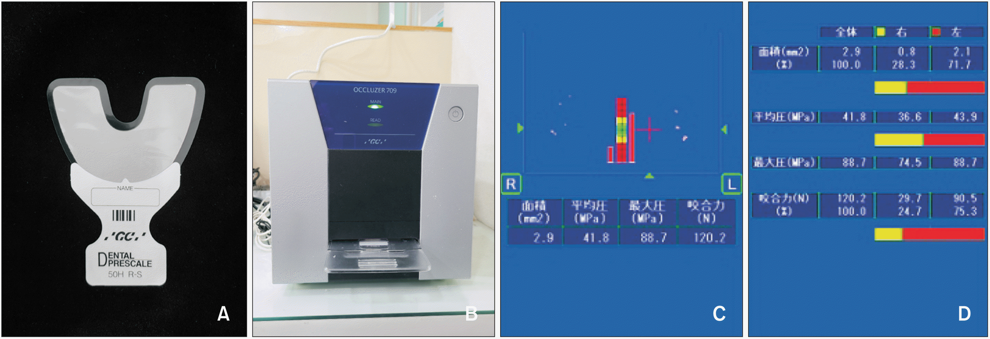
Lateral and posteroanterior cephalograms were taken before treatment (T0) and 1 month (T2), and 2 years after surgery (T6) for skeletal analysis. OCA and BF were assessed before presurgical orthodontic treatment (T0), within 1 month before surgery (T1) and 1 month (T2), 3 months (T3), 6 months (T4), 1 year (T5), and 2 years (T6) after surgery. As there were subjects with missing data at some time point, the number of subjects at each time point was as follows: in T0, T1, T2, T3, T4, T5, and T6, the numbers were 67, 59, 65, 66, 67, 65, and 58, respectively. OCA and BF were measured using the Dental Prescale System (FujiFilm Corp., Tokyo, Japan) (Figure 1). The device consists of a pressure-sensitive sheet and an analysis apparatus (Occluzer FPD-707). The accuracy and reliability of this device have been examined in previous studies.12,16,20 Before recording the measurements, all subjects were seated upright in an unsupported headrest and practiced clenching in the intercuspal position. The pressure-sensitive sheet was placed between the maxillary and mandibular teeth, and the center of the sheet coincided with the facial midline. The patient was instructed to bite the sheet forcefully during maximum intercuspation for 5 seconds. The sheets were scanned and analyzed using an analysis apparatus. The maximum occlusal pressure (unit, MPa), average occlusal pressure (unit, MPa), OCA (unit, mm2), and BF (unit, Newton) were displayed on the screen. The scanning procedures were performed by one examiner. Twenty sheets were randomly selected and re-analyzed by the same examiner at 2-week intervals. An intra-examiner error was evaluated using the intraclass correlation coefficient (ICC). The ICC values for the OCA and BF (97–99%) indicated good intra-examiner reliability. At T0, each patient was tested twice, and a paired t-test was performed. As a result, there was no statistically significant difference (p > 0.05); therefore, each patient was tested once after the T1 period.

Statistical analysis

The measured data were described as means and standard errors. Lateral cephalometric measurements were performed at T0, T2, and T6 (Table 1). A linear mixed model was applied to compare the measurements of OCA and BF over time. This method considers both intra- and inter-subject variabilities. The Bonferroni post-hoc test was applied to correct for type I errors related to multiple comparisons. A linear mixed model with random intercept was fitted with participants as a random effect to test for significant changes in the measurements. Among several cephalometric measurements, changes in the mandibular plane angle, mandibular body length, and chin deviation were selected as variables. The paired t-test was used to compare the differences in the measured values of OCA and BF on the deviated and non-deviated sides at each time point; the magnitudes of changes at each time point were also compared. The significance level was set at p < 0.05. SPSS software ver. 25 (IBM Corp., Armonk, NY, USA) was used for all statistical analyses.

RESULTS

The chin deviation decreased after the surgery (Table 1). The OCA and BF decreased gradually before surgery and increased after surgery on both sides. The OCA on both sides and the BF on the deviated side recovered to its initial state 2 years after surgery. Otherwise, the BF on the non-deviated side was restored to its original state 1 year after surgery; thereafter, it increased (Table 2).
The OCA and BF were significantly greater on the deviated side than on the non-deviated side at T0 and T1 (p < 0.05). There were no significant differences between the deviated and non-deviated sides after surgery except BF at T3 (p > 0.05, Table 3, Figure 2).
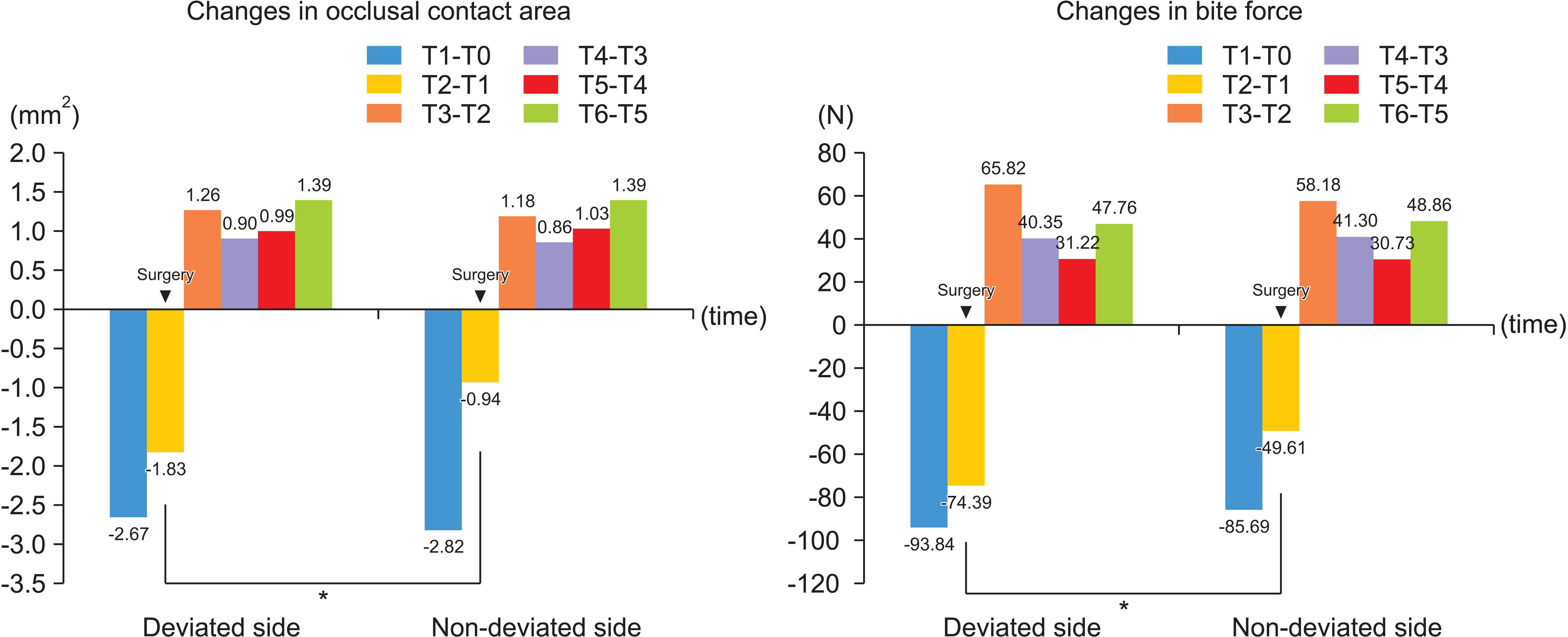
The difference in changes at each observation time point on the deviated and non-deviated sides was evaluated (Table 4, Figure 3). The patterns of changes were similar for each side. The biggest changes were observed during the pre-surgical orthodontic period (T1-T0). The difference between the deviated and non-deviated sides was observed only at T2-T1 (p < 0.05). After surgery, the OCA and BF gradually increased, although the amount of increase showed diverse pattern on both the deviated and non-deviated sides.
According to the linear mixed-effect model, time significantly affected the changes in the OCA and BF on both sides (p < 0.05). Only the changes in the mandibular plane angle (ΔSN to MP) had a significant effect on the BF (p < 0.05). The changes in the mandibular body length (ΔBody length) and the amount of chin deviation did not affect the OCA and BF (p > 0.05). When comparing the deviated and non-deviated sides, there were no significant differences but the non-deviated side was influenced by the change in mandibular plane angle in multi variable analysis (Table 5).

DISCUSSION

The OCA and BF decreased during the presurgical orthodontic period and 1 month after surgery and gradually increased after surgery (Figure 2). The pattern of changes in OCA and BF over time was similar on the deviated and non-deviated sides. Many studies have reported time-dependent changes in the OCA and BF in patients undergoing orthognathic surgery. In all studies, the OCA and BF gradually decreased until 4–6 weeks after surgery and steadily increased 2–3 years after surgery; which is consistent with our findings.7-9,11,13 The OCA and BF increased more than the initial level; however, the final values of the OCA and BF were still lower than those of the standard group like in previous studies.5,13,17 The average OCA and BF in the Korean normal occlusion group were 24.2 ± 8.2 mm2 and 744.5 ± 262.6 N, respectively.20 Choi et al.17 examined the OCA and BF in skeletal Class III patients who underwent orthognathic surgery. The patients showed lower OCA and BF than the standard occlusion group before surgery and at the end of the observational period (2 years after surgery). The measured values were similar to those obtained in our study.
When comparing the deviated and non-deviated sides, the deviated side had a greater OCA and higher BF than the non-deviated side at T0 and T1, and no differences were observed after surgery except BF at T3 (Figure 2). Goto et al.15 reported that the occlusal balance shifted to the deviated side in patients with asymmetry. The causes of these differences may be due to the difference in the musculoskeletal system on each side and/or the difference in the dental compensation pattern. However, they found that the morphology and orientation of the muscles did not show a linear correlation but a complex correlation with the BF.
Goto et al.21 investigated the size and orientation of the masticatory muscles, such as the masseter, medial, and lateral pterygoid muscles in patients with mandibular asymmetry using magnetic resonance imaging. On the deviated side, the length of the masseter muscle was shorter, the volume was smaller, and it was placed more vertically in the frontal view and more forward in the sagittal view. The differences in the orientation and morphology of the muscles may have caused different dental compensation on the deviated and non-deviated sides. After two-jaw orthognathic surgery, Lee and Yu22 also evaluated the masseter muscle morphology using computed tomographic images in skeletal Class III patients with facial asymmetry. They found a difference in the muscle orientation but not in the thickness and width of the masseter muscles on the deviated and non-deviated sides before surgery. Muscle orientation was closely related to the position of the gonion and the zygomatic arch. Therefore, when skeletal asymmetry was corrected after surgery, the muscle orientation showed no difference on the two sides. Iodice et al.23 performed a systematic review to investigate the association between unilateral posterior crossbite (UPCB) and muscle asymmetry. The studies, which used electromyography (EMG) to measure muscle activities, were selected. They found that the UPCB was related to asymmetric EMG activities of the masticatory muscles. However, it could not be concluded that there is a correlation between UPCB and skeletal asymmetry due to insufficient evidence.
More occlusal contacts on the deviated side could indicate that compensatory tooth movement was more evident on the deviated side than on the non-deviated side. Kim et al.24 observed the pattern of dental compensation in patients with facial asymmetry and compared the deviated and non-deviated sides. There was more buccolingual tipping movement on the deviated side and more vertical changes on the non-deviated side. They explained that buccolingual dental compensation was related to the difference in the mandibular body length, and the vertical dental compensation was related to the ramus length and inclination difference. In addition, the more the menton deviated from the center, the more vertical extrusion appeared on the non-deviated side. Kim et al.25 observed short-term changes in mandibular movement and chewing patterns in skeletal Class III adult patients with facial asymmetry. They found that the patients recovered their muscle activity and mandibular movement to their presurgical level 7–8 months after surgery. However, the asymmetric muscle activity between the deviated and non-deviated sides was maintained 7–8 months after surgery. Although this asymmetric muscle activity remained, it did not appear to have an asymmetric effect on the OCA and BF in our study.
The deviated side showed larger OCA and BF than the non-deviated side before surgery. The magnitudes of change in the OCA and BF were similar on both sides during the presurgical orthodontic period (T1-T0); the magnitude increased significantly on the deviated side within 1 month before and 1 month after surgery (T2-T1), and there was no difference between the two sides 1 month after surgery. Muscle weakness and atrophy due to maxillomandibular fixation are thought to be the leading causes of these decrements in OCA and BF.6 After two months, there was a large magnitude of change. Regional acceleratory phenomenon (RAP) could be one of the reasons for this change after surgery. The average duration of RAP on the bone is known to be 4–6 months.26 In our study, the magnitude of change was large immediately after surgery but decreased 3 months after surgery. The RAP may have lasted for approximately 3 months for our samples.
The BF on the non-deviated side reached its original level 1 year after surgery and increased further thereafter, whereas the OCA and BF on the deviated side recovered to their initial levels 2 years after surgery. Moroi et al.19 had reported that the asymmetry group showed delayed recovery. The duration of BF recovery to the preoperative level is known to be within 3 months.8,17,18 The magnitude of BF is positively correlated with the muscular environment, such as the thickness of the masseter muscle.27 This result showed that muscular rehabilitation was slower in patients with facial asymmetry than in those with symmetry. It is inferred that muscular function was the major factor in recovering BF rather than OCA. Because BF recovers before OCA, it can be inferred that the BF by the muscle affects the occlusal seating.
In our study, the change in the mandibular plane angle (ΔSN-MP) only affected the time-dependent changes in BF among other skeletal variables. No skeletal variables affected the time-dependent changes in OCA. This result was in accordance with previous studies showing that skeletal patterns were not the primary factor affecting BF.17,20
Based on these results, the first hypothesis that there would be no difference between the deviated and non-deviated sides during the entire observation period was rejected. There were differences in the OCA and BF between the deviated and non-deviated sides before surgery. The second hypothesis was rejected. The change in the mandibular plane angle affects the change, especially on the non-deviated side, during the observation period.
There are several limitations to this study. First, the samples were not classified according to their vertical facial pattern. Previous studies have shown that vertical facial patterns are closely related to occlusal function.28-30 In addition, the vertical facial pattern after surgery may affect the OCA and BF. Second, the differences in dental problems, such as crowding and spacing, between the deviated and non-deviated were not considered. The severity of dental problems could affect the rehabilitation of masticatory function.30 In our study, the magnitudes of arch length discrepancy in the maxilla and mandible were 1.01 ± 2.32 mm and 1.12 ± 1.89 mm, respectively. Since it was not severe, the magnitude of arch length discrepancy was not considered when selecting the sample. In the future, studies involving patients with various skeletal patterns should be conducted. Besides, it is recommended to consider EMG to evaluate the role of the muscles.

CONCLUSION

There was no significant difference in the pattern of change in the OCA and BF between the deviated and non-deviated sides. The OCA and BF were larger on the deviated side than on the non-deviated side before surgery, and no differences were observed after surgery. There was a significant difference in the magnitude of change over time on the deviated and non-deviated sides only 1 month before and 1 month after surgery. The BF on the non-deviated side recovered to its original state 1 year after the surgery, and the BF on the deviated side and OCA recovered 2 years after surgery. The change in the mandibular plane angle affected the time-dependent changes in BF.

ACKNOWLEDGEMENTS

The study was supported by a faculty research grant of Yonsei University College of Dentistry (6-2017-0022).

Notes

CONFLICTS OF INTEREST

No potential conflict of interest relevant to this article was reported.

REFERENCES

1. Bailey LJ, Haltiwanger LH, Blakey GH, Proffit WR. 2001; Who seeks surgical-orthodontic treatment: a current review. Int J Adult Orthodon Orthognath Surg. 16:280–92.
2. Severt TR, Proffit WR. 1997; The prevalence of facial asymmetry in the dentofacial deformities population at the University of North Carolina. Int J Adult Orthodon Orthognath Surg. 12:171–6.
3. Anistoroaei D, Golovcencu L, Saveanu I, Zegan G. 2014; The prevalence of facial asymmetry in preorthodontic treatment. Int J Med Dent. 4:210–5.
4. Kobayashi T, Honma K, Nakajima T, Hanada K. 1993; Masticatory function in patients with mandibular prognathism before and after orthognathic surgery. J Oral Maxillofac Surg. 51:997–1001. discussion 1002–3. DOI: 10.1016/S0278-2391(10)80043-6. PMID: 8355107.
crossref
5. Ellis E 3rd, Throckmorton GS, Sinn DP. 1996; Bite forces before and after surgical correction of mandibular prognathism. J Oral Maxillofac Surg. 54:176–81. DOI: 10.1016/S0278-2391(96)90443-7. PMID: 8604066.
crossref
6. Kim YG, Oh SH. 1997; Effect of mandibular setback surgery on occlusal force. J Oral Maxillofac Surg. 55:121–6. discussion 126–8. DOI: 10.1016/S0278-2391(97)90224-X. PMID: 9024347.
crossref
7. Iwase M, Sugimori M, Kurachi Y, Nagumo M. 1998; Changes in bite force and occlusal contacts in patients treated for mandibular prognathism by orthognathic surgery. J Oral Maxillofac Surg. 56:850–5. discussion 855–6. DOI: 10.1016/S0278-2391(98)90013-1. PMID: 9663576.
crossref
8. Harada K, Watanabe M, Ohkura K, Enomoto S. 2000; Measure of bite force and occlusal contact area before and after bilateral sagittal split ramus osteotomy of the mandible using a new pressure-sensitive device: a preliminary report. J Oral Maxillofac Surg. 58:370–3. discussion 373–4. DOI: 10.1016/S0278-2391(00)90913-3. PMID: 10759115.
crossref
9. Kobayashi T, Honma K, Shingaki S, Nakajima T. 2001; Changes in masticatory function after orthognathic treatment in patients with mandibular prognathism. Br J Oral Maxillofac Surg. 39:260–5. DOI: 10.1054/bjom.2000.0576. PMID: 11437420.
crossref
10. Nagai I, Tanaka N, Noguchi M, Suda Y, Sonoda T, Kohama G. 2001; Changes in occlusal state of patients with mandibular prognathism after orthognathic surgery: a pilot study. Br J Oral Maxillofac Surg. 39:429–33. DOI: 10.1054/bjom.2001.0681. PMID: 11735137.
11. Ohkura K, Harada K, Morishima S, Enomoto S. 2001; Changes in bite force and occlusal contact area after orthognathic surgery for correction of mandibular prognathism. Oral Surg Oral Med Oral Pathol Oral Radiol Endod. 91:141–5. DOI: 10.1067/moe.2001.112334. PMID: 11174588.
crossref
12. Sultana MH, Yamada K, Hanada K. 2002; Changes in occlusal force and occlusal contact area after active orthodontic treatment: a pilot study using pressure-sensitive sheets. J Oral Rehabil. 29:484–91. DOI: 10.1046/j.1365-2842.2002.00849.x. PMID: 12028498.
crossref
13. Iwase M, Ohashi M, Tachibana H, Toyoshima T, Nagumo M. 2006; Bite force, occlusal contact area and masticatory efficiency before and after orthognathic surgical correction of mandibular prognathism. Int J Oral Maxillofac Surg. 35:1102–7. DOI: 10.1016/j.ijom.2006.08.014. PMID: 17097270.
crossref
14. Ueki K, Marukawa K, Shimada M, Nakagawa K, Yamamoto E. 2007; Changes in occlusal force after mandibular ramus osteotomy with and without Le Fort I osteotomy. Int J Oral Maxillofac Surg. 36:301–4. DOI: 10.1016/j.ijom.2006.09.022. PMID: 17239561.
crossref
15. Goto TK, Yamada T, Yoshiura K. 2008; Occlusal pressure, contact area, force and the correlation with the morphology of the jaw-closing muscles in patients with skeletal mandibular asymmetry. J Oral Rehabil. 35:594–603. DOI: 10.1111/j.1365-2842.2007.01837.x. PMID: 18482354.
crossref
16. Choi YJ, Chung C, Kim KH. 2010; Changes in occlusal force and occlusal contact area after orthodontic treatment. Korean J Orthod. 40:176–83. DOI: 10.4041/kjod.2010.40.3.176.
crossref
17. Choi YJ, Lim H, Chung CJ, Park KH, Kim KH. 2014; Two-year follow-up of changes in bite force and occlusal contact area after intraoral vertical ramus osteotomy with and without Le Fort I osteotomy. Int J Oral Maxillofac Surg. 43:742–7. DOI: 10.1016/j.ijom.2014.02.005. PMID: 24630069.
crossref
18. Islam I, Lim AAT, Wong RCW. 2017; Changes in bite force after orthognathic surgical correction of mandibular prognathism: a systematic review. Int J Oral Maxillofac Surg. 46:746–55. DOI: 10.1016/j.ijom.2017.01.012. PMID: 28209396.
crossref
19. Moroi A, Ishihara Y, Sotobori M, Iguchi R, Kosaka A, Ikawa H, et al. 2015; Changes in occlusal function after orthognathic surgery in mandibular prognathism with and without asymmetry. Int J Oral Maxillofac Surg. 44:971–6. DOI: 10.1016/j.ijom.2015.03.015. PMID: 25864000.
crossref
20. Yoon HR, Choi YJ, Kim KH, Chung C. 2010; Comparisons of occlusal force according to occlusal relationship, skeletal pattern, age and gender in Koreans. Korean J Orthod. 40:304–13. DOI: 10.4041/kjod.2010.40.5.304.
crossref
21. Goto TK, Nishida S, Yahagi M, Langenbach GE, Nakamura Y, Tokumori K, et al. 2006; Size and orientation of masticatory muscles in patients with mandibular laterognathism. J Dent Res. 85:552–6. DOI: 10.1177/154405910608500614. PMID: 16723654.
crossref
22. Lee DH, Yu HS. 2012; Masseter muscle changes following orthognathic surgery: a long-term three-dimensional computed tomography follow-up. Angle Orthod. 82:792–8. DOI: 10.2319/111911-717.1. PMID: 22390633. PMCID: PMC8823124.
23. Iodice G, Danzi G, Cimino R, Paduano S, Michelotti A. 2016; Association between posterior crossbite, skeletal, and muscle asymmetry: a systematic review. Eur J Orthod. 38:638–51. DOI: 10.1093/ejo/cjw003. PMID: 26823371.
crossref
24. Kim HJ, Hong M, Park HS. 2019; Analysis of dental compensation in patients with facial asymmetry using cone-beam computed tomography. Am J Orthod Dentofacial Orthop. 156:493–501. DOI: 10.1016/j.ajodo.2018.10.025. PMID: 31582121.
crossref
25. Kim KA, Park HS, Lee SY, Kim SJ, Baek SH, Ahn HW. 2019; Short-term changes in muscle activity and jaw movement patterns after orthognathic surgery in skeletal Class III patients with facial asymmetry. Korean J Orthod. 49:254–64. DOI: 10.4041/kjod.2019.49.4.254. PMID: 31367580. PMCID: PMC6658898.
crossref
26. Verna C. 2016; Regional acceleratory phenomenon. Front Oral Biol. 18:28–35. DOI: 10.1159/000351897. PMID: 26599115.
crossref
27. Raadsheer MC, van Eijden TM, van Ginkel FC, Prahl-Andersen B. 1999; Contribution of jaw muscle size and craniofacial morphology to human bite force magnitude. J Dent Res. 78:31–42. DOI: 10.1177/00220345990780010301. PMID: 10065943.
crossref
28. Proffit WR, Fields HW, Nixon WL. 1983; Occlusal forces in normal- and long-face adults. J Dent Res. 62:566–70. DOI: 10.1177/00220345830620051201. PMID: 6573373.
crossref
29. Braun S, Bantleon HP, Hnat WP, Freudenthaler JW, Marcotte MR, Johnson BE. 1995; A study of bite force, part 2: relationship to various cephalometric measurements. Angle Orthod. 65:373–7. DOI: 10.1043/0003-3219(1995)065<0373:ASOBFP>2.0.CO;2. PMID: 8526297.
30. Alkayyal MA, Turkistani KA, Al-Dharrab AA, Abbassy MA, Melis M, Zawawi KH. 2020; Occlusion time, occlusal balance and lateral occlusal scheme in subjects with various dental and skeletal characteristics: a prospective clinical study. J Oral Rehabil. 47:1503–10. DOI: 10.1111/joor.13095. PMID: 32966657.
crossref

Figure 1
The Dental Prescale System (FujiFilm Corp., Tokyo, Japan). A, Pressure-sensitive sheet (50H, type R-L). B, The image scanner (Occluzer FPD-707). C, D, An example of the results of the bite force and occlusal contact area. The results are presented on the screen comparing the left and the right sides.
kjod-52-3-172-f1.tif
Figure 2
Comparison of the occlusal contact area and bite force of the deviated side and non-deviated sides at each time point.
T0, before treatment; T1, 1 month before surgery; T2, T3, T4, T5, and T6 indicate 1, 3, 6, 12, and 24 months after surgery, respectively. The dashed line indicates the time when surgery was performed. The vertical bars indicate the 95% confidence intervals.
*p < 0.05; **p < 0.01.
kjod-52-3-172-f2.tif
Figure 3
Comparison of the magnitudes of change in occlusal contact area and bite force of the deviated and non-deviated sides at each time point.
T0, before treatment; T1, 1 month before surgery; T2, T3, T4, T5, and T6 indicate 1, 3, 6, 12, and 24 months after surgery, respectively. The time indicated by an inverted triangle is when surgery was performed.
*p < 0.05.
kjod-52-3-172-f3.tif
Table 1
Cephalometric measurements of the study subjects
Measurements Time
T0 T2 T6
SNA (°) 81.3 ± 3.6 83.4 ± 4.0 83.0 ± 3.8
SNB (°) 85.5 ± 10.8 79.5 ± 3.9 79.1 ± 3.8
ANB (°) −3.1 ± 2.6 3.9 ± 0.1 3.9 ± 0.1
SN to MP (°) 35.3 ± 5.9 37.2 ± 5.3 38.6 ± 6.0
Body length (mm) 80.5 ± 4.8 74.6 ± 4.1 73.0 ± 3.9
Chin deviation (mm) 6.0 ± 3.4 1.8 ± 0.8 2.0 ± 1.0

Values are presented as mean ± standard deviation.

T0, before treatment; T2, 1 month after surgery; T6, 2 years after surgery; SNA, sella-nasion-A point; SNA, sella-nasion-B point; ANB, A point-nasion-B point; SN, sella-nasion; MP, mandibular plane.

Table 2
Time-dependent changes in the occlusal contact area and the bite force on deviated side and non-deviated sides (N = 67)
Measurements Sides T0 T1 T2 T3 T4 T5 T6 p-value
Occlusal contact area (unit: mm2) Deviated side 5.61 (0.24) 2.75 (0.25) 0.89 (0.24) 2.11 (0.24) 3.01 (0.24) 4.01 (0.24) 5.32 (0.25) 0.001**
A BC D B C E A
Non-deviated side 4.61 (0.22) 1.74 (0.23) 0.83 (0.22) 1.99 (0.22) 2.86 (0.22) 3.88 (0.22) 5.26 (0.24) 0.001**
AB C D C E B A
Bite force (unit: N) Deviated side 219.02 (9.06) 119.67 (9.54) 44.50 (9.17) 109.71 (9.11) 150.02 (9.06) 180.40 (9.17) 225.53 (9.6) 0.001**
A BC D B CE E A
Non-deviated side 179.00 (8.71) 92.59 (9.18) 43.82 (8.82) 101.17 (8.76) 142.46 (8.71) 172.86 (8.82) 223.29 (9.25) 0.001**
A B C B D AD E

Data are presented as estimated mean (standard error).

T0, before treatment; T1, within 1 month before surgery; T2, T3, T4, T5, and T6 indicate 1, 3, 6, 12, and 24 months after surgery, respectively.

Comparison of the timings of different measurements of bite force and occlusal contact area with adjustment for age and sex using linear mixed models. The letters indicate the Bonferroni post-hoc results, with the different letters in each row representing statistically significant differences.

**p < 0.01.

Table 3
Comparison of the occlusal contact area and bite force of the deviated and non-deviated sides on each time point
Measurements Sides T0 (N = 67) T1 (N = 59) T2 (N = 65) T3 (N = 66) T4 (N = 67) T5 (N = 65) T6 (N = 58)
Occlusal contact area
(unit: mm2)
Deviated side 5.61 (0.42) 2.75 (0.21) 0.87 (0.08) 2.13 (0.14) 3.01 (0.17) 4.02 (0.20) 5.30 (0.30)
Non-deviated side 4.61 (0.42) 1.75 (0.17) 0.82 (0.07) 2.01 (0.11) 2.86 (0.15) 3.89 (0.18) 5.26 (0.29)
p-value 0.001** < 0.001*** 0.420 0.199 0.232 0.353 0.813
Bite force (unit: N) Deviated side 218.92 (14.47) 119.82 (9.07) 44.44 (3.71) 110.51(6.57) 149.93 (7.01) 180.72 (8.84) 225.63 (11.20)
Non-deviated side 178.99 (14.44) 93.16 (7.45) 43.93 (3.45) 101.79 (6.11) 142.46 (6.89) 173.19 (8.43) 223.48 (11.42)
p-value 0.001** < 0.001*** 0.862 0.037* 0.133 0.343 0.767

Data are presented as mean (standard error).

T0, before treatment; T1, within 1 month before surgery; T2, T3, T4, T5, and T6 indicate 1, 3, 6, 12, and 24 months after surgery, respectively.

Comparison of the occlusal contact area and bite force of the deviated and non-deviated sides using paired t-tests.

*p < 0.05; **p < 0.01; ***p < 0.001.

Table 4
Comparison of the changes in the occlusal contact area and bite force of the deviated and non-deviated sides for each time point
Measurements Period T1-T0 T2-T1 T3-T2 T4-T3 T5-T4 T6-T5
Occlusal contact area
(unit: mm2)
Deviated side −2.67 (0.38) −1.83 (0.22) 1.26 (0.16) 0.90 (0.12) 0.99 (0.11) 1.39 (0.17)
Non-deviated side −2.82 (0.37) −0.94 (0.19) 1.18 (0.11) 0.86 (0.11) 1.03 (0.11) 1.39 (0.17)
p-value 0.567 < 0.001*** 0.376 0.768 0.781 0.961
Bite force (unit: N) Deviated side −93.84 (12.51) −74.39 (9.45) 65.82 (6.28) 40.35 (5.7) 31.22 (6.19) 47.76 (9.82)
Non-deviated side −85.69 (12.52) −49.61 (8.16) 58.18 (5.34) 41.30 (5.67) 30.73 (5.88) 48.86 (8.7)
p-value 0.402 0.001** 0.104 0.862 0.943 0.932

Data are presented as mean (standard error).

T0, before treatment; T1, within 1 month before surgery; T2, T3, T4, T5, and T6 indicate 1, 3, 6, 12, and 24 months after surgery, respectively.

Comparison of the changes in the occlusal contact area and bite force for the deviated and non-deviated sides using paired t-tests.

**p < 0.01; ***p < 0.001.

Table 5
p-values for the correlation coefficients using linear mixed effect models showing factors affecting time-dependent changes in bite force and occlusal contact area
Side Variable Bite force Occlusal contact area
Univariable Multivariable Univariable Multivariable
Deviated side Occlusal contact area < 0.0001*** < 0.0001*** - -
Bite force - - < 0.0001*** < 0.0001***
Time 0.0002*** < 0.0001*** 0.0702 0.0001***
Sex 0.2717 0.2141 0.6594 0.2482
Age 0.8673 0.2583 0.8752 0.4184
Chin deviation 0.2143 0.1882 0.5791 0.2831
ΔSN-MP 0.0089** 0.0543 0.0603 0.3671
ΔBody length 0.2673 0.3411 0.5367 0.2337
Non-deviated side Occlusal contact area < 0.0001*** < 0.0001*** - -
Bite force - - < 0.0001*** < 0.0001***
Time < 0.0001*** < 0.0001*** < 0.0001*** 0.0102*
Sex 0.0819 0.4609 0.1035 0.7949
Age 0.3981 0.3471 0.8063 0.2808
Chin deviation 0.2987 0.8710 0.4653 0.0867
ΔSN-MP 0.0407* 0.0196* 0.1356 0.7521
ΔBody length 0.0638 0.5186 0.1742 0.9495

SN, sella-nasion; MP, mandibular plane.

*p < 0.05; **p < 0.01; ***p < 0.001.

TOOLS
Similar articles