Journal List > Ann Lab Med > v.42(5) > 1516078631

Lee, Kim, Kim, Yun, Jeong, Lee, Roh, and Lee: Clinical and Genomic Profiles of Korean Patients with MECOM Rearrangement and the t(3;21)(q26.2;q22.1) Translocation

Abstract

The translocation (3;21)(q26.2;q22.1) is a unique cytogenetic aberration that characterizes acute myeloid leukemia with myelodysplasia-related changes (AML-MRC) in patients with AML and myelodysplastic syndrome (MDS) or a therapy-related myeloid neoplasm. Using multigene target sequencing and FISH, we investigated the clinical and genomic profiles of patients with t(3;21) over the past 10 years. The frequency of t(3;21) among myeloid malignancies was very low (0.2%). Half of the patients had a history of cancer treatment and the remaining patients had de novo MDS. Twenty-one somatic variants were detected in patients with t(3;21), including in CBL, GATA2, and SF3B1. Recurrent variants in RUNX1 (c.1184A>C, p.Glu395Ala) at the same site were detected in two patients. None of the patients with t(3;21) harbored germline predisposition mutations for myeloid neoplasms. MECOM rearrangement was detected at a higher rate using FISH than using G-banding, suggesting that FISH is preferable for monitoring. Although survival of patients with t(3;21) is reportedly poor, the survival of patients with t(3;21) in this study was not poor when compared with that of other AML patients in Korea.

Acute myeloid leukemia (AML) with inv(3)(q21.3q26.2) or t(3;3)(q21.3q26.2) was added to the 2016 WHO classification as a distinct entity categorized within AML with recurrent genetic abnormalities [1]. The translocation t(3;21) is regarded as an myelodysplastic syndrome (MDS)-related cytogenetic abnormality occurring after chemotherapy or radiation therapy that suggests a poor prognosis and rapid disease progression [2]. Detection of t(3;21) is clinically important because of the grave prognostic implications [3]. The WHO distinguishes AML with t(3;21)(q26.2;q22.1) from AML with inv(3) or t(3;3), which is typical of therapy-related neoplasms (t-MN) [1]. Without a history of cytotoxic or radiation treatment, t(3;21)(q26.2;q22.1) is included in the cytogenetic abnormalities within the diagnostic criteria for AML with myelodysplasia-related changes (AML-MRC) [1]. The t(3;21) (q26.2;q22.1) translocation involves gene rearrangement in the MDS1-EVI1 complex (MECOM) locus on chromosome 3q26 [4]. Although inv(3)(q21.3q26.2), t(3;3)(q21.3q26.2), and t(3;21)(q26.2;q22.1) commonly involve 3q26.2, hematologic neoplasms with t(3;21)(q26.2;q22.1) are classified as AML-MRC or t-MN. We attempted to determine the clinical signatures of patients with t(3;21)(q26.2;q22.1) using multigene target sequencing.
Based on a retrospective review of 1,945 patients diagnosed as having a myeloid neoplasm (928 patients with AML, 811 patients with MDS, 127 patients with AML-MRC, and 79 patients with t-MN) over the past 10 years (January 2010 to December 2019), four patients had the chromosome aberration t(3;21)(q26.2;q22.1) based on G-banding analysis. To detect hidden t(3;21), which was not detected using G-banding in follow-up samples, we performed FISH for MECOM rearrangement using XL MECOM (3q26) Dual Color Break Apart Rearrangement Probe (MetaSystems, Altlussheim, Germany). To find unique gene variants associated with t(3;21), we sequenced a 506- or 650-gene panel for hematologic malignancies using the Illumina NextSeq550 platform (Illumina, San Diego, CA, USA). The institutional review boards of Seoul National University Hospital and Seoul National University Boramae Medical Center in Korea approved this study (Nos. 2008-068-1147 and 20-2020-149, respectively).
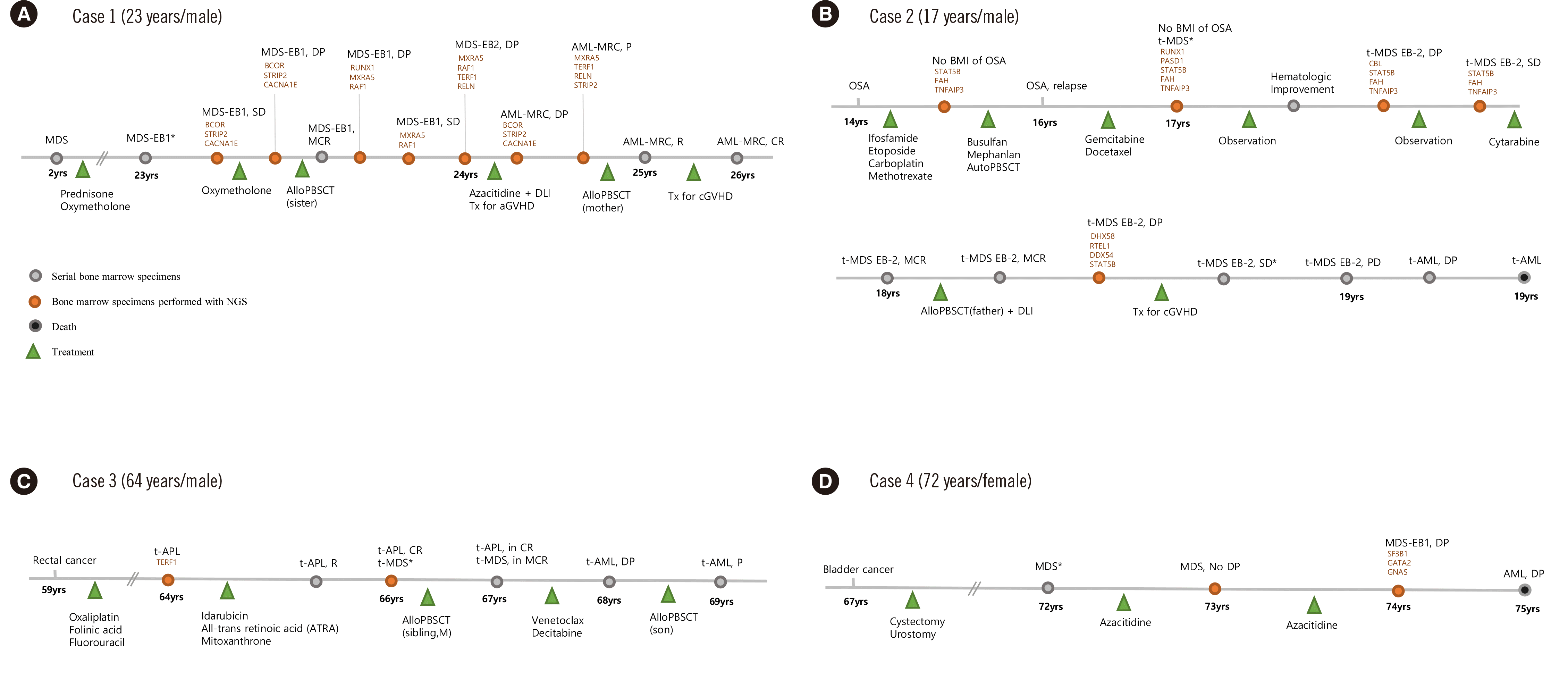
Case 1 (23-year-old male) was diagnosed as having hypoplastic MDS at the age of two years (Table 1, Fig. 1). Prednisolone and oxymetholone were administered without chemotherapy. At 23 years of age, the patient developed pancytopenia (Hb, 45 g/L; white blood cell [WBC] count, 1,290×106/L; platelet [PLT] count, 10×109/L), and he was diagnosed as having MDS with excess blasts 1 (MDS-EB1). The bone marrow (BM) was markedly hypocellular (cellularity, 1%–10%) with blasts (7.5%). A peripheral blood smear showed a dysgranulopoietic feature in the neutrophils. G-banding revealed the cytogenetic aberration 46,XY,t(3;21) (q26;q22)[8]/46,XY[15]. MECOM rearrangement was detected in 49% of the BM nucleated cells (Supplemental Data Figure S1). Multigene sequencing revealed eight somatic variants in RUNX1 (c.1184A>C, p.Glu395Ala), BCOR (c.4071+1G>A, p?), MXRA5 (c.6508G>T, p.Ala2170Ser), RAF1 (c.353A>G, p.Tyr118Cys), TERF1 (c.186_188del, p.Glu62del), RELN (c.3513G>C, p.Met1171Ile), STRIP2 (c.560G>A, p.Arg187Gln), and CACNA1E (c.598C>G, p.Leu200Val). The patient underwent two peripheral blood stem cell transplantations (PBSCTs) from his sister and from his mother, respectively. The disease subsequently progressed to AML-MRC and remission was achieved after chemotherapy. He is currently planning to undergo lung transplantation for chronic graft-versus-host disease (GVHD).
Case 2 (17-year-old male) was previously diagnosed as having osteosarcoma (OSA). Nine months after chemotherapy with alkylating agents (methotrexate, busulfan, and melphalan), the patient developed pancytopenia (Hb, 119 g/L; WBC, 2,980×106/L; PLT, 73×109/L), and he was diagnosed as having t-MDS. G-banding revealed the cytogenetic aberration 45,XY,t(3;21)(q26;q11.2),–7[1]/51,idem,+8,+9,+13,+14,+20,+mar[4]/46,XY[17]. MECOM rearrangement was present in 7% of the BM nucleated cells. Nine somatic variants were detected in RUNX1 (c.1184A>C, p.Glu395Ala), DHX58 (c.1613C>T, p.Ala538Val), RTEL1 (c.2395C>G, p.Leu799Val), DDX54 (c.1529G>A, p.Arg510His), CBL (c.122_127dup, p.His41_His42dup), PASD1 (c.706_708del, p.Ala236del), STAT5B (c.881G>A, p.Arg294His), FAH (c.391C>T, p.Arg131Trp), and TNFAIP3 (c.991G>C, p.Asp331His). He died 31 months after PBSCT from his father.
Case 3 (66-year-old male) was diagnosed as having rectal cancer at 59 years of age and was administered chemotherapy (oxaliplatin, folinic acid, and fluorouracil). He was diagnosed as having t-AML 5 years later. The BM was hypercellular (cellularity 81%–90%), with 32.2% blasts. G-banding revealed the cytogenetic aberration 46,XY,t(15;17)(q24;q21)[12]/46,XY[8] and FISH revealed 99% PML/RARA rearrangement. Two years after the patient achieved remission, he was diagnosed as having t-MDS, and FISH revealed 9% MECOM rearrangement, without PML/RARA rearrangement. A somatic variant in TERF1 (c.186_188del, p.Glu62del) was detected.
Case 4 (72-year-old female) was diagnosed as having bladder cancer 5 years earlier. She was diagnosed as having MDS, unclassifiable (Hb, 74 g/L; WBC, 900×106/L; PLT, 48×109/L). G-banding revealed the cytogenetic aberration 46,XX,t(3;21)(q26.2;q22), and FISH was not performed because of poor sample quality. Three somatic variants were detected in SF3B1 (c.2098A>G, p.Lys700Glu), GATA2 (c.99C>G, p.Tyr33*), and GNAS (c.107C>G, p.Ala36Gly). The disease progressed to AML after 36 months and the patient died of AML.
The frequency of the t(3;21)(q26.2;q22.1) MECOM rearrangement was 0.2% among AML and MDS patients (4/1,945). Two patients with de novo MDS had no history of chemotherapy or radiotherapy (cases 1 and 4). The other two patients, with t-MN, had a history of OSA as the primary cancer (case 2) and a history of chemotherapy due to rectal cancer and subsequent therapy-related acute promyelocytic leukemia (APL) (case 3), respectively.
Ninety consecutive FISH analyses for MECOM rearrangement were performed in cases 1, 2, and 3. The G-banding and FISH results were 100% concordant at initial diagnosis, whereas the concordance was 83.3% at follow-up when 16.7% of the samples were analyzed only using the FISH probe for MECOM, which was not detected using G-banding. Dysmegakaryopoietic features were observed in all four patients, with a percentage of dyspoietic megakaryocytes ranging from 10% to 75.0% (mean, 52.3%). Dysmegakaryopoietic features were determined using Wright–Giemsa staining of BM aspirates and immunohistochemical staining for CD61 (CD61 Mouse Monoclonal Antibody, Roche, Indianapolis, IN, USA) in BM sections, based on WHO criteria [5].
The overall survival (OS) was 78 months (case 1), 31 months (case 2), 37 months (case 3), and 36 months (case 4) (mean OS, 45.5 months). Two-year survival was 100% and 3-year survival was 75%, whereas 5-year survival was 25%. Case 4 was the oldest patient, who died 36 months after the initial diagnosis. Case 2 showed the shortest OS; this patient harbored monosomy 7 in the context of t(3;21) at initial karyotyping, whereas the other patients had t(3;21) only. Summerer, et al. [6] reported poor outcomes in patients with MECOM rearrangement and multiple cytogenetic alterations, especially in chromosome 7, compared to those of patients with a single aberration. Case 2 showed a poor prognostic implication of monosomy 7 in a patient with t(3;21). In case 1, the patient was still alive after 78 months. The survival of these patients was not as poor as expected for patients with t(3;21), with a reported median OS for AML and MDS in Korea of 15.7 and 17.7 months, respectively [7, 8].
Targeted multigene sequencing was performed using a 356- or 507-gene panel including known leukemia-related genes and WHO 2016 genetic predisposition genes. The variant-calling strategy is described in Supplemental Data Figure S2, and pathogenicity was assessed according to the 2015 American College of Medical Genetics (ACMG) guidelines [9]. Variant calling revealed 21 somatic variants that were sorted into tier groups (Table 2) [10]. Somatic variants in RUNX1 and CBL are strongly associated with a short OS in MDS patients [11]. RUNX1 (c.1184A>C, p.Glu395Ala) was detected at the same site in two patients (cases 1 and 2) and CBL (c.122_127dup, p.His41_His42dup) was detected in one patient (case 2). None of the patients with t(3;21) harbored germline predisposition mutations to myeloid neoplasms. Ripperger, et al. [12] suggested the MECOM locus as a novel candidate gene for hereditary hematological malignancies, and their literature review revealed that constitutional MECOM variants include mutations and microdeletions. Reported variants in MECOM are p.His751Arg (missense), p.Arg750Trp (missense), and p.Cys766Gly (missense), with the latter as the most frequently reported MECOM variant [12]. Inherited predisposition genes related to myeloid neoplasms and MECOM variants were not detected in patients with t(3;21)(q26.2;q22.1) in this study.
The limitation of this study is that the germline analysis results could not be confirmed using saliva samples. Alternatively, the detected variants from serial BM samples in the same patients were reviewed based on clinical associations and correlated with the patient’s clinical course. As a small number of patients were enrolled because t(3;21)(q26.2;q22.1) is rare, we compared the survival length of MDS and AML patients who received intensive treatment in Korea. To consider the Korean ethnicity, we filtered out the variants observed in healthy Korean controls [13].
In conclusion, the frequency of t(3;21) is very low (0.2%), and the association between t(3;21) and t-MN is 50%. Targeted multigene sequencing revealed 21 somatic variants in patients with MECOM rearrangement with t(3;21), including in CBL, GATA2, and SF3B1. RUNX1 (c.1184A>C, p.Glu395Ala) was detected in half of the patients. The detection rate of t(3;21) by FISH was higher than that by G-banding at follow-up; thus, FISH is recommended for monitoring and should be considered a routine evaluation for patients with MECOM rearrangements.

Supplemental Materials

ACKNOWLEDGEMENTS

None.

Notes

AUTHOR CONTRIBUTIONS

Lee DS and Lee J designed the study and wrote the manuscript. Lee DS and Roh EY collected the samples. Lee DS, Lee J, and Yun J reviewed the medical records of the patients. Kim S performed the cytogenetic analyses. Kim SM processed the data. Yun J, Jeong D, and Lee Y interpreted the data. Lee DS contributed to the revision of the manuscript. All authors approved the final manuscript to be published.

CONFLICTS OF INTEREST

None.

RESEARCH FUNDING

This study was funded by a National Research Foundation of Korea (NRF) grant by the Korean government (MSIT) (NRF-2017R1A2A1A17069780).

REFERENCES

1. Arber DA, Orazi A, Hasserjian R, Thiele J, Borowitz MJ, Le Beau MM, et al. 2016; The 2016 revision to the World Health Organization classification of myeloid neoplasms and acute leukemia. Blood. 127:2391–405. DOI: 10.1182/blood-2016-03-643544. PMID: 27069254.
crossref
2. Bejar R. 2014; Clinical and genetic predictors of prognosis in myelodysplastic syndromes. Haematologica. 99:956–64. DOI: 10.3324/haematol.2013.085217. PMID: 24881041. PMCID: PMC4040892.
crossref
3. Glass C, Wilson M, Gonzalez R, Zhang Y, Perkins AS. 2014; The role of EVI1 in myeloid malignancies. Blood Cells Mol Dis. 53:67–76. DOI: 10.1016/j.bcmd.2014.01.002. PMID: 24495476.
crossref
4. De Braekeleer M, Le Bris MJ, De Braekeleer E, Basinko A, Morel F, Douet-Guilbert N. 2015; 3q26/EVI1 rearrangements in myeloid hemopathies: a cytogenetic review. Future Oncol. 11:1675–86. DOI: 10.2217/fon.15.64. PMID: 26043219.
5. Barbui T, Thiele J, Gisslinger H, Kvasnicka HM, Vannucchi AM, Guglielmelli P, et al. 2018; The 2016 WHO classification and diagnostic criteria for myeloproliferative neoplasms: document summary and in-depth discussion. Blood Cancer J. 8:15. DOI: 10.1038/s41408-018-0054-y. PMID: 29426921. PMCID: PMC5807384.
crossref
6. Summerer I, Haferlach C, Meggendorfer M, Kern W, Haferlach T, Stengel A. 2020; Prognosis of MECOM (EVI1)-rearranged MDS and AML patients rather depends on accompanying molecular mutations than on blast count. Leuk Lymphoma. 61:1756–9. DOI: 10.1080/10428194.2020.1737689. PMID: 32189545.
7. Kwon J, Kim SY, Yeob KE, Han HS, Lee KH, Shin DW, et al. 2020; Differences in diagnosis, treatment, and survival rate of acute myeloid leukemia with or without disabilities: A national cohort study in the Republic of Korea. Cancer Med. 9:5335–44. DOI: 10.1002/cam4.3179. PMID: 32491262. PMCID: PMC7402831.
crossref
8. Lee JH, Jang JH, Park J, Park S, Joo YD, Kim YK, et al. 2011; A prospective multicenter observational study of decitabine treatment in Korean patients with myelodysplastic syndrome. Haematologica. 96:1441–7. DOI: 10.3324/haematol.2011.046078. PMID: 21659363. PMCID: PMC3186304.
crossref
9. Richards S, Aziz N, Bale S, Bick D, Das S, Gastier-Foster J, et al. 2015; Standards and guidelines for the interpretation of sequence variants: a joint consensus recommendation of the American College of Medical Genetics and Genomics and the Association for Molecular Pathology. Genet Med. 17:405–24. DOI: 10.1038/gim.2015.30. PMID: 25741868. PMCID: PMC4544753.
crossref
10. Li MM, Datto M, Duncavage EJ, Kulkarni S, Lindeman NI, Roy S, et al. 2017; Standards and guidelines for the interpretation and reporting of sequence variants in cancer: A joint consensus recommendation of the Association for Molecular Pathology, American Society of Clinical Oncology, and College of American Pathologists. J Mol Diagn. 19:4–23. DOI: 10.1016/j.jmoldx.2016.10.002. PMID: 27993330. PMCID: PMC5707196.
crossref
11. Bejar R, Papaemmanuil E, Haferlach T, Garcia-Manero G, Maciejewski JP, Sekeres MA, et al. 2015; Somatic mutations in MDS patients are associated with clinical features and predict prognosis independent of the IPSS-R: analysis of combined datasets from the International Working Group for Prognosis in MDS-Molecular Committee. Blood. 126:907. DOI: 10.1182/blood.V126.23.907.907.
crossref
12. Ripperger T, Hofmann W, Koch JC, Shirneshan K, Haase D, Wulf G, et al. 2018; MDS1 and EVI1 complex locus (MECOM): a novel candidate gene for hereditary hematological malignancies. Haematologica. 103:e55–8. DOI: 10.3324/haematol.2017.178723. PMID: 29097497. PMCID: PMC5792286.
13. Lee S, Seo J, Park J, Nam JY, Choi A, Ignatius JS, et al. 2017; Korean Variant Archive (KOVA): a reference database of genetic variations in the Korean population. Sci Rep. 7:4287. DOI: 10.1038/s41598-017-04642-4. PMID: 28655895. PMCID: PMC5487339.
crossref

Fig. 1
Detailed flow charts of clinical and genomic events in four patients with t(3;21). Serial BM analyses of cases 1 (A) to 4 (D) are indicated as round circles in the charts. Orange and gray circles represent BM samples analyzed and not analyzed using multigene target sequencing, respectively. All detected somatic variants are indicated in brown letters. Black circles indicate the death of a patient. Diagnosis is indicated above the timeline and treatments are indicated below the timeline as green triangles. *BM with initial detection of MECOM rearrangement.
Abbreviations: M, male; F, female; OSA, osteosarcoma; MDS-EB, myelodysplastic syndrome with excess blasts; AML-MRC, acute myeloid leukemia with myelodysplasia-related changes; t-MDS, therapy-related myelodysplastic syndrome; t-AML, therapy-related acute myeloid leukemia; APL, acute promyelocytic leukemia; MDS-U, myelodysplastic syndrome, unclassifiable; AlloPBSCT, allogenic peripheral blood stem cell transplantation; MCR, marrow complete remission; DLI, donor leukocyte infusion; GVHD, graft-versus-host disease; d/t, due to; SD, stable disease; R, remission; CR, continuous remission; DP, disease progression; P, persistent; Tx, treatment; BMI, bone marrow involvement.
alm-42-5-590-f1.tif
Table 1
Clinical characteristics of patients with a hematologic diagnosis with t(3;21)
Characteristics Case 1 Case 2 Case 3 Case 4
Diagnosis* MDS-EB1 t-MDS t-MDS MDS-U
Age (yr)/sex 23/male 17/male 66/male 72/female
Underlying disease (age, yr) MDS (2) Osteosarcoma (16) Rectal cancer (59) Bladder cancer (67)
Chemotherapy or RT None Methotrexate, ifosfamide, etoposide, carboplatin, busulfan, melphalan Oxaliplatin, folinic acid, fluorouracil None
Survival 78 months (alive) 31 months 37 months (alive) 36 months
CBC (Hb, WBC, PLT) 60 g/L, 1,800 × 106/L, 60 × 109/L 119 g/L, 2,980 × 106/L, 73 × 109/L 117 g/L, 2,130 × 106/L, 47 × 109/L 74 g/L, 900 × 106/L, 48 × 109/L
Blast count in BM§ 9.0% < 5% < 5% < 5%
Dysplasia Dysgranulopoiesis Dyserythropoiesis, dysmegakaryopoiesis Dysmegakaryopoiesis N/A
Chromosome (G-banding)ll 46,XY,t(3;21)(q26.2;q22) 45,XY,t(3;21)(q26.2;q22),–7 46,XY,t(3;21)(q26.2;q22) 46,XX,t(3;21)(q26.2;q22)
MECOM FISH positivityll Positive (52.7%) Positive (46%) Positive (50%) N/A
Somatic variant genes (VAF, %) RUNX1 (16.1) RUNX1 (43.6) SF3B1 (23.9)
BCOR (62.1) DHX58 (13.0) TERF1 (17.3) GATA2 (27.9)
MXRA5 (48.9) RTEL1 (44.1) GNAS (22.6)
RAF1 (38.8) DDX54 (57.3)
TERF1 (12.3) CBL (57.7)
RELN (22.5) PASD1 (14.5)
STRIP2 (49.5) STAT5B (73.5)
CACNA1E (39.4) FAH (50.0)
TNFAIP3 (46.4)

*Initial hematologic diagnosis in the presence of a MECOM rearrangement; Age at initial hematologic diagnosis with MECOM rearrangement; Survival time from initial hematologic diagnosis to April 2021 for patients who are still alive; §Blast count observed on BM aspiration or BM section at initial diagnosis; llChromosome and MECOM FISH results at AML transformation.

Abbreviations: MDS, myelodysplastic syndrome; MDS-EB1, myelodysplastic syndrome with excess blasts 1; t-MDS, treatment-related myelodysplastic syndrome; MDS-U, myelodysplastic syndrome, unclassifiable; RT, radiotherapy; CBC, complete blood count; BM, bone marrow; N/A, not available due to poor quality; VAF, variant allele frequency; WBC, white blood cell; PLT, platelet.

Table 2
Somatic variants in four patients with MECOM rearrangement with t(3;21)
Case No. Chr Start End Ref Variant Gene Type Accession No. Base change AA change SIFT Polyphen2 CADD Tier [10]
1 1 21 36,164,610 36,164,610 T G RUNX1* Substitution NM_001001890 c.1184A >C p.Glu395Ala D B 23.5 2
2 X 39,921,998 39,921,998 C T BCOR Substitution NM_001123383 c.4071+1G >A p? . . 25.2 2
3 X 3,235,214 3,235,214 C A MXRA5 Substitution NM_015419 c.6508G >T p.Ala2170Ser D D 25.8 3
4 3 12,645,774 12,645,774 T C RAF1 Substitution NM_001354695 c.353A >G p.Tyr118Cys T P 14.16 3
5 8 73,921,284 73,921,286 GAG - TERF1 Deletion NM_003218 c.186_188del p.Glu62del . . . 3
6 7 103,236,929 103,236,929 C G RELN Substitution NM_005045 c.3513G >C p.Met1171Ile T P 25.4 3
7 7 129,094,012 129,094,012 G A STRIP2 Substitution NM_001134336 c.560G >A p.Arg187Gln D D 35 3
8 1 181,546,987 181,546,987 C G CACNA1E Substitution NM_000721 c.598C >G p.Leu200Val D D 28.3 3
2 9 21 36,164,610 36,164,610 T G RUNX1* Substitution NM_001001890 c.1184A >C p.Glu395Ala D B 23.5 2
10 17 40,255,767 40,255,767 G A DHX58 Substitution NM_024119 c.1613C >T p.Ala538Val T P 11.83 3
11 20 62,325,796 62,325,796 C G RTEL1 Substitution NM_001283010 c.2395C >G p.Leu799Val D D 26.1 3
12 12 113,603,723 113,603,723 C T DDX54 Substitution NM_001111322 c.1529G >A p.Arg510His T P 16.74 3
13 11 119,077,232 119,077,232 - CACCAC CBL Duplication NM_005188 c.122_127dup p.His41_His42dup . . . 3
14 X 150,817,142 150,817,144 GCT - PASD1 Deletion NM_173493 c.706_708del p.Ala236del . . . 3
15 17 40,370,849 40,370,849 C T STAT5B Substitution NM_012448 c.881G >A p.Arg294His D D 34 3
16 15 80,454,614 80,454,614 C T FAH Substitution NM_000137 c.391C >T p.Arg131Trp D P 24.2 3
17 6 138,199,573 138,199,573 G C TNFAIP3 Substitution NM_001270507 c.991G >C p.Asp331His D D 24.7 3
3 18 8 73,921,284 73,921,286 GAG - TERF1 Deletion NM_003218 c.186_188del p.Glu62del . . . 3
4 19 2 198,266,834 198,266,834 T C SF3B1 Substitution NM_012433 c.2098A >G p.Lys700Glu D D 28 1
20 3 128,205,776 128,205,776 G C GATA2 Substitution NM_001145661 c.99C >G p.Tyr33* . . 37 1
21 20 57,428,427 57,428,427 C G GNAS Substitution NM_080425 c.107C >G p.Ala36Gly D B 23.5 3

*RUNX1 (c.1184A>C, p.Glu395Ala) was detected in cases 1 and 2; Protein-level prediction algorithms (SIFT, Polyphen2) are presented for the nonsynonymous variants. Tolerated and deleterious variants found in the SIFT prediction algorithm are annotated as T and D, respectively, and benign, possibly damaging, and probably damaging variants identified from Polyphen2 prediction are annotated as B, P, and D, respectively; The prediction algorithm CADD can score human single nucleotide variants and short insertion/deletions. Variants with score above 10 to 20 indicate potential deleteriousness in CADD prediction.

Abbreviations: Chr, chromosome; Ref, reference sequence; AA, amino acid; SIFT, sorting intolerant from tolerant; Polyphen2, polymorphism phenotyping version 2; T, tolerated; D, deleterious; B, benign; P, possibly damaging; CADD, combined annotation-dependent depletion.

TOOLS
Similar articles