Journal List > Ann Lab Med > v.42(5) > 1516078627

Gulati, Uppal, and Gong: Unreliable Automated Complete Blood Count Results: Causes, Recognition, and Resolution

Abstract

Automated hematology analyzers generate accurate complete blood counts (CBC) results on nearly all specimens. However, every laboratory encounters, at times, some specimens that yield no or inaccurate result(s) for one or more CBC parameters even when the analyzer is functioning properly and the manufacturer’s instructions are followed to the letter. Inaccurate results, which may adversely affect patient care, are clinically unreliable and require the attention of laboratory professionals. Laboratory professionals must recognize unreliable results, determine the possible cause(s), and be acquainted with the ways to obtain reliable results on such specimens. We present a concise overview of the known causes of unreliable automated CBC results, ways to recognize them, and means commonly utilized to obtain reliable results. Some examples of unreliable automated CBC results are also illustrated. Pertinent analyzer-specific information can be found in the manufacturers’ operating manuals.

INTRODUCTION

Automated analyzers have become the mainstay of clinical laboratories globally. Hematology laboratories routinely utilize these analyzers to obtain complete blood count (CBC) results with or without differential white blood cell (WBC) counts (DIFFs) on EDTA-anticoagulated blood specimens. Automated CBC, also known as hemogram in some parts of the world, typically includes nine parameters: WBC count, red blood cell (RBC) count, Hb, Hct, mean RBC volume (MCV), mean corpuscular Hb content (MCH), mean corpuscular Hb concentration (MCHC), red cell distribution width (RDW), and platelet (PLT) count. Some laboratories also include mean PLT volume (MPV) and/or DIFF in the CBC. Further discussion here is limited to the eight basic CBC parameters as in our opinion, RDW is irrelevant to the subject matter of the article. Appropriately calibrated and quality-controlled automated hematology analyzers operated according to the manufacturer’s instructions generate accurate CBC results on nearly all specimens. However, every laboratory sometimes encounters specimens that yield no or inaccurate result(s) for one or more CBC parameters even when the analyzer is functioning properly and the manufacturer’s instructions are followed religiously. Inaccurate results, which may adversely affect patient care, are clinically unreliable and require the attention of laboratory professionals. Laboratory professionals are expected to recognize unreliable results, identify the potential cause(s), and be acquainted with the ways to obtain reliable results on such specimens. We provide an overview of the current knowledge about the causes of unreliable automated CBC results, ways to recognize them, and means to obtain clinically reliable results.

CAUSES

Unreliable automated CBC results may be caused by (i) specimen characteristics that interfere with the measurement of one or more CBC parameters, (ii) abnormal cells and/or cellular phenomena that mimic other abnormal or normal cells and therefore are misidentified and miscounted, or (iii) a combination of (i) and (ii). Interferents generating inaccurate CBC results include lipemia, hemolysis, hyperbilirubinemia, RBC agglutinins, WBC agglutinins, PLT agglutinins, hyperproteinemia/paraproteinemia, cryoproteinemia, microorganisms (marked bacteremia, fungemia, and possibly, malaria), hyperglycemia (glucose concentration ≥600 mg/dL or ≥33 mmol/L), dilution with intravenous (IV) fluid infusion(s), adipose tissue fragments/fat globules, fibrin clumps, and small clots [1-5]. Abnormal cells and/or cellular phenomena that may adversely affect one or more CBC parameters include RBC fragments/schistocytes (including microspherocytes), extremely microcytic RBCs (MCV <60 fL, particularly, if MCV <50 fL), lysis-resistant RBCs (e.g., RBCs containing Hb C), hyperleukocytosis, giant PLTs, cytoplasmic fragments of leukemic cells, PLT satellitosis, nucleated RBCs (NRBCs), megakaryocytes, and non-hematopoietic cells/carcinoma cells [1-5]. Yet unknown factors that may adversely affect automated CBC results may be recognized in the future. Among the listed causes of unreliable automated CBC results, some are encountered more commonly than others. The adverse effect(s) of various causative agents may be observed in the CBC results generated by some analyzers and not others, depending on the technologies and reagents utilized by analyzers from various manufacturers. Moreover, the effect(s) may be observed in some models and not in other models even from the same manufacturer. The manufacturers’ operating manuals for analyzers often contain specific information on handling problematic specimens for laboratory professionals.

RECOGNITION AND RESOLUTION

There are multiple avenues by which unreliable CBC results can be recognized, and it is often necessary to utilize as many of these as possible, practically feasible, and effective to ensure that unreliable results are dealt with appropriately and timely. The available avenues include:
  • 1. Automated or manual review of analyzer-generated flags (specific and non-specific), histograms (RBC count, PLT, and WBC count if available), and scattergrams (WBC count DIFF).

  • 2. Automated or manual review of delta check failures.

  • 3. Automated or manual review of the analyzer-generated results for validity based on expectation (clinical or otherwise) and/or predefined quality-control rules, such as,

  • a. Hb and Hct discrepancy or failure of one or more of the so-called “three rules of three”

  • Hct=Hb×3±3 (often referred to as the Hct and Hb rule)

  • Hb=RBC×3±0.3 (Hb and RBC rule)

  • RBC=Hb÷3±0.3 (RBC and Hb rule)

  • The “three rules of three” work well when the RBCs are normocytic and normochromic but not so well when the RBCs are microcytic or macrocytic.

  • b. Elevated MCHC, typically >36 g/dL or >360 g/L, but the elevation threshold may vary with the analyzer; based on the authors’ personal experience, it is >37.5 g/dL or >375 g/L for Sysmex hematology analyzers (Sysmex Corp., Kobe, Japan).

  • c. Decreased MCHC, typically <28 g/dL or <280 g/L, but the threshold may vary with the analyzer.

  • 4. Visual inspection of the settled or spun blood specimen tube for lipemia, hemolysis, icterus, possible RBC clumps/agglutinates, cryoprotein precipitate, and/or clot(s). Alternatively, for lipemia, hemolysis, and icterus, the respective indices, if available from chemistry analysis results, may be utilized.

  • 5. Blood smear examination for the validation of automated results (suspect or otherwise) and/or for detecting and/or confirming the presence of NRBCs, particularly if missed by the analyzer, megakaryocytes, non-hematopoietic cells (carcinoma cells), giant PLTs, PLT clumps, RBC agglutinates, WBC clumps, organisms (bacteria, fungi, and malarial parasites), RBC fragments/schistocytes (including microspherocytes), sickle cells, spherocytes, acanthocytes, Hb C crystals, cytoplasmic fragments of leukemic cells, PLT satellitosis, cryoprotein precipitates and/or crystals, fat globules, and fibrin strands.

Examples of unreliable automated CBC results and the means utilized to obtain reliable results are illustrated in Figs. 1-12.

Characteristics of specimens with lipemia interference

An increased concentration of lipids (triglycerides consisting of chylomicrons and very-low-density lipoproteins) in the blood is referred to as hyperlipidemia or lipemia. It is not uncommon to observe lipemia in postprandial specimens and in specimens drawn from patients with diabetes or those receiving parenteral nutrition with intralipid emulsion [6]. Lipemia interferes primarily with Hb measurement and results in falsely higher Hb, MCH, and MCHC [7]. A discrepancy between Hb and Hct with an elevated MCHC concentration is a useful indicator of the effect of lipemia. An analyzer-generated flag of turbidity or Hb interference is also helpful. Lipemic blood appears milky turbid upon visual inspection of the specimen that had time to settle or was centrifuged (Fig. 1). Microscopic examination of the blood smear is helpful in ruling out the cause(s) of truly elevated MCHC (marked drepanocytosis [sickle cells], marked spherocytosis, and/or possibly marked acanthocytosis). Smears from lipemic blood often reveal some hazy RBCs and some damaged WBCs (primarily neutrophils, bands, and eosinophils) appearing as cells without a cell wall [8].
Although there are a few ways to obtain reliable results on such specimens, in our opinion, a simple and practical way is to reanalyze the specimen in the analyzer after replacing the plasma with an equal amount of an isotonic solution (preferably the diluent utilized in the analyzer) [6, 7, 9, 10]. We describe the plasma replacement procedure utilized by the clinical laboratory at Thomas Jefferson University Hospital (Philadelphia, PA, USA) below.

Plasma replacement procedure

Centrifuge the blood specimen at 3,500 rpm for 10 min. Manually mark the plasma meniscus level on the centrifuged specimen tube, and using a disposable pipette, aspirate out as much of the plasma as possible without disturbing the buffy coat and transfer it into an empty tube. Substitute the plasma in the specimen tube with an isotonic solution (the diluent used in the analyzer or normal saline) up to the plasma meniscus level mark. Mix well manually and/or on a rotator and rerun the analysis. The CBC results obtained from the rerun after plasma replacement are considered reliable if the WBC, RBC, and PLT counts match with those of the initial run (within between-run reproducibility limits). In case of a discrepancy between the rerun and initial results of WBC, RBC, and/or PLT counts, the reliable results from the initial run (WBC, RBC, and PLT counts, Hct, MCV, and RDW) may be reported along with the rerun results of Hb, MCH, and MCHC.
Another approach utilized by some laboratories to obtain reliable results is to measure the plasma Hb concentration and calculate the correct blood Hb concentration using the following formula [1]:
Correct Hb=lipemic blood Hb−(1−Hct)×lipemic plasma Hb, and then recalculate the MCH and MCHC using the standard formulas:
MCH (pg/cell)=Hb (g/dL)×10÷RBC(106/μL)MCHC(g/dL)=Hb (g/dL)×100÷Hct(%)
Alternatively, one may measure the Hb concentration using a point-of-care analyzer (HemoCue, Ängelholm, Sweden), which is unaffected by lipemia, and then recalculate the MCH and MCHC [7].
Another simple approach for lipemic specimens, though not yet validated or reported in the literature, is to dilute an aliquot of the specimen with an isotonic diluent by an appropriate factor (e.g., 1 : 5) and rerun the analysis to obtain reliable results. This approach may or may not yield reliable results, depending on the degree of hyperlipidemia. If it works, the WBC, RBC, Hb, Hct, and PLT results should be appropriately corrected to account for the dilution. The MCV, MCH, MCHC, and RDW values are unaffected by the dilution and do not require correction.
If for some reason neither of the above approaches can be applied, one may choose to report only the reliable results, i.e., WBC, RBC, and PLT counts, Hct, MCV, and RDW and append a comment “accurate Hb, MCH, and MCHC are not obtained due to lipemia” [7].
Although never observed by the authors, lipemia-associated pseudoerythrocytosis has been reported [11].

Hemolysis

RBCs may lyse in vivo in cases of autoimmune hemolytic anemia, hereditary spherocytosis, and sickle-cell disease or in vitro due to an inappropriate specimen collection process, inappropriate storage, and/or transport conditions [12, 13]. The indicators for in vivo hemolysis include a clinical diagnosis of hemolytic anemia, increased indirect bilirubin, decreased or absent haptoglobin, reticulocytosis, and microscopic results of increased polychromasia and/or the presence of spherocytes, schistocytes, and/or sickle RBCs. The indicators for in vitro hemolysis include normal reticulocyte count and parallel increases in potassium, lactate dehydrogenase, and AST concentrations corresponding to the serum or plasma Hb concentration. Blood specimens from patients with in vivo hemolysis yield correct automated CBC results but with higher than normal MCH and MCHC, because the Hb concentration represents the sum of cellular and plasma Hb concentrations. In vitro hemolysis may cause a falsely lower RBC count and Hct concentration and falsely higher MCH and MCHC [14]. A falsely higher PLT count may also be generated if RBC ghosts remaining after hemolysis are counted as PLTs by the analyzer [14]. Mildly elevated MCHC (typically <39 g/dL or <390 g/L) is considered suspicious for hemolysis (in vivo and in vitro). In our opinion, the automated CBC results may be reported without any correction/modification but with a comment “specimen hemolyzed, results may be affected” appended to at least one of the affected parameters (e.g., MCHC) only in cases where moderate or marked hemolysis is evident in the centrifuged or settled specimen tube or the spun micro-Hct. Alternatively, one may choose to report only the reliable results of WBC count, Hb, MCV, RDW, and blood smear-verified or estimated PLT count.

Hyperbilirubinemia

The total bilirubin concentration may be increased in several conditions, including liver disease (e.g., viral hepatitis or cirrhosis), hemolytic anemia, Gilbert’s syndrome, and gallstones. Bilirubin concentrations ≤25 mg/dL (≤425 μmol/L) generally do not adversely affect the results of any of the automated CBC parameters [15]. However, bilirubin concentrations of 25–35 mg/dL (425–600 μmol/L) may cause spectral interference in Hb measurement and result in falsely higher Hb, MCH, and MCHC. Plasma with an increased bilirubin concentration appears icteric (bright yellow). The analyzer may generate an Hb interference flag along with the elevated MCHC. To obtain reliable results, one may dilute the specimen by an appropriate factor (generally in the range of 1 : 2 to 1 : 5) with an isotonic solution (preferably the diluent used in the analyzer) and rerun the analysis. Before reporting, the results of WBC count, RBC count, Hb, Hct, and PLT count from the rerun should be appropriately corrected to account for the dilution. The MCV, MCH, MCHC, and RDW values do not require correction.

RBC agglutinins (RBC agglutination)

Cold-reactive (i.e., reactive at temperatures <37°C) IgM antibodies are typically associated with cytomegalovirus infection, mycoplasma pneumonia, and cold agglutinin disease. These antibodies generally cause RBC autoagglutination (RBC clumping), which interferes with the measurement of RBC-associated CBC parameters and yields falsely lower RBC count and Hct and falsely higher MCV, MCH, and MCHC [16]. Sometimes, WBCs may get trapped in the RBC agglutinates and therefore not be counted by the analyzer, resulting in a falsely lower WBC count. RBC agglutination is generally noted upon microscopic examination of the blood smear (Fig. 2) but may also be visually evident on the interior wall of the specimen tube. Among the various approaches recommended for obtaining reliable results, a common practice is to incubate the blood specimen at 37°C, typically for 10–15 min, and immediately rerun the analysis [1618]. This approach generally yields accurate results of all CBC parameters. A blood smear made promptly after the incubation should be examined to confirm the absence of RBC agglutination. If this approach fails to yield accurate results, one may choose to request a new specimen to be collected and maintained at 37°C until analyzed or perform a micro-Hct and calculate all other RBC parameters (Hb, RBC count, MCV, MCH, and MCHC) using the standard formulas based on conventional units of measurement (Hb=Hct÷3, RBC=Hb÷3 or Hct÷9, MCV=Hct ×10÷RBC, MCH=Hb×10÷RBC, and MCHC=Hb×100÷Hct), particularly if the blood smear reveals normocytic and normochromic RBCs.

WBC agglutinins (WBC clumping)

WBC clumping is an infrequently observed phenomenon that may involve one or more cell types [19-22]. The cell clumps are often too large to be counted, yielding a falsely lower WBC count. Clumps of granulocytes (Fig. 3) have occasionally been observed in the blood smears of patients with infection, cirrhosis, autoimmune disorder, uremia, immunosuppression, or various malignancies [21-26]. Clumps of lymphocytes have occasionally been observed in the blood smears of patients with lymphoproliferative disorders [21, 22]. The cause of WBC clumping may be multifactorial, including the presence of leukoagglutinins, which may be cold-reactive (at temperatures <37°C) and/or EDTA-dependent [19, 22-27]. To obtain reliable results, one may attempt any or all of the following approaches because none of them yields reliable results in all cases [19-27].
  • a. If available, add kanamycin (an aminoglycoside drug) to the specimen or to an aliquot of the specimen and rerun the analysis [26].

  • b. Vortex the EDTA-anticoagulated blood specimen for 1–2 min and rerun the analysis (personal observation).

  • c. Incubate the EDTA-anticoagulated blood specimen at 37°C for 10 min and rerun the analysis [20, 22, 23].

  • d. Obtain a citrated blood specimen (blue-capped tube as used for coagulation tests) and rerun the analysis. Multiply the WBC, RBC, Hb, Hct, and PLT results obtained by 1.1 to account for the dilution of the blood with the citrate solution [24, 27]. The MCV, MCH, MCHC, and RDW values do not require correction. The use of alternative anticoagulants, such as acid citrate dextrose and citrate-pyridoxal 5´-phosphate-Tris, to collect blood specimens has also been recommended [23].

Irrespective of the method selected to obtain reliable results, it is important to examine a blood smear prepared after the rerun to ensure the absence of WBC clumping before reporting. The results are then reported with an appropriate comment (e.g., “WBC clumps noted,” “kanamycin added,” “vortexed,” “incubated,” or “citrated blood specimen”) appended to the WBC count. If all attempts fail, one may have to report “unable to obtain reliable WBC count due to WBC clumping.”

PLT agglutinins (PLT clumping)

A falsely low PLT count, referred to as pseudothrombocytopenia, is not uncommon in clinical laboratories processing EDTA-anticoagulated blood specimens for CBC [28, 29]. The most common cause of PLT clumping is the presence of EDTA-dependent PLT antibodies/agglutinins [28-31]. A review of the analyzer-generated flag(s) and PLT and WBC histograms often points to the possibility of PLT clumps, but microscopic examination of a blood smear will confirm their presence (Fig. 4) [31, 32]. The PLT clumps may vary in size but often are outside the PLT counting threshold settings of the analyzer and therefore are excluded from the PLT counts, resulting in a falsely lower PLT count. Medium- and large-sized PLT clumps may be counted as WBCs, resulting in a falsely higher WBC count [33, 34]. One way to obtain reliable counts on such a specimen is to vortex the specimen for 1–2 min immediately before rerunning the analysis. Vortexing breaks up the PLT clumps in approximately 50% of such specimens and does not interfere with the measurement of any of the other basic CBC parameters [35]. Examining a blood smear prepared immediately after the vortexing is important to confirm the absence of clumps before reporting the results. Another commonly employed approach to obtaining reliable results is to request a new specimen properly collected in a citrated tube (blue-capped tube used for coagulation tests) [31]. Before reporting the result obtained with the citrated tube, multiply it with 1.1 to account for the dilution factor (based on the ratio of nine parts blood and one part anticoagulant in liquid form in the citrated tube), and prepare and examine a blood smear to ensure the absence of PLT clumps. A CBC can also be performed on the citrated blood specimen, and the results can be reported after multiplying the selected parameter results (WBC count, RBC count, Hb, Hct, and PLT count) by the dilution factor of 1.1. The MCV, MCH, MCHC, and RDW values do not require correction. PLT clumping may also occur in citrated specimens in some cases. Other means utilized by some laboratories to obtain reliable PLT counts in such cases include (i) collecting and maintaining the anticoagulated blood at 37°C until analysis, (ii) using an alternative anticoagulant, such as acid citrate dextrose, citrate-pyrophosphate-Tris mixture, or magnesium sulfate, to collect blood, and (iii) adding an aminoglycoside drug (amikacin or kanamycin) to the EDTA-anticoagulated blood specimen before performing the CBC [32]. Concomitant pseudoleukocytosis, if caused by PLT clumps, will also resolve with the approach used to resolve the pseudothrombocytopenia owing to PLT clumping/agglutination [36]. A reliable WBC count estimate can be obtained from the blood smear.

Hyper/paraproteinemia

Increased concentrations of paraproteins (IgM, IgG, and IgA), as observed in cases of plasma cell and lymphoplasmacytic disorders, may interfere with Hb measurement and result in falsely higher Hb, MCH, and MCHC [37]. Analyzer-generated flag(s), delta check failures, elevated MCHC, and an unexpected Hb result are helpful indicators. To obtain reliable results, one can perform plasma replacement before rerunning the analysis or measure the plasma Hb and calculate the true Hb, MCH, and MCHC as described above for lipemic specimens.
Although never observed by the authors, paraprotein-associated falsely higher WBC and PLT counts have been reported [38]. Plasma replacement before rerunning the analysis will yield reliable cell counts.

Cryoproteinemia

Cryoglobulins and cryofibrinogen are plasma proteins that precipitate on cooling (to temperatures <37°C) and redissolve upon warming (to 37°C). Clinical conditions associated with cryoglobulinemia and/or cryofibrinogenemia include various benign and malignant conditions, such as infections (particularly, hepatitis C), autoimmune disorders, plasma cell disorders, lymphoproliferative disorders, and carcinomas [39]. Their presence may be suspected from analyzer-generated flag(s), on observing turbid plasma (Fig. 5) in the specimen tube kept at room temperature or in the refrigerator, and/or upon noticing pale amorphous precipitates (Fig. 6), globules, and/or crystalline material on microscopic examination of a blood smear [40]. Cryoproteins may result in falsely higher WBC and/or PLT counts in various analyzers [40-46]. To obtain reliable results, one may incubate the blood specimen at 37°C for 10-20 min immediately before rerunning the analysis or perform plasma replacement using a warm isotonic diluent maintained at 37°C. Some laboratories choose to draw a new blood specimen and maintain it at 37°C until analysis to obtain reliable results.

Microorganisms

Bacterial, fungal (yeast), and malarial parasites, when present in the blood at very high concentrations, may cause falsely higher PLT and/or WBC counts on various analyzers [47-52]. The presence of individual bacterial cells (particularly, extracellular bacteria) is more likely to cause a falsely higher PLT count, whereas the presence of bacterial or fungal cell clusters or large fungal cells is more likely to cause a falsely higher WBC count because of their respective sizes. Large fungal cells (e.g., certain Candida and cryptococcal species) may mimic small lymphocytes and thus adversely affect automated DIFF. Although not observed by the authors, RBCs infected with Plasmodium falciparum trophozoites caused falsely higher PLT counts in some analyzers [52]. Organisms may be present either as a contaminant (bacteria and yeast) in the specimen tube or due to an actual infection (bacteremia, fungemia, and malaria). Analyzer-generated flag(s), histograms, and scattergrams, delta check failures, unexpected results, and clinical diagnosis are helpful indicators. Microscopic examination of a blood smear and/or blood culture report will confirm the presence of microorganisms. To obtain reliable results, one may choose to report estimated WBC and PLT counts obtained from a blood smear or request a new, properly collected specimen, particularly if contamination is suspected.

Hyperglycemia

Blood glucose concentrations ≥600 mg/dL (≥33 mmol/L) may cause falsely higher MCV and Hct and falsely lower MCHC [53]. Such high glucose concentrations, though rarely observed in patients with diabetes, is generally the result of contamination of a blood specimen with glucose-containing intravenous fluid. The combination of elevated MCV and decreased MCHC is a reliable indicator of a high blood glucose concentration affecting the CBC results. Reviewing the chemistry test results, if available, will confirm the high glucose concentration. Microscopic examination of a blood smear will reveal macrocytic, normochromic RBCs (Fig. 7). To obtain reliable results, one may dilute a small aliquot of the specimen with the isotonic diluent and incubate it at room temperature, typically for 10 min, and then rerun the analysis. The results of the rerun are to be corrected to account for the dilution before reporting. Alternatively, one can run a micro-Hct, which is not affected by a high blood glucose concentration, and recalculate the MCV and MCHC using standard formulas:
MCV(fL)=micro-Hct(%)×10÷RBC(106/μL)MCHC(g/dL)=Hb(g/dL)×100÷Hct(%)
Falsely higher MCV and Hct along with falsely lower MCHC have been obtained on blood specimens left at room temperature for one to four days before being processed for CBC on an automated analyzer [54]. One may choose to reject such specimens or report only the WBC, RBC, Hb, MCH, and PLT results after validating the WBC and PLT by smear review. To the best of our knowledge, there exists only one report of an in vitro study of blood specimens collected from healthy humans that revealed falsely low WBC counts associated with increasing blood glucose concentrations [55].

Dilution with IV fluid infusion

Blood specimens drawn from a site above the intravenous line without stopping the infusion are often diluted with the transfusion fluid [56]. The dilution effect will manifest in the form of falsely low WBC, RBC, Hb, Hct, and PLT counts. Such low counts are accurate for the specimen but unreliable for patient care because of the dilution effect. Delta check failures, unexpected results, and chemistry results, if available, often point to the dilution effect. To obtain reliable results, it is important to request a new, properly collected specimen.

Adipose tissue fragments/fat globules

The contamination of blood with subcutaneous fat tissue during a difficult/traumatic venipuncture, though rare, may cause a falsely higher WBC count besides affecting the DIFF in some analyzers [57, 58]. Analyzer-generated flag(s) and the WBC scattergram, delta check failures, and unexpected result(s) are helpful indicators. Microscopic examination of a blood smear stained with a Romanowski stain may reveal fat globules in the form of round/ovoid empty spaces. These globules stain orange-red with a fat stain, such as Sudan III. To obtain reliable results, one may choose to replace the automated WBC count with an estimated WBC count obtained from a blood smear and the automated DIFF results with manual DIFF results or request a new, properly collected specimen.

Fibrin clumps

If a blood specimen that contains fibrin clumps is inadvertently run in the analyzer, it may yield a falsely higher WBC count or clog the counting aperture or flow cell, yielding no or erroneous CBC results [59, 60]. Analyzer-generated flag(s) and histograms may provide a clue to the presence of fibrin clumps, but microscopic examination of a blood smear will confirm their presence. Such a blood specimen is generally considered unsuitable for performing a CBC, and one often chooses to request a new, properly collected blood specimen. The authors have experienced that, sometimes, only the PLT count is adversely affected, and the results of all other CBC parameters are comparable to previously obtained results.

Small clots in the specimen tube

The presence of small clots in the specimen may cause inaccurate result(s) for any one or more CBC parameters, depending on the number of small clots aspirated by the sampling probe of the analyzer, or no results due to clogging of the counting aperture or flow cell. Typically, all cell counts and Hb are falsely lower [1]. Delta check failures, unexpected results, and review of analyzer-generated flags, histograms, and scattergrams are helpful in recognizing such unreliable CBC results. A properly collected new blood specimen is needed to obtain reliable CBC results.

ABNORMAL CELLS AND/OR CELLULAR PHENOMENA

RBC fragments/schistocytes (including microspherocytes)

Fragments of RBCs, commonly referred to as schistocytes, may take various forms, including helmet cells, bite cells, horn cells, triangular cells, and microspherocytes. Clinical conditions associated with the presence of RBC fragments include disseminated intravascular coagulation, thrombotic thrombocytopenic purpura, malignant hypertension, prosthetic heart valve, severe burns, metastatic carcinoma, HELLP syndrome (hemolysis, elevated liver enzymes, and low PLTs), hereditary pyropoikilocytosis, and march hemoglobinuria. The fragment size is variable but often falls in the PLT counting range of many analyzers. Consequently, they are frequently counted as PLTs, resulting in a falsely higher PLT count [61-64]. One of the authors (GG) has observed a case of pseudothrombocytosis associated with marked RBC fragmentation in a blood specimen inadvertently left over a heat radiator immediately after collection, presumably for a short period (few min). Analyzer-generated flag(s), RBC and PLT histograms, and unexpected PLT results are helpful indicators of falsely high PLT count. The presence of RBC fragments (Fig. 8) is confirmed by a blood smear microscopic examination. For specimens revealing a high number of schistocytes (graded as ≥2+) on the blood smear, one may choose to replace the automated PLT count with an estimated PLT count obtained from the blood smear utilizing the predefined laboratory or standard criteria or perform a manual PLT count using phase microscopy. Optical PLT counting with or without fluorescence may also yield a reliable PLT count in some cases.

Extremely microcytic RBCs

Blood specimens revealing extreme microcytosis (MCV <60 fL, particularly if MCV <50 fL) may contain some RBCs that are too small (below the RBC counting threshold of the analyzer) to be counted as RBCs but are within the PLT counting threshold and hence are counted as PLTs, particularly in analyzers utilizing impedance technology [65, 66]. The outcome is a falsely higher PLT count and possibly, a falsely higher MPV. The effects on RBC parameters (falsely lower RBC count and Hct and falsely normal MCV) are often not clinically significant and may be reported without any correction but with a comment “extremely small RBCs present, result may be affected” appended to one of the affected parameters, preferably the MCV, only after confirming their presence in a blood smear. Combined presence of extremely small RBCs and RBC fragments (Fig. 9) may, at times, result in a falsely higher RBC count, depending on the size and relative proportion of the RBC fragments. The effects on the PLT count and MPV may be clinically significant and require resolution. The PLT count estimated from a blood smear should replace the unreliable automated PLT count or one may choose to perform the PLT count using an alternative automated approach (such as an optical method with or without fluorescence), if available [67]. The PLT count obtained by the optical method may or may not be reliable and therefore requires verification by a PLT estimate from a blood smear. The MPV and RBC count should either not be reported or reported with an appropriate comment.

Lysis-resistant RBCs

Although not observed by the authors, it has been reported that some abnormal RBCs, i.e., those containing Hb C or S, may not lyse well with the lysing reagent utilized in some analyzers [68]. The non-lysed RBCs may be counted as WBCs and create turbidity causing interference in Hb measurement, ultimately leading to falsely higher WBC count, Hb, MCH, and MCHC. Analyzer-generated flag(s) and the WBC scattergram, microscopic examination of a blood smear, clinical diagnosis, and results of Hb variant analysis are helpful indicators of this phenomenon. The ways to obtain reliable results include increasing the amount of lysing reagent, prolonging the lysis time, and/or preferably diluting the blood specimen and rerunning the analysis.

Hyperleukocytosis

A WBC count of ≥100,000/µL is generally referred to as hyperleukocytosis. Clinical conditions associated with hyperleukocytosis include chronic leukemias, acute leukemias, and occasionally, severe reactive conditions. Hyperleukocytosis may cause turbidity interference in Hb measurement and result in falsely higher Hb, MCH, and MCHC. Depending on the degree of hyperleukocytosis, falsely higher results may also be observed for the RBC count, MCV, and Hct, all of which may be clinically significant or insignificant [69, 70]. To obtain reliable results, one may dilute the blood specimen with an isotonic fluid (preferably the diluent used in the analyzer) by an appropriate factor and rerun the analysis. Before reporting, the Hb, Hct, and WBC, RBC, PLT count results from the rerun should be appropriately corrected to account for the dilution. The MCV, MCH, MCHC, and RDW values do not require correction.

Giant PLTs

Giant PLTs are PLTs of the size of normal RBCs or larger (≥8 µm). Clinical conditions associated with the presence of giant PLTs in the blood include chronic myeloproliferative neoplasms (particularly, primary myelofibrosis and essential thrombocythemia) and various inherited macrothrombocytopenias, such as May–Hegglin anomaly and Bernard–Soulier syndrome. When present in high numbers, giant PLTs frequently result in a falsely lower PLT count and occasionally may result in a falsely higher WBC count [71, 72]. The RBC parameters (RBC count, MCV, Hct, MCH, and MCHC) may also be affected, but these changes are generally not clinically significant and not worth spending time to correct. The presence of giant PLTs is often suspected by review of analyzer-generated flag(s), histograms, and scattergrams and confirmed by microscopic examination of a blood smear (Fig. 10). To obtain a reliable PLT count, one may resort to manual counting in a hemocytometer using phase microscopy or preferably, report an estimated PLT count obtained from a blood smear according to the generally recommended or laboratory’s predefined criteria. Similarly, an estimated WBC count is reported in case of an unreliable automated WBC count as evidenced by a discrepancy between the automated and estimated counts.

Cytoplasmic fragments of WBCs

Cytoplasmic fragments of leukemic cells are occasionally observed on microscopic examination of the peripheral blood smears of some patients diagnosed as having acute leukemia or hairy cell leukemia [73-76]. These may mimic PLTs, RBCs, and/or WBCs depending on their size, and when present in high numbers (graded as ≥2+), may result in falsely higher respective automated counts. Often, they are small and counted as PLTs, resulting in a falsely higher automated PLT count [73-76]. Their effect on the RBC count, if present, is generally clinically insignificant, and their effect on the WBC count may be significant in some cases. Although the presence of cytoplasmic fragments of leukemic cells is generally detected during the microscopic examination of a blood smear, one may suspect the false nature of the automated counts by reviewing the analyzer-generated flags and histograms, delta check failures, and/or by observing a discrepancy between the obtained and expected results. The PLT and WBC counts estimated from a blood smear according to the generally recommended or laboratory’s predefined criteria should replace the respective automated cell counts. The criteria used in the clinical laboratory at Thomas Jefferson University Hospital are provided below:
Estimated PLT count (×103/μL or ×109/L of blood) = average numbera of PLTs per field under a 100× oil objective lens multiplied by 15Estimated WBC count (×103/μL or 109/L of blood) = average numbera of WBCs per filed under a 50× oil objective lens multiplied by 3
aTo obtain the average number of cells per field, one generally counts the respective cells in each of 10 microscopic fields in different parts of the readable area of the smear and then divides the total number by 10. Reliable estimated counts can only be obtained if the blood smear is of acceptable quality and is devoid of clumps of the respective cells.
An estimated RBC count is neither recommended nor necessary.

PLT satellitosis

Adherence of PLTs to WBCs, primarily neutrophils and bands, is generally referred to as PLT satellitosis or PLT satellitism [77]. PLT satellitosis may be an immunologic or non-immunologic phenomenon that occurs mainly in EDTA-anticoagulated blood specimens and may cause a falsely lower PLT count [78, 79]. Analyzer-generated flag(s), histograms, and scattergrams, delta check failures, and unexpected results are helpful indicators, but microscopic examination of a blood smear will confirm the presence of satellitosis and its adverse effects on the cell counts. To obtain reliable results, one may choose to obtain citrated blood to perform the CBC or vortex the EDTA-anticoagulated specimen before rerunning the analysis [77-80].

NRBCs

NRBCs may be present in the blood of premature newborns and in several clinical conditions, including hemolytic disease of the newborn, hemolytic anemias, pure erythroid leukemia, chronic myeloproliferative neoplasms, and myelodysplastic syndromes [81]. NRBCs are counted along with WBCs by many, if not all, automated analyzers; the outcome is a falsely higher WBC count. Analyzer-generated flag(s) and the WBC scattergram often highlight the possible presence of NRBCs, which may be confirmed by microscopic examination of a blood smear (Fig. 11). Many analyzers are capable of reporting a WBC count that has automatically been corrected for the presence of NRBCs [82, 83]. If not, the uncorrected automated WBC count may be manually corrected using the following formula:
Corrected WBC count = automated uncorrected WBC count × 100100+number of NRBCs per 100 WBCs

Megakaryocytes

A variable number of megakaryocytes (megakaryoblasts, naked nuclei of megakaryocytes, and/or micromegakaryocytes) may be seen in the blood smears of patients with suspected or confirmed diagnosis of chronic myeloproliferative neoplasms, myelodysplastic syndromes, and acute megakaryoblastic leukemia [84, 85]. Rarely, an occasional megakaryocyte may be seen in peripheral blood smears of premature newborns or adults with non-malignant conditions [86]. When present, megakaryocytes will be included in the WBC count by the analyzer, resulting in a falsely higher WBC count. To the best of our knowledge, none of the current CBC analyzers can differentiate megakaryocytes from WBCs and consequently, the automated WBC count has to be manually corrected for their presence using the below formula. In the clinical laboratory at Thomas Jefferson University, manual correction is conducted only if megakaryocytes are present in high numbers (arbitrarily defined as more than 10 per 100 WBCs).
Corrected WBC count = automated uncorrected WBC count ×100100+number of megakaryocytes per 100 WBCs

Combination of NRBCs and megakaryocytes

If both NRBCs and megakaryocytes are present in the blood, both will be included in the automated WBC count. Upon confirming their presence in a blood smear, one may correct the uncorrected automated WBC count for their presence, particularly when present in high numbers (arbitrarily defined as more than 10 NRBCs and megakaryocytes per 100 WBCs) using the following formula:
Corrected WBC count = automated uncorrected WBC count ×100100+number of NRBCs plus megakaryocytes per 100 WBCs

Non-hematopoietic cells (carcinoma cells)

The presence of carcinoma cells in blood (carcinocythemia) is a rarely encountered phenomenon. As none of the current automated analyzers can differentiate carcinoma cells from WBCs, the former will be counted along with the latter in the total WBC. The outcome will be a falsely higher WBC count, which may be clinically significant or insignificant depending on the degree of carcinocythemia. A reliable WBC count may be obtained by correcting the automated uncorrected WBC count using the following formula:
Corrected WBC count = automated uncorrected WBC count ×100100+number of carcinoma cells per 100 WBCs

CONCLUSIONS

This review provides an overview of the possible causes of unreliable automated CBC results, means to recognize them, and ways to obtain reliable results. A summary of all the results is presented in Table 1. For analyzer-specific information about which factors may adversely affect CBC results, what are the associated indicators/alerts, and suggested means to obtain reliable results, laboratory professionals should consult the manufacturer’s operating manual.

ACKNOWLEDGEMENTS

We thank the laboratory professionals working at Thomas Jefferson University Hospital in Philadelphia, PA, USA, and Cooper University Hospital in Camden, NJ, USA, for their help in collecting example cases.

Notes

AUTHORS CONTRIBUTIONS

Gulati G prepared the initial draft. Uppal G and Gong J reviewed and revised the initial draft. All three authors reviewed and approved the final draft.

CONFLICTS OF INTEREST

None of the authors have any conflicts of interest to declare.

RESEARCH FUNDING

None declared.

REFERENCES

1. Cornbleet J. 1983; Spurious results from automated hematology cell counters. Lab Med. 14:509–14. DOI: 10.1093/labmed/14.8.509.
crossref
2. Zandecki M, Genevieve F, Gerard J, Godon A. 2007; Spurious counts and spurious results on haematology analysers: a review. Part I: platelets. Int J Lab Hematol. 29:4–20. DOI: 10.1111/j.1365-2257.2006.00870.x. PMID: 17224004.
crossref
3. Zandecki M, Genevieve F, Gerard J, Godon A. 2007; Spurious counts and spurious results on haematology analyzers: a review. Part II: white blood cells, red blood cells, hemoglobin, red cell indices and reticulocytes. Int J Lab Hematol. 29:21–41. DOI: 10.1111/j.1365-2257.2006.00871.x. PMID: 17224005.
crossref
4. Godon A, Genevieve F, Marteau-Tessier A, Zandecki M. 2012; Anomalies et erreurs de détermination de l'hémogramme avec les automates d'hématologie cellulaire Partie 3. Hémoglobine, hématies, indices érythrocytaires, réticulocytes [Automated hematology analyzers and spurious counts Part 3. Hemoglobin, red blood cells, cell count and indices, reticulocytes]. Ann Biol Clin (Paris). 70:155–68. DOI: 10.1684/abc.2012.0685. PMID: 22484526.
5. De la Salle B. 2019; Pre- and postanalytical errors in haematology. Int J Lab Hematol. 41(S1):170–6. DOI: 10.1111/ijlh.13007. PMID: 31069968.
crossref
6. Nicholls PD. 1983; An improved method for obtaining reliable blood counts with hyperlipaemic samples on an automated particle counter. Med Lab Sci. 40:397–400.
7. Novak R. CAP Today. In: Savage RA, ed. Q & A column. http://www.captodayonline.com/Archives/q_and_a/qa_07_04.html. Updated on July 2004.
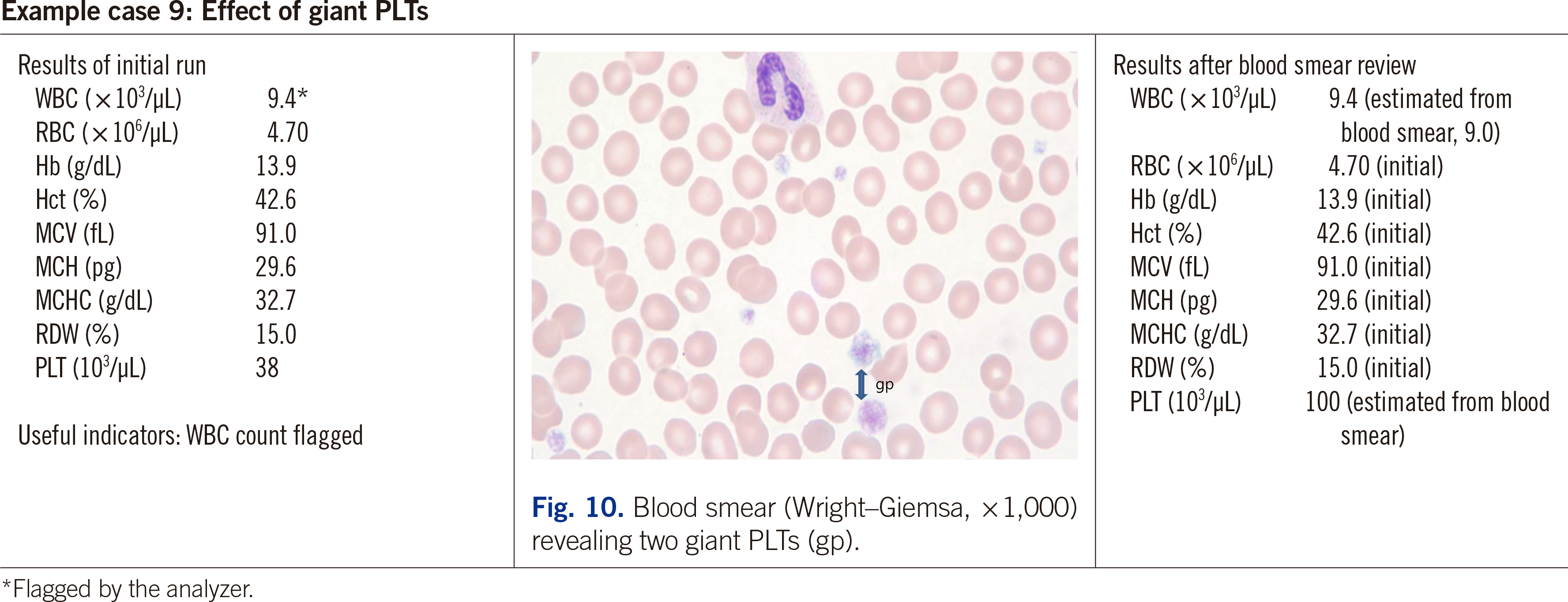
8. Gulati GL. 2021. Blood cells: morphology and clinical relevance. 3rd ed. American Society of Consultant Pharmacists Press;Chicago, IL: p. 13. p. image 2.17.
9. Zeng SG, Zeng TT, Jiang H, Wang LL, Tang SQ, Sun YM, et al. 2013; A simple, fast correction method of triglyceride interference in blood hemoglobin automated measurement. J Clin Lab Anal. 27:341–5. DOI: 10.1002/jcla.21568. PMID: 24038218. PMCID: PMC6807499.
crossref
10. Kurec A. What CBC parameters are affected when the specimen is lipemic? https://www.mlo-online.com/home/article/13008628/lipemia-and-the-cbc-immunohematology-results. Updated on Feb 2016.
11. Zhao XC, Ju B, Wei N, Ding J, Men FJ, Zhao HG. 2020; Severe hyperlipemia-induced pseudoerythrocytosis - implication for misdiagnosis and blood transfusion: A case report and literature review. World J Clin Cases. 8:4595–602. DOI: 10.12998/wjcc.v8.i19.4595. PMID: 33083423. PMCID: PMC7559684.
crossref
12. Lippi G, Blanckaert N, Bonini P, Green S, Kitchen S, Palicka V, et al. 2008; Haemolysis: an overview of the leading cause of unsuitable specimens in clinical laboratories. Clin Chem Lab Med. 46:764–72. DOI: 10.1515/CCLM.2008.170. PMID: 18601596.
crossref
13. Lippi G, Musa R, Avanzini P, Aloe R, Pipitone S, Sandei F. 2012; Influence of in vitro hemolysis on hematological testing on Advia 2120. Int J Lab Hematol. 34:179–84. DOI: 10.1111/j.1751-553X.2011.01378.x. PMID: 22051137.
14. de Jonge G, Dos Santos TL, Cruz BR, Simionatto M, Bittencourt JIM, Krum EA, et al. 2018; Interference of in vitro hemolysis complete blood count. J Clin Lab Anal. 32:e22396. DOI: 10.1002/jcla.22396. PMID: 29396875. PMCID: PMC6817011.
15. Vajpayee N. Interference in lab assays. https://labmedicineblog.com/2017/03/30/interference-in-lab-assays/. Updated on March 2017.
16. Topic A, Milevoj Kopcinovic L, Bronic A, Pavic M. 2018; Effect of cold agglutinins on red blood cell parameters in a trauma patient: a case report. Biochem Med (Zagreb). 28:031001. DOI: 10.11613/BM.2018.031001. PMID: 30429683. PMCID: PMC6214689.
crossref
17. Rim JH, Chang MH, Oh J, Gee HY, Kim JH, Yoo J. 2018; Effects of cold agglutinin on the accuracy of complete blood count results and optimal sample pretreatment protocols for eliminating such effects. Ann Lab Med. 38:371–4. DOI: 10.3343/alm.2018.38.4.371. PMID: 29611389. PMCID: PMC5895868.
crossref
18. La Gioia A. 2019; Eliminating or minimizing the effects of cold agglutinins on the accuracy of complete blood count results. Ann Lab Med. 39:499–500. DOI: 10.3343/alm.2019.39.5.499. PMID: 31037871. PMCID: PMC6502948.
crossref
19. Hillyer CD, Knopf AN, Berkman EM. 1990; EDTA-dependent leukoagglutination. Am J Clin Pathol. 94:458–61. DOI: 10.1093/ajcp/94.4.458. PMID: 2121021.
crossref
20. Yang D, Guo X, Chen Y, Xu G. 2008; Leukocyte aggregation in vitro as a cause of pseudoleukopenia. Lab Med. 39:89–91. DOI: 10.1309/RHN7D0FRW6W0QKWR.
21. Lesesve JF, Fediuk T, Merseille JM, Braun F. 2007; EDTA-dependent lymphoagglutination in a patient with non-Hodgkin lymphoma. Int J Lab Hematol. 29:221–4. DOI: 10.1111/j.1751-553X.2006.00840.x. PMID: 17474901.
crossref
22. Walradth EA. 2021; Lymphocyte aggregation in low-grade B-cell lymphoma. Lab Med. 52:e133–6. DOI: 10.1093/labmed/lmab010. PMID: 33900388.
crossref
23. Goyal P, Agrawal D, Kailash J, Singh S. Cold-induced pseudoneutropenia in human immunodeficiency virus infection: first case report and review of related articles. Indian J Hematol Blood Transfus. 2014; 30(suppl. 1):148–50. DOI: 10.1007/s12288-013-0300-1. PMID: 25332564. PMCID: PMC4192267.
crossref
24. Sultan S, Irfan SM. 2017; In vitro leukoagglutination: A rare hematological cause of spurious leukopenia. Acta Med Iran. 55:408–10.
25. Chen L, Xu X, Zhang Y, Peng J, Zhou K, Wang J, et al. 2018; Detection of EDTA-induced pseudo-leukopenia using automated hematology analyzer with VCS technology. Clin Chem Lab Med. 56:e204–6. DOI: 10.1515/cclm-2017-1173. PMID: 29455187.
crossref
26. Hoffmann JJ. 2001; EDTA-induced pseudo-neutropenia resolved with kanamycin. Clin Lab Haematol. 23:193–6. DOI: 10.1046/j.1365-2257.2001.00343.x. PMID: 11553062.
crossref
27. Anand M, Gulati HK, Joshi AR. 2012; Pseudoleukopenia due to ethylenediaminetetraacetate- induced leukoagglutination in a case of hypovolemic shock. Indian J Crit Care Med. 16:113–4. DOI: 10.4103/0972-5229.99140. PMID: 22988370. PMCID: PMC3439775.
crossref
28. Schuff-Werner P, Mansour J, Gropp A. 2020; Pseudo-thrombocytopenia (PTCP). A challenge in the daily laboratory routine? J Lab Med. 44:295–304. DOI: 10.1515/labmed-2020-0099.
crossref
29. Lardinois B, Favresse J, Chatelain B, Lippi G, Mullier F. 2021; Pseudothrombocytopenia-a review on causes, occurrence and clinical implications. J Clin Med. 10:594. DOI: 
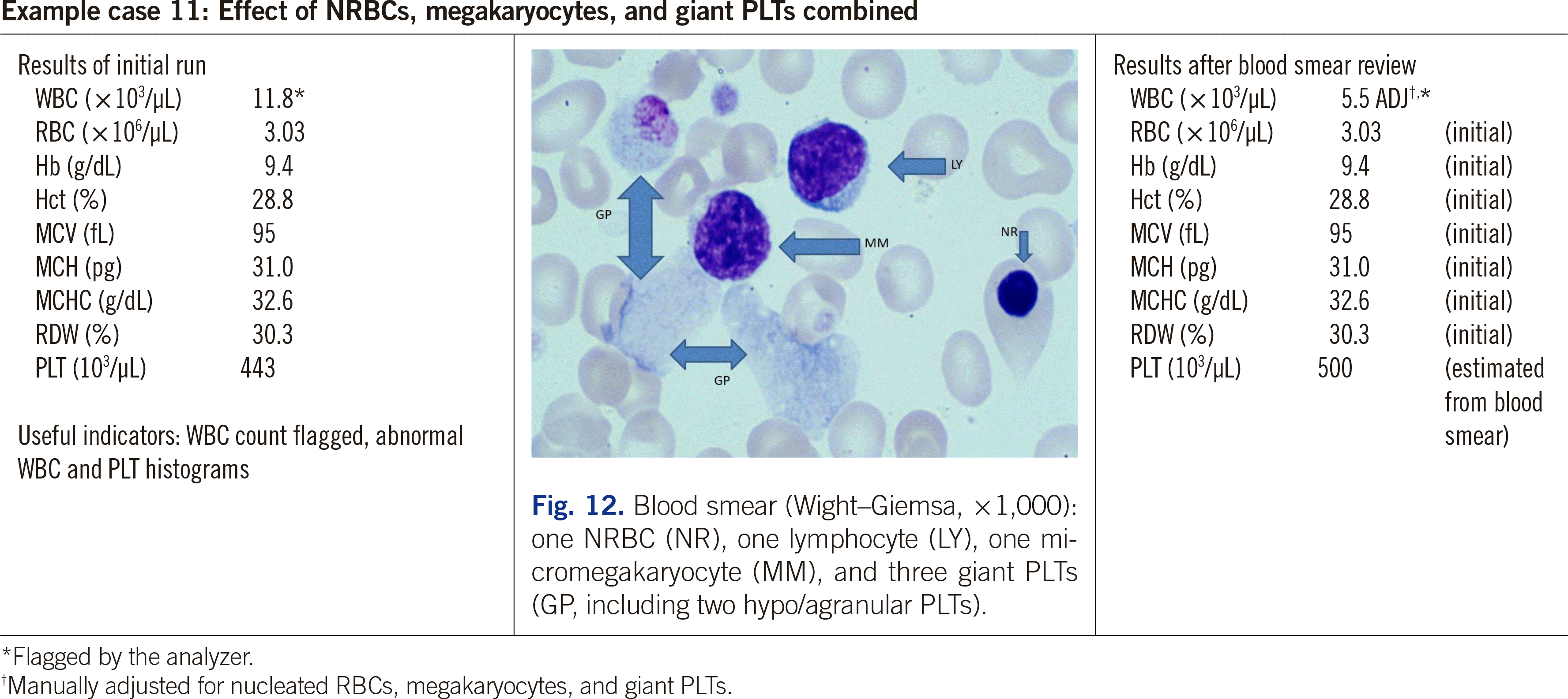
10.3390/jcm10040594. PMID: 33557431. PMCID: PMC7915523.
crossref
30. Abal CC, Calviño LR, Manso LR, Domínguez MCF, Lorenzo MJL, Balboa CR, et al. 2020; Pseudothrombocytopenia by ethylenediaminetetraacetic acid can jeopardize patient safety-report. EJIFCC. 31:65–9.
31. Bartels PC, Schoorl M, Lombarts AJ. 1997; Screening for EDTA-dependent deviations in platelet counts and abnormalities in platelet distribution histograms in pseudothrombocytopenia. Scand J Clin Lab Invest. 57:629–36. DOI: 10.3109/00365519709055287. PMID: 9397495.
crossref
32. Baccini V, Geneviève F, Jacqmin H, Chatelain B, Girard S, Wuilleme S, et al. 2020; Platelet counting: ugly traps and good advice. Proposals from the French-Speaking Cellular Hematology Group (GFHC). J Clin Med. 9:808. DOI: 10.3390/jcm9030808. PMID:
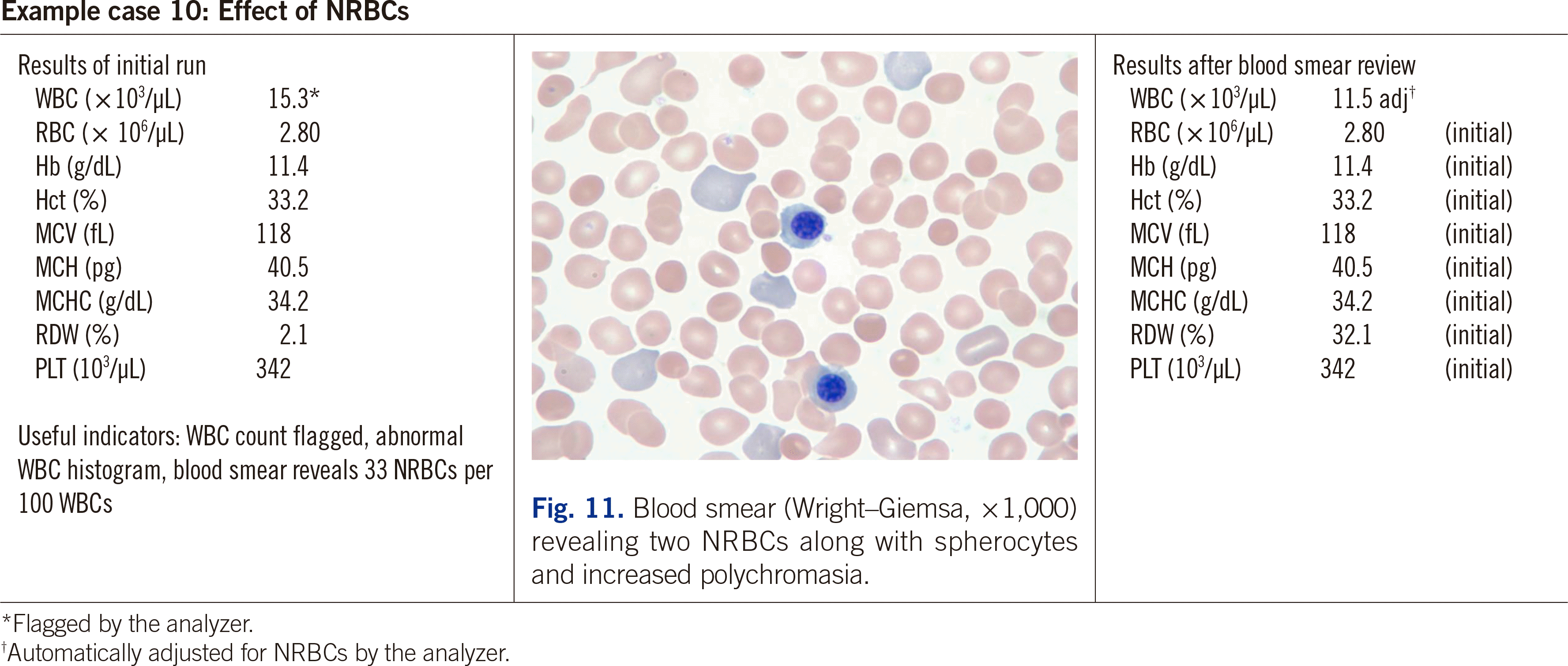
 32188124. PMCID: PMC7141345.
crossref
33. Solanki DL, Blackburn BC. 1983; Spurious leukocytosis and thrombocytopenia. A dual phenomenon caused by clumping of platelets in vitro. JAMA. 250:2514–5. DOI: 10.1001/jama.1983.03340180068032. PMID: 6632146.
34. Savage RA. 1984; Pseudoleukocytosis due to EDTA-induced platelet clumping. Am J Clin Pathol. 81:317–22. DOI: 10.1093/ajcp/81.3.317. PMID: 6422738.
crossref
35. Gulati GL, Asselta A, Chen C. 1997; Using a vortex to disaggregate platelet clumps. Lab Med. 28:665–7. DOI: 10.1093/labmed/28.10.665.
crossref
36. Lombarts AJ, de Kieviet W. 1988; Recognition and prevention of pseudothrombocytopenia and concomitant pseudoleukocytosis. Am J Clin Pathol. 89:634–9. DOI: 10.1093/ajcp/89.5.634. PMID: 3128946.
crossref
37. Roberts WL, Fontenot JD, Lehman CM. 2000; Overestimation of hemoglobin in a patient with an IgA-kappa monoclonal gammopathy. Arch Pathol Lab Med. 124:616–8. DOI: 10.5858/2000-124-0616-OOHIAP. PMID: 10747323.
38. Shastry IK, Nayak DM, Manohar C, Prabhu R, Thomas J. 2014; Paraprotein-induced factitious results on an automated hematology analyzer. Br Biomed Bull. 2:187–91.
39. Brouet JC, Clauvel JP, Danon F, Klein M, Seligmann M. 1974; Biologic and clinical significance of cryoglobulins. A report of 86 cases. Am J Med. 57:775–88. DOI: 10.1016/0002-9343(74)90852-3. PMID: 4216269.
40. Fohlen-Walter A, Jacob C, Lecompte T, Lesesve JF. 2002; Laboratory identification of cryoglobulinemia from automated blood cell counts, fresh blood samples, and blood films. Am J Clin Pathol. 117:606–14. DOI: 10.1309/QXPP-DC4X-N3Q8-KW62. PMID: 11939736.
crossref
41. Patel KJ, Hughes CG, Parapia LA. 1987; Pseudoleucocytosis and pseudothrombocytosis due to cryoglobulinaemia. J Clin Pathol. 40:120–1. DOI: 10.1136/jcp.40.1.120. PMID: 3818970. PMCID: PMC1140844.
crossref
42. Gulliani GL, Hyun BH, Gabaldon H. 1977; Falsely elevated automated leukocyte counts on cryoglobulinemic and/or cryofibrinogenemic blood samples. Lab Med. 8:14–6. DOI: 10.1093/labmed/8.12.14.
crossref
43. Abela M, McArdle B, Qureshi M. 1980; Pseudoleucocytosis due to cryoglobulinaemia. J Clin Pathol. 33:796. DOI: 10.1136/jcp.33.8.796. PMID: 7430391. PMCID: PMC1146221.
crossref
44. Keshgegian AA, Van Tran N. 1985; (Lankenau Hospital case conference) Mixed cryoglobulinemia causing pseudoleukocytosis. Clin Chem. 31:769–73. DOI: 10.1093/clinchem/31.5.769. PMID: 3987007.
crossref
45. Gulati GL, Piao YF, Song AS, Kim WB, Hyun BH. 1995; Interference by cryoproteins in the blood with automated CBCs. Lab Med. 26:138–42. DOI: 10.1093/labmed/26.2.138.
crossref
46. Geara A, El-Imad B, Baz W, Odaimi M, El-Sayegh S. 2009; Pseudoleukocytosis secondary to hepatitis C-associated cryoglobulinemia: a case report. J Med Case Rep. 3:91. DOI: 10.1186/1752-1947-3-91. PMID: 19946507. PMCID: PMC2783090.
crossref
47. Kakkar N. 2004; Spurious rise in the automated platelet count because of bacteria. J Clin Pathol. 57:1096–7. DOI: 10.1136/jcp.2004.016758. PMID: 15452168. PMCID: PMC1770466.
crossref
48. Latif S, Veillon DM, Brown D, Kaltenbach J, Curry S, Linscott AJ, et al. 2003; Spurious automated platelet count. Enumeration of yeast forms as platelets by the cell-DYN 4000. Am J Clin Pathol. 120:882–5. DOI: 10.1309/H2K6E59LYFE63GPB. PMID: 14671977.
49. Kim HR, Park BRG, Lee MK. 2008; Effects of bacteria and yeast on WBC counting in three automated hematology counters. Ann Hematol. 87:557–62. DOI: 10.1007/s00277-008-0464-1. PMID: 18301891.
crossref
50. Branda JA, Kratz A. 2006; Effects of yeast on automated cell counting. Am J Clin Pathol. 126:248–54. DOI: 10.1309/1G5HB7PAK8V0KAR7. PMID: 16891201.
crossref
51. Lesesve JF, Khalifa MA, Denoyes R, Braun F. 2009; Peripheral blood candidosis infection leading to spurious platelet and white blood cell counts. Int J Lab Hematol. 31:572–6. DOI: 10.1111/j.1751-553X.2008.01069.x. PMID: 18498388.
crossref
52. Crabbe G, Van Poucke M, Cantinieaux B. 2002; Artefactually-normal automated platelet counts due to malaria-infected RBC. Clin Lab Haematol. 24:179–82. DOI: 10.1046/j.1365-2257.2002.00414.x. PMID: 12067284.
crossref
53. Morse EE, Kalache G, Germino W, Stockwell R. 1981; Increased electronic mean corpuscular volume induced by marked hyperglycemia. Ann Clin Lab Sci. 11:184–7. PMID: 7259094.
54. de Baca ME, Gulati GL, Kocher W, Schwarting R. 2006; Effects of storage of blood at room temperature on hematologic parameters measured on Sysmex XE-2100. Lab Med. 37:28–36. DOI: 10.1309/1EERK1M02QFJRX6P.
crossref
55. Buonocore R, Picanza A, Gennari D, Pipitone S, Lippi G. 2015; Spurious hyperglycaemia impairs automated leucocyte counting. A pilot study with two different haematological analysers. Blood Transfus. 13:656–61. DOI: 10.2450/2015.0104-15. PMID: 26513771. PMCID: PMC4624544.
56. Watson KR, O'Kell RT, Joyce JT. 1983; Data regarding blood drawing sites in patients receiving intravenous fluids. Am J Clin Pathol. 79:119–21. DOI: 10.1093/ajcp/79.1.119. PMID: 6849287.
crossref
57. Gulati GL, Hyun BH. 1996; An unusual WBC scattergram and its possible causes. Lab Med. 27:398–402. DOI: 10.1093/labmed/27.6.398.
crossref
58. Whiteway AJ, Bain BJ. 1999; Artefactual elevation of an automated white cell count following femoral vein puncture. Clin Lab Haematol. 21:65–8. DOI: 10.1046/j.1365-2257.1999.00178.x. PMID: 10197267.
crossref
59. Maeda K, Pohlad R. 1980; Pseudoleukocytosis due to fibrin strands. Am J Clin Pathol. 74:497.
60. Kumar A, Goswami R, Masih M, Kakkar N. 2018; Spurious leukocyte counts and an abnormal histogram pattern due to fibrin clumps. Indian J Hematol Blood Transfus. 34:785–7. DOI: 10.1007/s12288-018-0998-x. PMID: 30369770. PMCID: PMC6186246.
crossref
61. Akwari AM, Ross DW, Stass SA. 1982; Spuriously elevated platelet counts due to microspherocytosis. Am J Clin Pathol. 77:220–1. DOI: 10.1093/ajcp/77.2.220. PMID: 7064920.
crossref
62. Sapanara NL, Tanev SS, Swami V. 2004; Spurious thrombocytosis in a burn patient. Lab Med. 35:98–100. DOI: 10.1309/DCPND5J6FL59UCD3.
crossref
63. Tantanate C. 2015; Spuriously high platelet counts by various automated hematology analyzers in a patient with disseminated intravascular coagulation. Clin Chem Lab Med. 53:e257–9. DOI: 10.1515/cclm-2015-0055. PMID: 25830905.
crossref
64. Zahid MF, Alsammak MS. 2018; Spurious thrombocytosis in the setting of hemolytic anemia and microcytosis secondary to extensive burn injury. Turk J Haematol. 35:205–6. DOI: 10.4274/tjh.2017.0466. PMID: 29391327. PMCID: PMC6110454.
crossref
65. Velizarova M, Tsvetkova G, Hadjiev E, Yacheva T, Tomov T. 2018; Pseudothrombocytosis in a patient with heterozygous beta-thalassemia. Int J Health Sci Res. 8:297–9.
66. Slota AA, Malik D, Hall D. 2020; Pseudo-thrombocytosis caused by extreme microcytosis in a patient with alpha thalassemia trait. Indian J Hematol Blood Transfus. 36:779–80. DOI: 10.1007/s12288-020-01297-6. PMID: 33100731. PMCID: PMC7573008.
crossref
67. Pan LL, Chen CM, Huang WT, Sun CK. 2014; Enhanced accuracy of optical platelet counts in microcytic anemia. Lab Med. 45:32–6. DOI: 10.1309/LM7QPULDM5IHBO3L. PMID: 24719982.
crossref
68. Booth F, Mead SV. 1983; Resistance to lysis of erythrocytes containing haemoglobin C-detected in a differential white cell counting system. J Clin Pathol. 36:816–8. DOI: 10.1136/jcp.36.7.816. PMID: 6863574. PMCID: PMC498395.
crossref
69. Seigneurin D, Passe D. 1983; Interference of hyperleukocytosis on Coulter Counter Model S blood counts: methods for correction. Biomed Pharmacother. 37:401–4.
70. Lipemia and hyperleukocytosis can lead to CBC errors. 2016; MLO Med Lab Obs. 48:64.
71. Hatzipantelis ES, Tsantali H, Athanassiou-Metaxa M, Avramidou K, Zambul D, Gombakis N. 2001; Hereditary giant platelet disorder presented as pseudothrombocytopenia. Eur J Haematol. 67:330–1. DOI: 10.1034/j.1600-0609.2001.00604.x. PMID: 11872083.
crossref
72. Wang L, Wang H, Liang H, Ren Z, Gao L. 2019; Giant platelets induce double interference in complete blood count. Ann Clin Lab Sci. 49:554–6.
73. Ballard HS, Sidhu G. 1981; Cytoplasmic fragments causing spurious platelet counts in hairy cell leukemia: ultrastructural characterization. Arch Intern Med. 141:942–4. DOI: 10.1001/archinte.1981.00340070122027. PMID: 7235818.
crossref
74. Hammerstrøm J. 1992; Spurious platelet counts in acute leukaemia with DIC due to cell fragmentation. Clin Lab Haematol. 14:239–43. DOI: 10.1111/j.1365-2257.1992.tb00370.x. PMID: 1451403.
crossref
75. van der Meer W, MacKenzie MA, Dinnissen JWB, de Keijzer MH. 2003; Pseudoplatelets: a retrospective study of their incidence and interference with platelet counting. J Clin Pathol. 56:772–4. DOI: 10.1136/jcp.56.10.772. PMID: 14514782. PMCID: PMC1770090.
crossref
76. Kakkar N, Garg G. 2005; Cytoplasmic fragments of leukaemic cells masquerading as platelets in an automated haematology analyser. J Clin Pathol. 58:224. PMID: 15677550. PMCID: PMC1770578.
77. Gulati GL, Hyun BH, Woodall M. 1979; Platelet satellitosis: a case report with laboratory characterization and quantitative evaluation. Ann Clin Lab Sci. 9:109–15.
78. Bobba RK, Doll DC. 2012; Platelet satellitism as a cause of spurious thrombocytopenia. Blood. 119:4100. DOI: 10.1182/blood-2011-08-369173. PMID: 22701880.
crossref
79. Kishore M, Nayak D, Manohar C. 2014; Platelet satellitism: A culprit for spurious thrombocytopenia. Br Biomed Bull. 2:230–4.
80. Tantanate C. 2021; Vortex mixing to alleviate pseudothrombocytopenia in a blood specimen with platelet satellitism and platelet clumps. Clin Chem Lab Med. 59:e189–91. DOI: 10.1515/cclm-2020-1432. PMID: 33554527.
crossref
81. Constantino BT, Cogionis B. 2000; Nucleated RBCs-significance in the peripheral blood film. Lab Med. 31:223–9. DOI: 10.1309/D70F-HCC1-XX1T-4ETE.
crossref
82. de Keijzer MH, van der Meer W. 2002; Automated counting of nucleated red blood cells in blood samples of newborns. Clin Lab Haematol. 24:343–5. DOI: 10.1046/j.1365-2257.2002.00477.x. PMID: 12452814.
crossref
83. Pipitone S, Buonocore R, Gennari D, Lippi G. 2015; Comparison of nucleated red blood cell count with four commercial hematological analyzers. Clin Chem Lab Med. 53:e315–8. DOI: 10.1515/cclm-2014-1211. PMID: 25720099.
crossref
84. Whitby L. 1948; The significance of megakaryocytes in the peripheral circulation. Blood. 3:934–8. DOI: 10.1182/blood.V3.8.934.934. PMID: 18873540.
crossref
85. Ku NK, Rashidi H. 2017; Unusual finding of a megakaryocyte in a peripheral blood smear. Blood. 130:2573. DOI: 10.1182/blood-2017-08-803635. PMID: 29217700.
crossref
86. Garg N, Gupta RJ, Kumar S. 2019; Megakaryocytes in peripheral blood smears. Turk J Haematol. 36:212–3. DOI: 10.4274/tjh.galenos.2019.2019.0022. PMID: 30859799. PMCID: PMC6682780.

alm-42-5-515-f1.tif
alm-42-5-515-f2.tif
alm-42-5-515-f3.tif
alm-42-5-515-f4.tif
alm-42-5-515-f5.tif
alm-42-5-515-f6.tif
alm-42-5-515-f7.tif
alm-42-5-515-f8.tif
alm-42-5-515-f9.tif
alm-42-5-515-f10.tif
alm-42-5-515-f11.tif
alm-42-5-515-f12.tif
Table 1
Summary of the results associated with unreliable automated CBC results
Cause Affected CBC parameters Typical resolution method(s)
Lipemia Falsely higher Hb, MCH, and MCHC Perform plasma replacement and rerun
In vitro hemolysis May cause falsely lower RBC count and Hct and falsely higher MCH, and MCHC,
May or may not cause falsely higher PLT count
Report all CBC results or only WBC count, Hb, MCV, RDW, and smear-verified PLT count
Bilirubin ≥25-35 mg/dL or 250-350 mg/L May cause falsely higher Hb, MCH, and MCHC Dilute the specimen, rerun, and correct the results for dilution
RBC agglutination Falsely lower RBC count and Hctand falsely higher MCV, MCH, and MCHC
May or may not cause falsely lower WBC count
Incubate the blood specimen at 37°C for 10–15 min and rerun
WBC clumping Falsely lower WBC count Vortex and rerun, or incubate at 37°C for 10–15 min and rerun, or use citrated blood and correct the result for dilution
Platelet clumping Falsely lower PLT count
May or may not cause falsely higher WBC count
Vortex and rerun, or incubate at 37°C and rerun, or use citrated blood and correct the result(s) for dilution
Hyper/paraproteinemia May cause falsely higher Hb, MCH, and MCHC Perform plasma replacement and rerun
Cryoproteinemia May cause falsely higher WBC count and/or PLT count Incubate at 37°C for 10–20 min and rerun
Organisms May cause falsely higher PLT count
May or may not cause falsely higher WBC count
Estimate PLT count and if needed, WBC count from blood smear
Glucose ≥ 600 mg/dL or ≥ 33 mmol/L May cause falsely higher MCV and Hct and falsely lower MCHC Dilute the specimen and incubate at RT for 10 min, rerun, and correct the results for dilution or perform a micro-Hct and recalculate MCV and MCHC
Blood stored at RT for 1–4 days Falsely higher MCV and Hct and falsely lower MCHC Reject specimen or report only WBC count, RBC count, Hb, MCH, and smear-verified PLT count
Blood diluted with IV fluid infusion Falsely lower WBC count, RBC count, Hb, Hct, and PLT count Request a properly collected specimen
Adipose tissue (fat globules) May cause falsely higher WBC count Estimate WBC count from blood smear
Fibrin clumps May cause falsely higher WBC count or no results Request a properly collected specimen
Small clot(s) Inaccurate or no results Request a properly collected specimen
RBC fragments
MCV < 60 fL, particularly if MCV < 50 fL
Falsely higher PLT count Estimate PLT count from blood smear or perform optical PLT count and verify by smear review
Lysis-resistant RBCs May cause falsely higher WBC count, Hb, MCH, and MCHC Dilute the specimen, rerun, and correct the results for dilution
Hyperleukocytosis May cause falsely higher Hb, MCH, MCHC and may or may not cause falsely higher RBC count, MCV, and Hct Dilute the specimen, rerun, and correct the results for dilution
High number of giant PLTs Falsely lower PLT count
May or may not cause falsely higher WBC count
Estimate PLT and if needed, WBC count(s) from blood smear
Cytoplasmic fragments May cause falsely higher PLT, may or may not cause falsely higher WBC count, and/or RBC count Estimate PLT count and if needed, WBC count from blood smear
PLT satellitosis May cause falsely lower PLT count Vortex and rerun or use citrated blood and correct the result for dilution
NRBCs, megakaryocytes, carcinoma cells Falsely higher uncorrected WBC count Manually correct the uncorrected WBC, as needed

Abbreviations: WBC, white blood cell; RBC, red blood cell; MCV, mean RBC volume; MCH, mean corpuscular Hb content; MCHC, mean corpuscular Hb concentration; PLT, platelet; NRBCs, nucleated red blood cells; RT, room temperature; IV, intravenous.

TOOLS
Similar articles