

 PDF
PDF Citation
 Print
Print


INTRODUCTION
The survival of patients with multiple myeloma has improved with therapeutic advances over recent decades, and highly sensitive methods capable of monitoring deeper treatment responses are becoming increasingly important [1]. Patient treatment goals are changing from simply delaying progression to achieving the best possible response [2].
Minimal residual disease (MRD) has been assessed using multicolor flow cytometry (MFC), allele-specific oligonucleotide quantitative PCR, and next-generation sequencing (NGS) techniques [3, 4]. MFC and NGS have mainly been used, and the International Myeloma Working Group (IMWG) stated that MRD negativity may be detected in the bone marrow (BM) using MFC or NGS, with a sensitivity of at least 10−5 [5].
MFC has several advantages over NGS, including high applicability, rapid turnaround time, no need for a patient baseline sample, and cost-effectiveness. However, there are concerns about its reproducibility and sensitivity when compared with those of molecular techniques. To overcome these issues, MFC has been progressively improved, resulting in the so-called next-generation flow (NGF) [3]. The EuroFlow Consortium developed and standardized the NGF-based MRD detection method, including sample preparation, antibody panel construction, and automatic identification of plasma cells [6]. EuroFlow-based NGF showed comparable results to NGS, with high sensitivity of 2×10⁻⁶. However, this method involves an eight-color two-tube panel, which is expensive and labor-intensive due to multiple antibody duplications, potentially hindering broad clinical applicability. There is considerable heterogeneity in the real-world clinical application and interpretation of results [7], posing a challenge for sharing and accumulating experience and data from various laboratories. We investigated the clinical utility of NGF-based MRD assessment in a heterogeneous population of patients with multiple myeloma (MM) at the Samsung Medical Center in Korea, focusing on response status, cytogenetic risk, and sustained MRD status.
MATERIALS AND METHODS
Patients
Patients with suspected morphological remission (<5% of plasma cells in the BM) after or during MM treatment were prospectively enrolled for MRD assessment between February 2019 and October 2020. BM samples were obtained for morphological and flow-cytometric evaluations. Patients without morphological remission were excluded. In total, 108 BM samples from 90 patients were included, excluding one BM sample that did not achieve morphological remission. Clinical and laboratory information, including protein electrophoresis, immunofixation, free light chain, and cytogenetic data, was obtained from electronic medical records. The disease response at the time of MRD assessment was determined as stringent complete remission (sCR), complete remission (CR), and very good partial response (VGPR), according to the consensus criteria of the IMWG [5]. Cytogenetic abnormalities were assessed using both conventional karyotyping and fluorescence in situ hybridization as previously described [8]. High-risk cytogenetic abnormalities were defined as the presence of at least one of the following abnormalities: del(17p), t(4;14)(p16;q32), or t(14;16)(q32;q23). Written informed consent was obtained from all patients, and the study was approved by the Institutional Review Board of Samsung Medical Center, Seoul, Korea (SMC-2018-09-054).
NGF-based MRD detection
NGF was performed according to the EuroFlow standardization protocol for MRD detection in MM [9]. BM-EDTA samples were processed within 6 hrs of sampling. After red blood cell bulk lysis, BM samples were stained in two eight-colored tubes: tube 1 for surface staining comprised CD45-PerCPCy5.5 (Cytognos, Salamanca, Spain), CD38-FITC (Cytognos), CD138-BV421 (Becton Dickinson [BD], San Jose, CA, USA), CyIgKappa-APC (Cytognos), CyIgLambda-APCC750 (Cytognos), CD19-PECy7 (Cytognos), CD27-BV510 (BD), and CD56-PE (Cytognos) antibodies, and tube 2 for surface and intracellular staining comprised CD45-PerCPCy5.5, CD38-FITC, CD138-BV421, CD117-APC (Cytognos), CD81-APCC750 (Cytognos), CD19-PECy7, CD27-BV510, and CD56-PE antibodies. A minimum of 5×10⁶ cells per tube (i.e., 107 cells per sample) were analyzed using a FACSLyric flow cytometer (BD). Data were analyzed using the Infinicyt software (version 1.8; Cytognos). The limit of detection (LOD) and limit of quantitation (LOQ) were determined as 20 and 50 cells among 107 events, respectively, resulting in a sensitivity of 2×10⁻⁶ (0.0002%) and 5×10⁻⁶ (0.0005%), respectively, according to the consensus guidelines of MRD reporting [10]. MRD positivity was defined as ≥10−5 (0.001%). Normal plasma cells were typically CD38+, CD138+, CD45+, CD19+, CD27+, CD56–, CD81+, CD117– with polyclonal CyIgKappa and CyIgLambda, and the representative immunophenotype of abnormal plasma cells was CD38+, CD138+, CD45−, CD19−, CD27−, CD56+, CD81−, CD117+ with monoclonal CyIgKappa or CyIgLambda [10].
Statistics
Statistical analyses were performed using the Statistical Software Package for Social Sciences (IBM SPSS Statistics version 25; IBM, Armonk, NY, USA). Categorical variables were compared using the chi-square test or Fisher’s exact test, as appropriate, whereas continuous variables were compared using the Mann–Whitney U-test, two-sample t-test, or Kruskal–Wallis test, as appropriate. The median follow-up duration was estimated using the reverse Kaplan–Meier (KM) method. Progression-free survival (PFS) was determined from the time of the last MRD assessment to disease progression or last follow-up. Survival analysis was performed using KM plots and differences in survival were compared using the log-rank test. Univariable and multivariable analyses were performed using Cox proportional hazards regression models. Values are expressed as the median with interquartile range (IQR). Statistical significance was set at P<0.05.
RESULTS
MRD status and patient characteristics
The median LOD and LOQ of NGF-based MRD assessment were 0.0003% (IQR, 0.0002%–0.0005%) and 0.0007% (IQR, 0.0006%–0.0011%), respectively. MRD was positive in 34 (31.5%) out of 108 samples. The median MRD level was 0.015% (IQR, 0.006%–0.072%), with MRD<0.01% in six (17.6%) samples. The frequencies of aberrant expression of individual markers in abnormal plasma cells were as follows: CD45−, 100%; CD19−, 100%; CD56+, 67.6%; CD27−, 94.1%; CD117+, 38.2%; CD81−, 94.1%; and monoclonal CyIgKappa or CyIgLambda, 100%.
The patient characteristics according to MRD status are summarized in Table 1. For patients who underwent MRD assessment twice, the MRD status was based on the results of the latest assessment to reflect the most recent MRD status in the survival analysis. There were no significant differences in clinical characteristics between MRD-negative and -positive patients, except that high-risk cytogenetic abnormalities were more frequent in MRD-positive patients (P=0.039). The median follow-up duration after MRD assessment was six months (95% confidence interval [CI], 4.4–7.6 months) and nine months (95% CI, 7.5–10.5 months) in MRD-negative and -positive patients, respectively.
Table 1
Characteristics of patients with MM according to MRD status
| Total | MRD status* | |||
|---|---|---|---|---|
|
|
||||
| Negative | Positive | P | ||
| Patients (N) | 90 | 59 | 31 | |
| Sex, male | 52 (57.8%) | 34 (57.6%) | 18 (58.1%) | 0.968 |
| Age (yr) | 61 (55–67) | 61 (54–67) | 62 (57–67) | 0.425 |
| Myeloma type | ||||
| IgG | 44 (48.9%) | 27 (45.8%) | 17 (54.8%) | 0.887 |
| IgA | 16 (17.8%) | 11 (18.6%) | 5 (16.1%) | |
| IgD | 3 (3.3%) | 3 (5.1%) | 0 (0%) | |
| IgM | 1 (1.1%) | 1 (1.7%) | 0 (0%) | |
| Light chain only | 25 (27.8%) | 16 (27.1%) | 9 (29.0%) | |
| Non-secretary | 1 (1.1%) | 1 (1.7%) | 0 (0%) | |
| Light chain type | ||||
| Kappa | 54 (60.7%) | 34 (58.6%) | 20 (64.5%) | 0.587 |
| Lambda | 35 (39.3%) | 24 (41.4%) | 11 (35.5%) | |
| International staging system (N = 85) | ||||
| I | 25 (29.4%) | 16 (28.6%) | 9 (31.0%) | 0.957 |
| II | 29 (34.1%) | 19 (33.9%) | 10 (34.5%) | |
| III | 31 (36.5%) | 21 (37.5%) | 10 (34.5%) | |
| Cytogenetics (N = 84) | ||||
| Standard risk† | 64 (76.2%) | 45 (83.3%) | 19 (63.3%) | 0.039 |
| High risk‡ | 20 (23.8%) | 9 (16.7%) | 11 (36.7%) | |
| Treatment | ||||
| VTD | 59 (65.6%) | 38 (64.4%) | 21 (67.7%) | 0.847 |
| VMP | 12 (13.3%) | 9 (15.3%) | 3 (9.7%) | |
| Others | 19 (21.1%) | 12 (20.3%) | 7 (22.6%) | |
| ASCT | 70 (77.8%) | 46 (78%) | 24 (77.4%) | 1.000 |
| Response status at the time of MRD assessment (N = 89) | ||||
| sCR | 62 (69.7%) | 44 (71.0%) | 18 (29.0%) | 0.176 |
| CR | 12 (13.5%) | 7 (58.3%) | 5 (41.7%) | |
| VGPR | 15 (16.9%) | 7 (46.7%) | 8 (53.3%) | |
| Median follow-up duration after MRD assessment, months | 7 (3–12) | 6 (2–12) | 9 (3–14) | 0.993 |
| Progressive disease after MRD assessment | 15 (16.7%) | 5 (8.5%) | 10 (32.3%) | 0.004 |
*MRD status was based on the results of the last MRD assessment; †Other than high-risk cytogenetics; ‡del(17p), t(4;14)(p16;q32), and/or t(14;16)(q32;q23).
Abbreviations: MM, multiple myeloma; MRD, minimal residual disease; VTD, bortezomib-thalidomide-dexamethasone; VMP, bortezomib-melphalan-prednisone; ASCT, autologous stem cell transplantation; sCR, stringent complete remission; CR, complete remission; VGPR, very good partial response; IQR, interquartile range.
![]()
MRD status according to clinical response
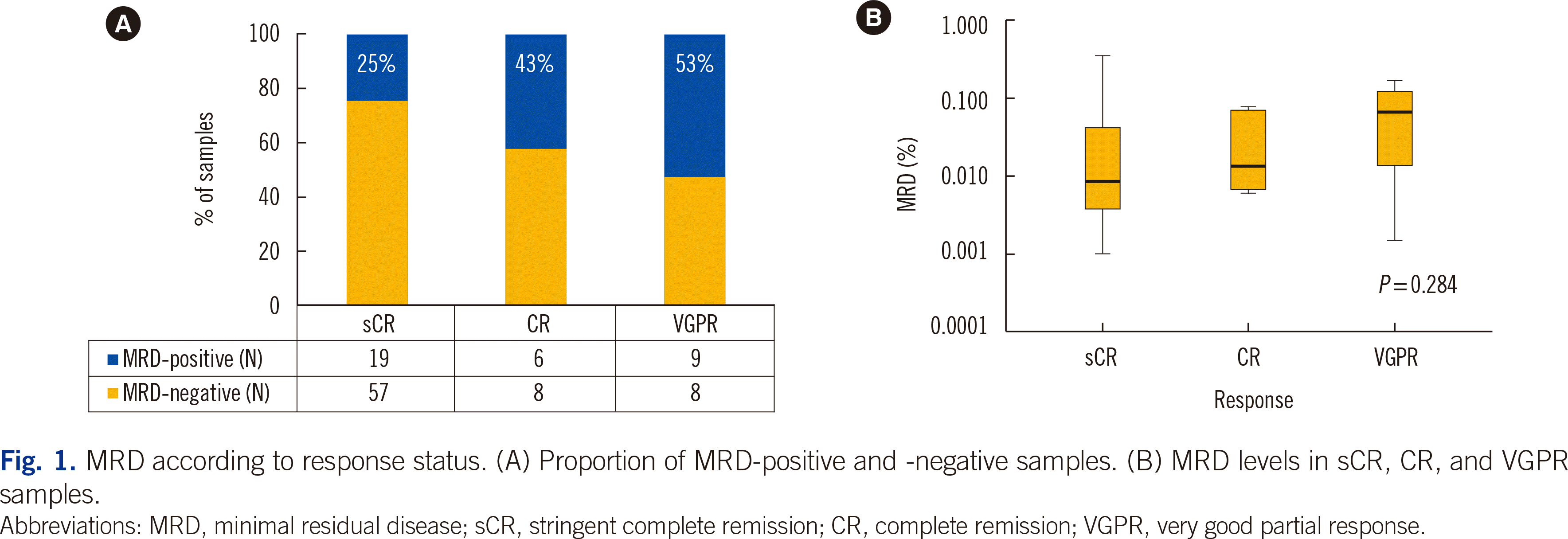
sCR samples showed a lower MRD-positive rate (25%) than CR (43%) and VGPR (53%) samples, although the difference was not significant (P=0.051) (Fig. 1A). The median MRD levels tended to increase to 0.009% (IQR, 0.004%–0.046%), 0.014% (0.007%–0.072%), and 0.066% (0.009%–0.135%) for samples from patients that achieved sCR, CR, and VGPR, respectively (P=0.284) (Fig. 1B).
Survival analysis according to clinical response and MRD
PFS in VGPR patients was lower than that in sCR/CR patients (P<0.001) (Fig. 2A), whereas there was no significant difference in PFS between sCR and CR patients (P=0.543) (Fig. 2B).
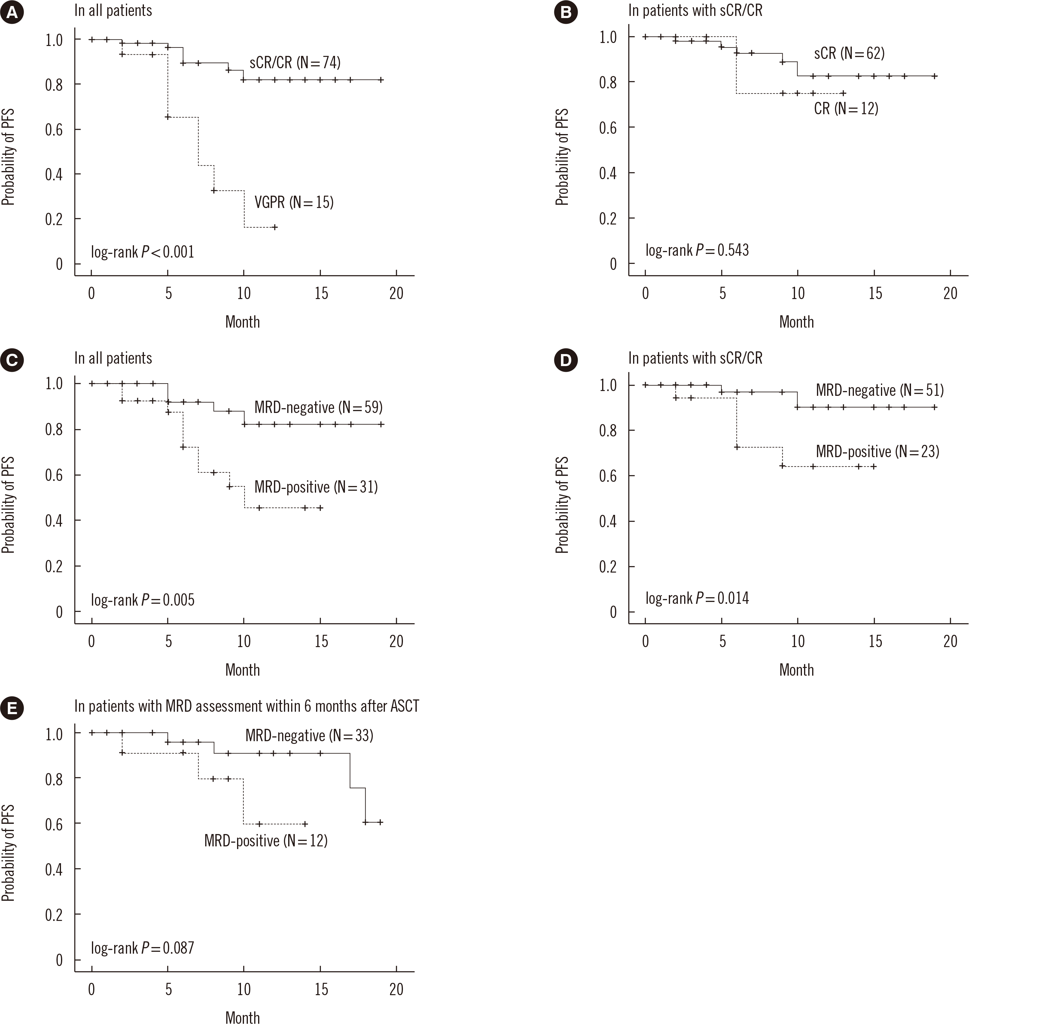
Fig. 2
PFS according to response status: (A) VGPR vs. sCR/CR patients and (B) CR vs. sCR patients. PFS according to MRD status: (C) MRD-positive vs. -negative in all patients, (D) MRD-positive vs. -negative in sCR/CR patients, and (E) MRD-positive vs. -negative in patients with MRD assessment within six months after ASCT. Response status was not evaluated in one patient.
Abbreviations: PFS, progression-free survival; MRD, minimal residual disease; sCR, stringent complete remission; CR, complete remission; VGPR, very good partial response; ASCT, autologous stem cell transplantation.
![]()
PFS was significantly lower in MRD-positive patients than in MRD-negative patients (P=0.005) (Fig. 2C). In VGPR patients, there was no significant difference in PFS according to MRD status (P=0.796). However, inferior PFS was persistently observed in MRD-positive sCR/CR patients (P=0.014) (Fig. 2D). In multivariable analysis, VGPR (hazard ratio [HR]=4.96, 95% CI=1.47–16.72; P=0.010) and MRD positivity (HR=3.23, 95% CI=1.01–10.34; P=0.048) were significantly associated with inferior PFS (Table 2).
Table 2
Univariable and multivariable analyses of PFS
![]()
We further evaluated the impact of MRD in 45 patients who underwent MRD assessment within six months after autologous stem cell transplantation (ASCT), demonstrating that MRD-positive patients showed a trend toward an inferior PFS (P=0.087) (Fig. 2E).
Survival analysis according to cytogenetic risk and MRD
There was no significant difference in PFS between patients with high-risk and standard-risk cytogenetics (P=0.222) (Fig. 3A). Further analysis according to MRD status also revealed no significant difference in PFS in patients with standard-risk cytogenetics (P=0.246) (Fig. 3B); however, among patients with high-risk cytogenetics, MRD-positive patients showed lower PFS than MRD-negative patients (P=0.016) (Fig. 3C).
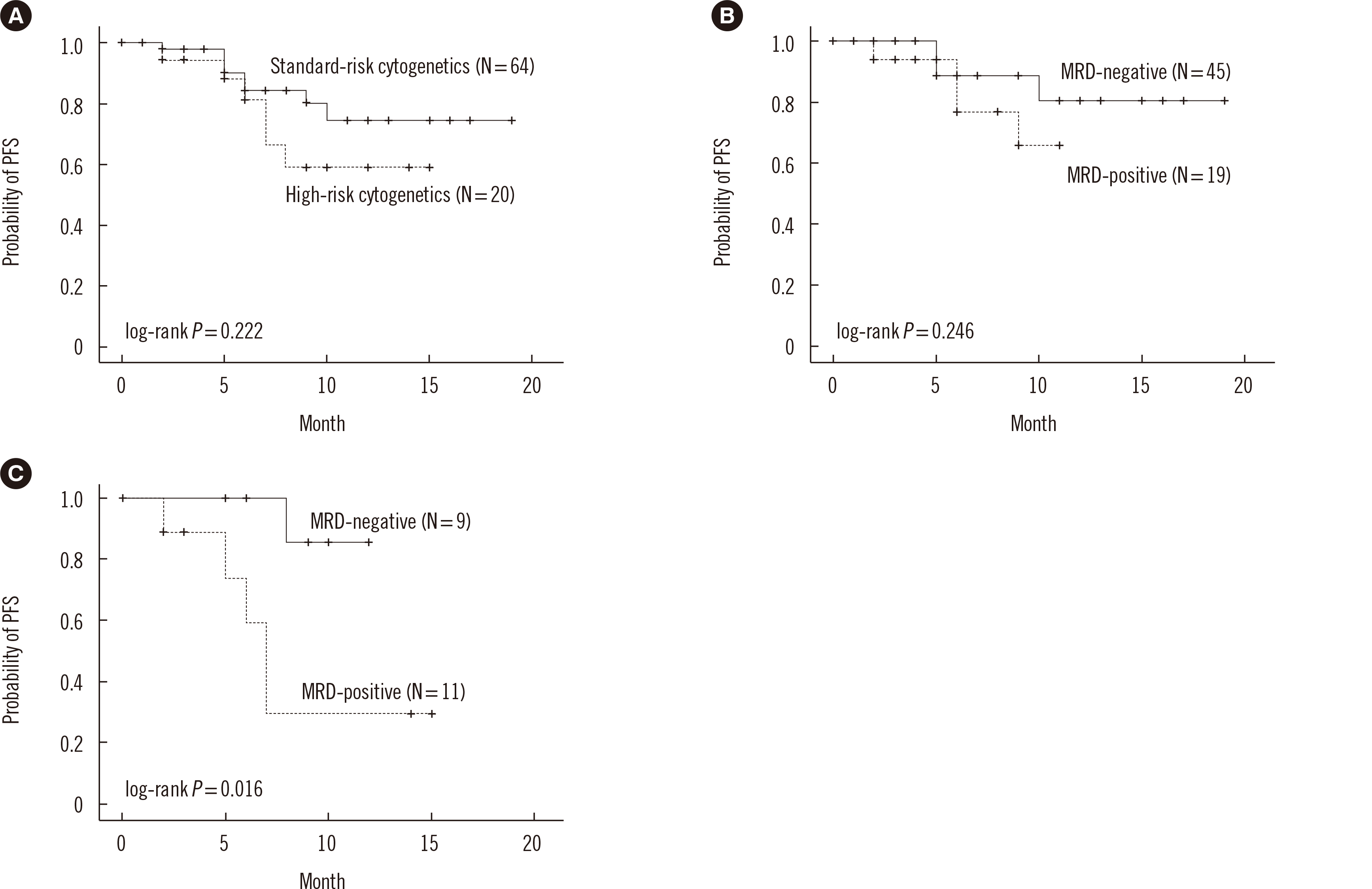
Fig. 3
PFS according to cytogenetic risk: (A) patients with high-risk vs. standard-risk cytogenetics. PFS according to MRD status: (B) MRD-positive vs. -negative in patients with standard-risk cytogenetics and (C) MRD-positive vs. -negative in patients with high-risk cytogenetics.
Abbreviations: PFS, progression-free survival; MRD, minimal residual disease.
![]()
Patient characteristics and survival according to sustained MRD status
MRD was assessed twice in 18 patients, with a median interval of 12 months (IQR, 11–12 months) (Fig. 4A). Sustained MRD negativity was observed only in patients with sustained sCR, and not in patients who showed CR or VGPR status at least once during follow-up (P=0.002) (Supplemental Data Table S1). None of the 11 patients with sustained MRD negativity showed disease progression during follow-up, and their PFS was superior to that of patients who were not MRD-negative (P=0.035) (Fig. 4B). Two patients who did not achieve sustained MRD negativity progressed at 17 and 18 months from initial MRD assessment, respectively.
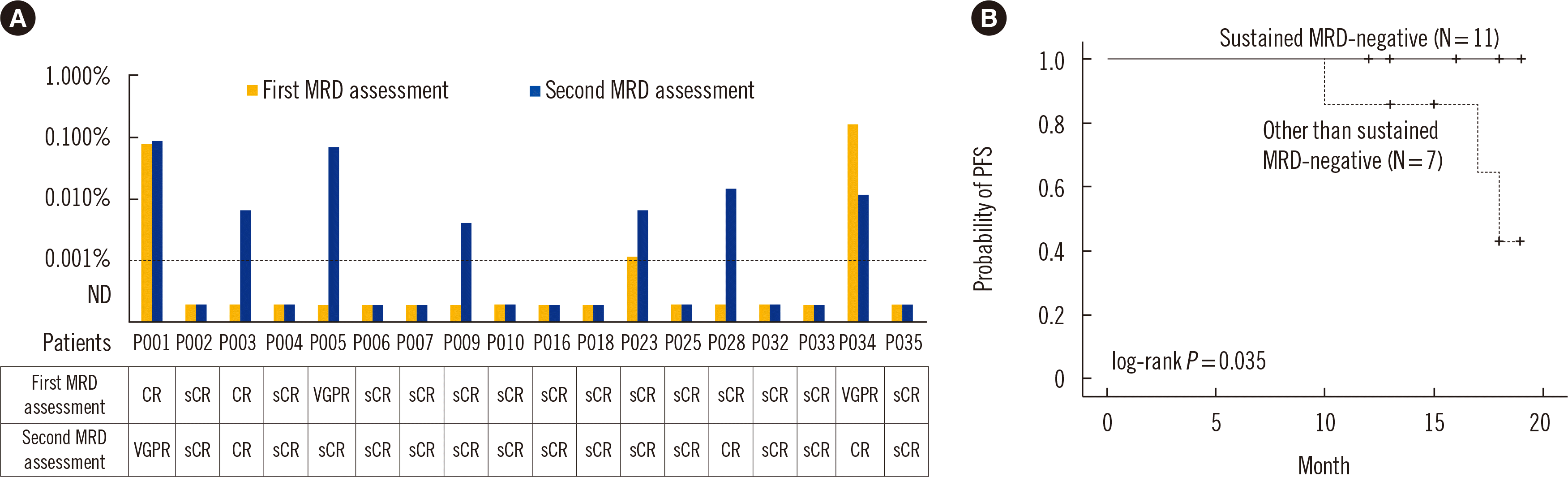
Fig. 4
(A) MRD changes between the first and second assessment. The median follow-up interval of MRD assessment was 12 months, and response statuses at the time of first and second MRD assessment are indicated below the plot. (B) PFS according to sustained MRD status.
Abbreviations: PFS, progression-free survival; MRD, minimal residual disease; ND, not detected; sCR, stringent complete remission; CR, complete remission; VGPR, very good partial response.
![]()
DISCUSSION
MRD assessment is becoming increasingly important for risk assessment in patients with MM. However, it remains difficult to implement high-sensitivity MRD tests in clinical laboratories [1]. We successfully performed NGF-based MRD assessment, achieving a sensitivity of 10−5 (0.001%), and demonstrated its clinical utility.
Highly variable MRD-positive rates have been reported, ranging from 16% to 93.8% [2], which may be explained by the treatment regimen, timing of MRD assessment, and MRD detection method used. There is no consensus on the timing of MRD assessment (e.g., post-induction/consolidation, post-ASCT). Perrot, et al. [11] reported MRD-positive rates of 50.5% and 68.7% at the beginning of maintenance therapy and of 40.3% and 79.5% after 12 months of maintenance therapy in patients with sCR/CR and VGPR, respectively. Kunacheewa, et al. [12] reported MRD-positive rates of 8.5% and 70.5% after initial therapy or ASCT in sCR/CR and VGPR patients, respectively. In our study, MRD-positive rates were 34% and 53% in patients with sCR/CR and VGPR, respectively.
The IMWG recommends assessing MRD at the time of a suspected CR [5]. However, it is unclear whether MRD should be evaluated in patients with VGPR [13]. Even after clonal plasma cells have completely disappeared, it takes several months for the paraprotein to be cleared; therefore, Landgren, et al. [14] recommended performing MRD tests in patients with VGPR in addition to those with CR. Lahuerta, et al. [6] demonstrated that MRD-negative patients with a near CR/partial response had similar PFS and overall survival to those of MRD-negative patients with a CR. We included patients with VGPR who showed lower PFS than sCR/CR patients; however, in a limited number of VGPR patients, MRD assessment did not provide additional prognostic information in terms of PFS. We cannot exclude the possibility of false-negatives to explain this result. As MM can exhibit spatial heterogeneity and patchiness, the extent to which the sample accurately represents the disease state may limit the performance of MRD assessment [1], potentially causing intrinsic false-negative MRD assessment in MM [14].
Among sCR/CR patients, MRD-positive patients showed inferior PFS to that of MRD-negative patients, which is well known [15], supporting the clinical utility of MRD assessment. There was no significant difference in PFS between patients with sCR and CR, which is also consistent with previous findings [16, 17].
Kunacheewa, et al. [12] reported that MRD-negative status did not mitigate the poor prognosis of high-risk cytogenetic patients. However, in our study, MRD-negative patients showed better PFS than MRD-positive patients among patients with high-risk cytogenetics, suggesting that MRD has a prognostic value even in high-risk cytogenetic patients. Long-term follow-up results will help resolve these conflicting findings.
We observed a strong effect of sustained MRD negativity on favorable PFS. Factors associated with sustained MRD negativity are unknown; however, sustained MRD negativity was observed only in patients who maintained sCR in this study, suggesting that maintaining sCR is a predictor of sustained MRD negativity.
This study had some limitations. We included a relatively small number of patients with various treatment regimens, heterogeneous MRD assessment timing, and short follow-up. Despite these limitations, this study demonstrated the clinical utility of NGF-based MRD assessment in predicting disease progression in patients with MM in an actual clinical setting. MRD can serve as a predictor of progression even in patients with high-risk cytogenetics. A follow-up study with a larger study population is warranted.
 XML Download