

 PDF
PDF Citation
 Print
Print


I. Introduction
Orofacial clefts are the most common congenital abnormalities of the head and neck region and can involve the jaws, lips, and hard and soft palates1. Complications such as dental anomalies, malocclusion, facial deformity, malnutrition, and respiratory-auditory-vocal dysfunction can accompany orofacial clefts. Hereditary and environmental factors have pivotal roles in the etiology of facial clefts. Thus, orofacial clefts are considered to have a multi-factorial etiology2. Environmental factors such as maternal hormonal disorders, neurologic drugs, vitamins and folic acid deficiency, hypoxia and smoking, obesity of the mother, and even season in incidence of cleft patients have been reported3-8.
Patients suffering from Tessier cleft types 2 and 3 require a special sequence of treatments from childhood to adolescence. Alveolar bone graft is one of the main reconstruction techniques in this sequence9. Advantages of alveolar bone grafting for reconstruction of the alveolar cleft include maxillary arch stability, elimination of oronasal fistula, ideal bone support for tooth eruption, reconstruction of undeveloped nasal piriform aperture, providing support for soft tissue of the nasal base, and creating better bone support for future implant placement9,10. Secondary alveolar bone graft, described by Boyne and Sands in 1970 for the first time, is currently the most popular method for alveolar bone graft treatment for cleft patients. This method provides an adequate amount of bone for eruption of maxillary canines without adverse effects in the alveolar process11.
The objective of this study was to compare the success rate and morbidities of bone formation in alveolar cleft treatment using anterior iliac crest bone versus chin symphysis bone graft in combination with allograft.
II. Patients and Methods
This randomized controlled trial was registered at the Iranian Registry of Clinical Trials (#IRCT20210515051308n1). This study was conducted on 22 patients referred to the Cleft Palate Center of Isfahan University of Medical Sciences from 2015 to 2017. The Ethical Committee of Isfahan University of Medical Sciences approved this study (IR.MUI.REC.1397.3.131).
The inclusion criteria were unilateral alveolar cleft patients without any systemic diseases or previous reconstructive treatment for alveolar cleft. Exclusion criteria were incidence of intraoperative emergencies or changes in treatment plan, as well as patient unwillingness to participate in the study at any stage. All patients were informed about each surgical procedure and its advantages/disadvantages. Thereafter, written consent forms were signed and recorded.
Patients were divided into two groups randomly through simple randomization using random allocation software such that patients with even numbers were allocated to Group A and patients with odd numbers were put in Group B. For Group A, a combination of allogenic bone material and symphysis corticocancellous bone was used for reconstruction of alveolar defects. For patients in Group B, an autogenous anterior iliac crest graft was used as the control reconstructive treatment.
Preparation of the defect area was achieved intraorally using an advanced gingival flap technique. The mucosal membrane of the defect was divided into nasal and oral segments. For nasal floor reconstruction, nasal mucosal flaps were separated from bony walls and reconnected by sutures. Then, palatal mucosal flaps were sutured to ready the substrate receiver.
1. Group A (allogeneic bone material+symphysis corticocancellous bone)
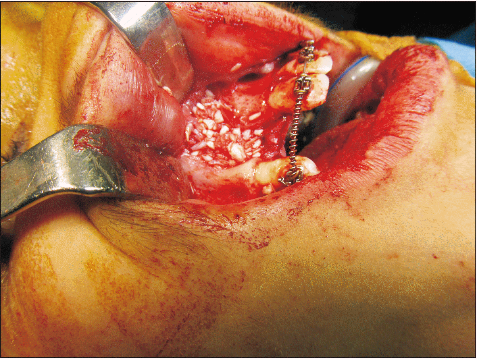
For patients in Group A, the maxillary crest mucosa was flapped using a size 15 scalpel. Then, using free mucosal tissue from the nasal bony walls and flaps suturing, the nasal floor was tightened using a watertight technique. Bony cortical pieces were extracted from the mental symphysis by embedding a sulcular incision from a canine (on one side) to a canine (on the other side), and 3-4 monocortical bone pieces with diameter of 6 milliliters were extracted using a milling trephine (Mesinger, Dusseldorf, Germany). Then, the soft tissue was sutured using polyglycolate 3.0 suture and 26 mm 3/8 reverse cutting needle (SUPA, Tehran, Iran). These pieces were divided into smaller cuts and blended with 2-5 mm allografts (Cenobone, Kish, Iran) in approximately equal amounts. Blended materials were packed in alveolar defects.(Fig. 1)
2. Group B (anterior iliac crest graft)
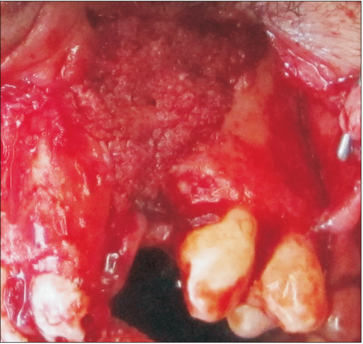
Patients in Group B were treated using autogenic anterior iliac crest graft. In this technique, an incision was made within 1 centimeter of the upper part of the anterior-superior iliac spine. As the iliac bone was exposed, monocortical bone pieces were extracted, and the soft tissue was closed. Cartilage and connective tissues were sutured using polyglycolate 3.0 suture, and skin was closed using nylon 3.0 suture and a 26 mm 3/8 reverse cutting needle (SUPA). Following packing grafts (Fig. 2), defects were closed using mucosal flaps by polyglycolate 3.0 suture. The duration of surgery was recorded for each session.
Patients in Group A were discharged after one day, while Group B patients were discharged two days after the procedure. Sutures embedded in iliac crest were removed within 10 days after surgery in Group B patients.
Patients were examined after one week, one month, three months, six months, and 12 months for assessment of surgical procedure complications.
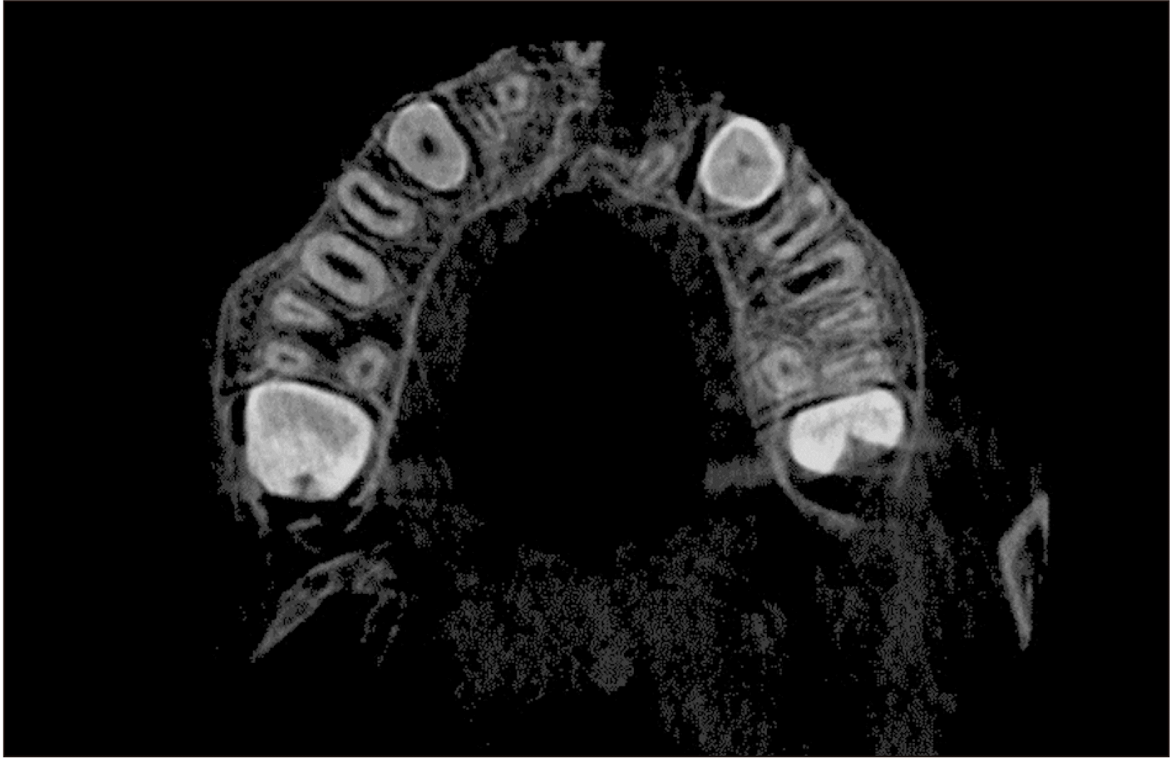
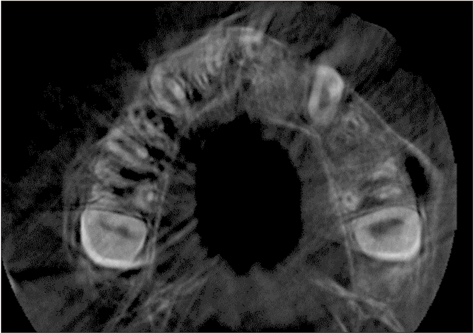
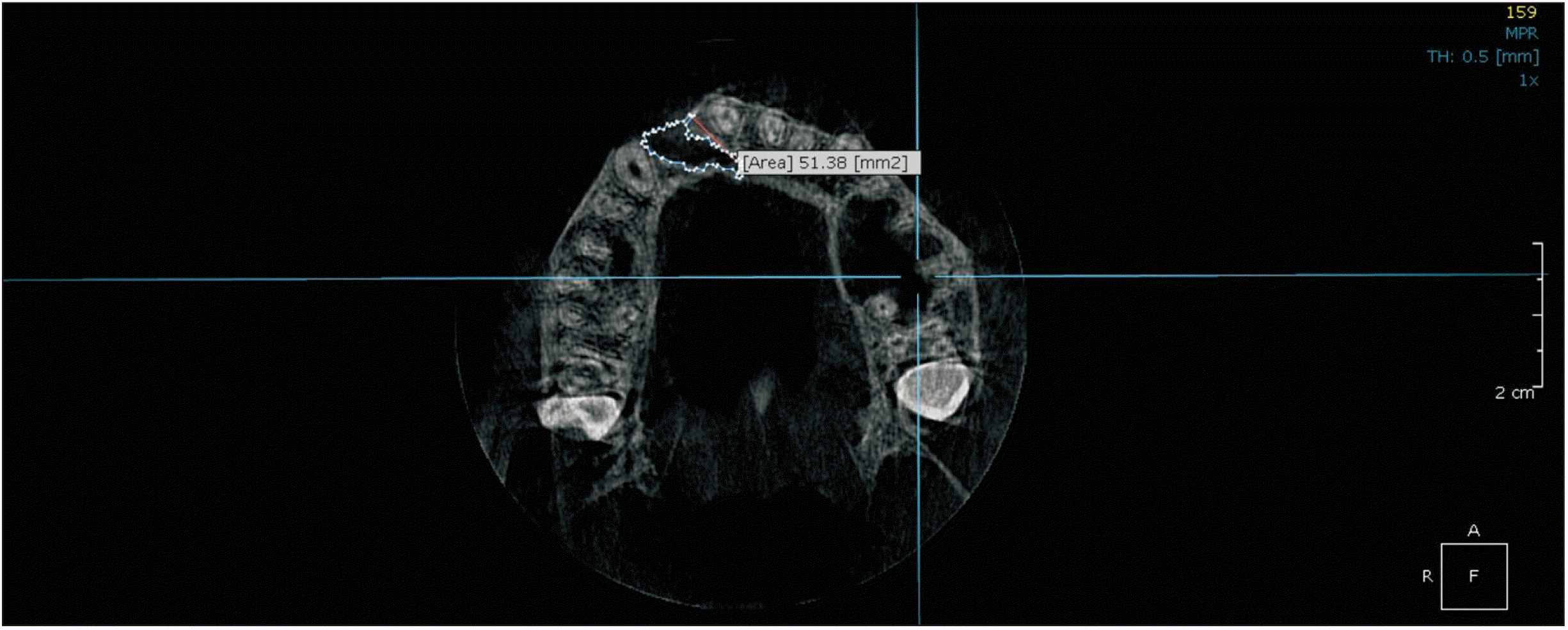
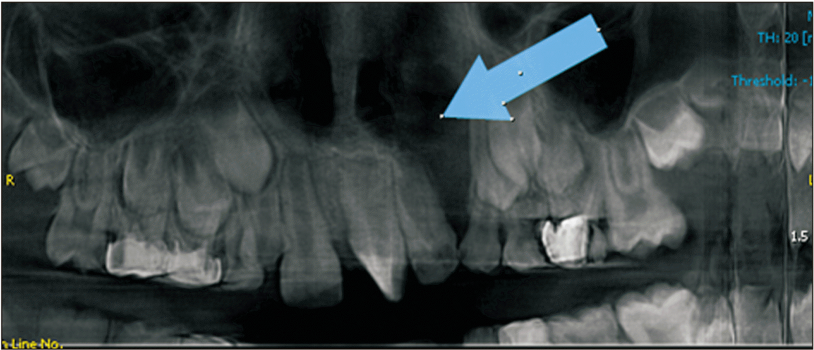
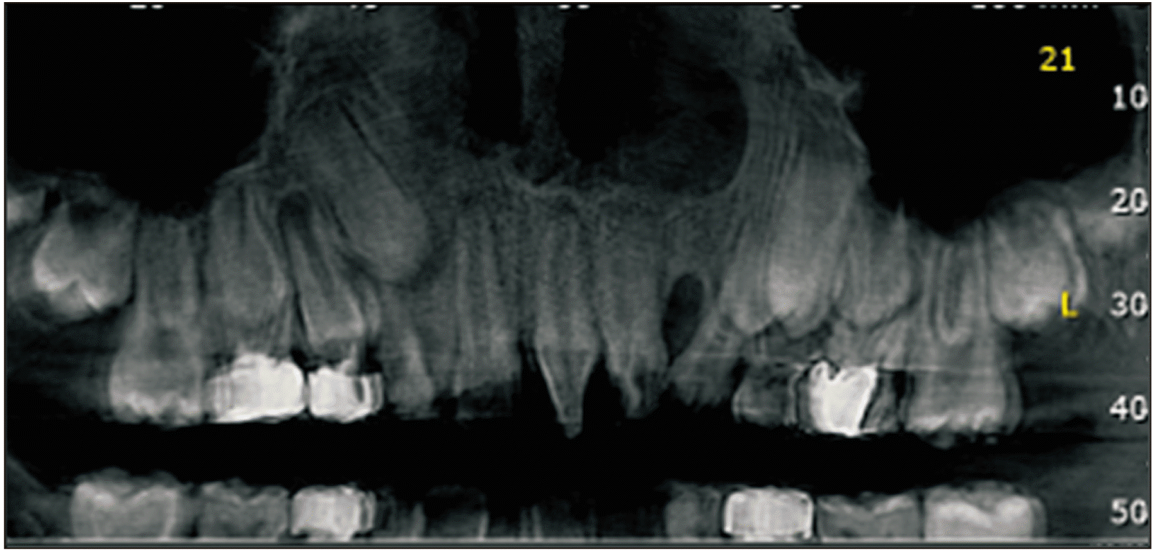
Cone-beam computed tomography (CBCT) was performed prior to and 12 months following the surgery to assess the status of the reconstructed alveolar clefts.(Fig. 3, 4) CBCT scans were obtained from the maxillary region using a Cranex 3D scanner (Soredex, Tuusula, Finland) with 90 kVp, 10 mA, voxel size of 85 µm, and field of view of 61 mm×41 mm. Images were displayed in three dimensions using OnDemand 3D software (Cybermed, Seoul, Korea). Alveolar defect diameter in the vertical plane was measured using coronal views. The lower bound of defect was considered as the cementoenamel junction adjacent to the cleft area. The superior border of the alveolar defect was considered as the base of the nasal cavity on the unaffected side. The defect volume was measured by multiplying the sum of cuts in the axial dimension by height of the defect in the coronal dimension divided by the number of assessed cuts.(Fig. 5)
A radiologist unaware of the study groups interpreted images both pre-surgically and post-surgically. CBCT images were evaluated by the radiologist again after one month to calculate intra-observer agreement.
Obtained data were statistically analyzed using IBM SPSS Statistics (ver. 23; IBM, Armonk, NY, USA) and described in mean values and percentages. The following tests were used for data analysis: intra-class correlation coefficient (ICC) to calculate intra-observer agreement, independent t-test to compare the duration of surgery and age, Fisher’s exact test to compare the sexes and sides, and multiple linear regression to compare the percentage of bone formation considering age and sex variables between the two groups. P<0.05 was considered statistically significant.
III. Results
In the present study, all included patients completed the 12-month follow-up period for monitoring of sequential treatment results in the Cleft Palate Center. A total of 10 males and 12 females with a mean age of 9.7±1.7 years were recruited.(Table 1) Patient mean age (P=0.645) and sex distribution (P=0.576) were not statistically different between Group A and Group B.
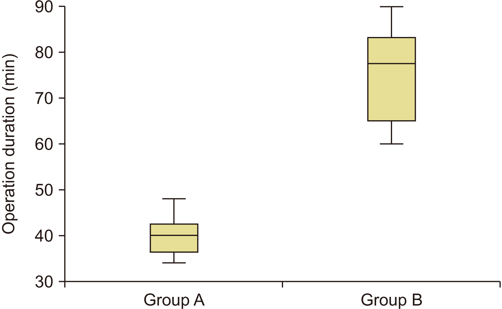
Mean operation duration was 40±4.2 minutes and 76±11 minutes for Group A and Group B, respectively, indicating a significant difference (P<0.001).
No dehiscence, infection, or flap necrosis was observed in the follow-up sessions. Oronasal fistula was closed in all the patients. Paresthesia of the lips or chin was not seen in any patients of Group A or Group B. Clinical and radiographic evaluation indicated normal dental roots and teeth buds. Bridge formation was distinguishable in the CBCT scans of all patients. All patients in Group A had normal gait one day after surgery, whereas 9.5±1.2 days were required for Group B patients to walk normally.
ICC revealed high intra-observer agreement (ICC=0.980), and average volumetric measurements were included in the analysis.
The mean preoperative defect volume was 1.56 cm3 in Group A and 0.95 cm3 in Group B (P=0.001), while the mean postoperative defect was 0.35 cm3 and 0.23 cm3, respectively (P=0.102). The bone formation percentage in the groups was calculated as 76.9% in Group A and 77.0% in Group B. Multiple linear regression analysis demonstrated that, with consideration of age and sex variants, there was no significant difference in bone formation between the two groups (P=0.941).(Table 2)
IV. Discussion
According to the findings of this study, bone formation between alveolar cleft patients treated with symphysis corticocancellous bone combined with allografts and those with iliac crest bone graft is not significantly different. However, patients treated with the anterior iliac crest graft method took longer to walk normally.
Different intra- and extra-oral donor sites have been suggested for alveolar bone graft in cleft patients:
1) Anterior iliac crest is currently considered the gold standard treatment for bilateral and large alveolar defects. It provides large quantities of cancellous bone, but it has disadvantages such as donor site morbidity, operation duration, and gait disturbance12.
2) Proximal side of tibia: The tibia is gracile in childhood, and the epiphyseal cartilage of the tibia is the growth center for the bone. Therefore, it is not a suitable donor site13. Additionally, proximal tibial fractures after bone harvesting have been reported14.
3) Cranial bone has a mesenchymal origin, and the surgical scar will be hidden after hair growth. Morbidities involve dura exposure, subdural hemorrhage, neurologic complications (rarely), and excessive surgical duration15.
4) Intraoral donor sites: The chin is the most popular site for intraoral bone harvesting16. Bone grafting from this region provides a conservative method with lower rate of pain and complication for patients17. In addition, the surgical process can be performed in a shorter duration. Some authors reported more satisfactory results compared with iliac graft, which have been attributed to its mesenchymal origin compared to endochondral origin of the iliac crest18.
Several extraoral and intraoral sites have been proposed for autologous bone harvesting for reconstructive treatments of the alveolar cleft. The average bone mass provided from mandibular bone block is about 2.3 mL19, which is inadequate for large or bilateral alveolar clefts20-22. Therefore, a combination of these bone blocks and bone substitute material has to be used for larger alveolar defects. In 2020, Mahardawi et al.23 measured the impact of certain factors on the success of alveolar bone grafting in cleft patients. They found that bilateral clefts and bone defects greater than 10 mm in transverse and vertical dimensions (evaluation by panoramic radiograph and Bergland scale) increase the risk of failure by four to six times. In our study, CBCT was used for three-dimensional evaluation and volumetric measurements of the cleft defects. The volume of defects in the present patients ranged from less than 0.4 to 2 mL. However, the size of bone defects did not affect treatment success. Further studies to specify the effect of defect volume on bone regeneration are suggested. Additionally, it can be suggested that, in bilateral alveolar clefts or for large defects requiring large bone volume, autogenous bone can be mixed with non-autogenous bone grafting materials to reduce complications and achieve suitable results. In this study, autogenous bone and non-autogenous bone material were combined at a 1:1 ratio. Further research can be performed to obtain the best combination ratio for reconstruction of large alveolar bone defects.
In 2018, Elbokle and Elsholkamy24 performed a study on 12 patients with unilateral cleft palate. Volumetric assessment of bone defects and available bone in the chin area as the donor site was conducted using CBCT imaging. After six months, another CBCT image was obtained to evaluate bone regeneration. The average bone formation was 79%, which is close to the rate of our study (76.9% in Group A). However, in our study, the volume of the defect was measured after one year, which is longer than the follow-up period of Elbokle and Elsholkamy24. In another study performed in our institution, the average bone formation percentage in patients treated with a combination of chin symphysis bone, allograft, and platelet-rich fibrin (PRF) was comparable to that of those treated with iliac bone graft. The authors recommended the first approach to be appropriate for small and moderate alveolar clefts25. In the present study, although PRF was not used, the percentage of bone formation was very similar between the two groups. In addition to PRF, other material can be used in combination with conventional bone grafts. These alternatives have their advantages and disadvantages. For instance, rhBMP (recombinant human bone morphogenetic protein), which has been reported for reconstruction of alveolar clefts26, can eliminate bone harvesting from the donor site but is not approved by the U.S. Food and Drug Administration for application in children, can cause massive edema in the surgical site, and is an expensive treatment27-29. In a systematic review and meta-analysis conducted by Kamal et al.30 in 2018, both methods of using autologous bone graft and tissue-engineered bone material were concluded to be successful in the treatment of alveolar clefts. The average percentage of bone formation was 62% and 68.7%, respectively, and the most widely used substance in the tissue-engineered bone group was BMP-2 (bone morphogenetic protein 2). In the study by Thuaksuban et al.31, treatments with iliac bone graft alone were compared to treatments with iliac bone graft and deproteinized bovine bone in alveolar cleft patients. The combined group showed a significant advantage in reducing morbidity (e.g., shorter hospitalization period and recovery time to walking, etc.), but the density and height of the bony bridge formed in the defect area were not significantly different between the groups. Weijs et al.32 in 2010 arranged an investigation on 47 patients with alveolar clefts and compared treatment outcomes between patients treated with chin symphysis graft and those treated with chin symphysis graft combined with beta tricalcium phosphate (TCP). The results were evaluated in two-dimensional occlusal radiographs. After one year, both groups showed satisfying treatment outcomes, and there was no significant difference between the new bone bridges heights between the groups. In 2019, Miyagawa et al.33 investigated the effect of beta TCP on the quality of bone regeneration. They evaluated cleft patients with CBCT and used bone structural index and trabecular bone parameter. Their findings suggested that utilization of chin symphysis bone graft in combination with beta TCP increases the quality and density of bone regeneration compared to chin symphysis bone graft alone. Further studies can be performed surveying the combination of beta TCP with other autogenous grafts such as iliac, cranium, and tibia in order to provide an appropriate combination for more favorable graft outcomes.
No subsequent morbidity was found in the mandibular symphysis region. For each patient in the symphysis bone graft group, three to four monocortical bone pieces were harvested using a 6 mm trephine bur with the furthest distance from the mental foramens, dental roots, and tooth buds. As a result of this conservative approach and careful sulcular incision, paresthesia (even temporary) or damage to the teeth was not seen in our patients. However, paresthesia is reported in some studies after bone harvesting from the mandibular symphysis34,35. The only complication in our study after chin bone graft was regional pain that resolved after three weeks in all patients.
Surgery duration was significantly shorter for bone harvesting from the chin.(Fig. 6) The same finding was observed by Movahedian et al.25 in 2016 when applying PRF combined with chin bone graft and allograft for repairing alveolar defects in cleft patients. The average duration of each surgical session for iliac bone graft harvesting is reported to be longer in other studies36,37. In addition, Bukhari et al.38 reported longer duration of surgery for iliac crest bone graft compared with chin bone graft. The length of surgical sessions depends on various parameters, including surgical technique, available instruments, and experience of the surgeon, as well as other members of the operating room team39. Shorter surgical durations are preferred for benefits such as less need for anesthetic drugs, decreased bleeding, and lower risk of subsequent infection.
Considering postsurgical complications, no particular event occurred in patients in either group. However, morbidity of the surgical site was more significant for the iliac crest graft group, as they were unable to walk normally for an average of about nine days. Upon investigation, the patients reported pain while walking. Fortunately, all patients eventually regained normal gait. In our investigation, we discharged all Group B patients 48 hours after surgery with oral analgesic drugs. In our opinion, it is unlikely that iliac crest or chin pain would affect the quantity and quality of bone regeneration in the alveolar cleft area; thus, we did not evaluate these items.
A previous study by Swan and Goodacre40 reported that children for whom an iliac crest bone graft was performed could not walk normally for 0 to 56 days due to morbidity at the iliac crest donor sites. The duration for which the patient has difficulty in walking is largely variable among studies. However, considerable morbidity seems to be present following bone harvesting procedures. In a study of 64 patients, Rawashdeh41 examined the side effects of bone removal from the iliac crest. They reported that 91% of patients were able to walk after 24 hours but not normally. In addition, 89% of patients were able to walk as before surgery after two weeks. In our study, all patients were able to walk normally by the 11th day. It seems the cause of difficulty in walking in the first days was donor site pain for Group B patients. Due to the young age of the patients, fear could have intensified limping. Further research is recommended to investigate the role of fear in postoperative walking and limping in patients with iliac crest bone grafts.
Albuquerque et al.42 in 2011 confirmed the accuracy and efficiency of CBCT in determining the volume of bony defects after several measurements on nine human skulls. They recommended CBCT for diagnosis and treatment assessment of cleft palate patients. In the present study, CBCT scans were used to evaluate the bone formation in the alveolar cleft region after 12 months. In the process of healing and inflammation, bony resorption will occur to a degree. Therefore, most of the time, it is practically not possible for the entire cleft volume to be filled. However, bridging of the alveolar bone and continuity in the bone in the region are signs of a successful treatment.(Fig. 7, 8)
 XML Download