

 PDF
PDF Citation
 Print
Print


I. Introduction
Infantile osteomyelitis is a rare infective disease and often goes misdiagnosed due to the myriad of presenting signs1. The condition is seen to occur in 1 to 3 infants in every 1,000 infants in neonatal intensive care units2. It is a serious disease that requires urgent treatment due to its quick progression to involve and further worsen the patients systemic condition3, and may also leave a permanent deficit. One of the distinct features of this disease is subperiosteal abscess formation. Other than its infective etiology, infantile osteomyelitis may also be caused by other factors, such as genetic, toxic, and environmental factors, with one of the most common being trauma during birth. It often presents with an array of symptoms such as pain, ardent fever, rapid pulse, dysphoria, and even emesis4. The complications include ophthalmological changes, airway involvement, infection of the cranial cavity, and death5,6. Most cases of osteomyelitis of maxilla in infants have been reported in the pre-antibiotic era and were mainly due to the extension of local infection from the paranasal sinuses, or were odontogenic in origin7,8. Lieberman and Brem described a “syndrome of acute osteomyelitis of superior maxilla in early infancy” presenting with a variation of signs and symptoms, usually ophthalmological or sino-nasal9. The wide spectrum of presenting signs and symptoms poses difficulty in diagnosis of the condition.
Considering the oddity of the disease and the quandary encountered in its diagnosis, we present a case in a 3 year old patient reporting with this disease.
II. Case Report
A 3-year-old female presented to the outpatient Department of Oral and Maxillofacial Surgery in our hospital with a right sided facial swelling and pain in the upper right back teeth that lasted for two weeks.
As per the history obtained from the parent, there was purulent discharge from the site and persistent fever for which a general practitioner prescribed analgesics and antibiotics.
Examination revealed a diffuse facial swelling on the right side of the face extending from the malar prominence to the inferior border of mandible and measured approximately 3 cm×3 cm.
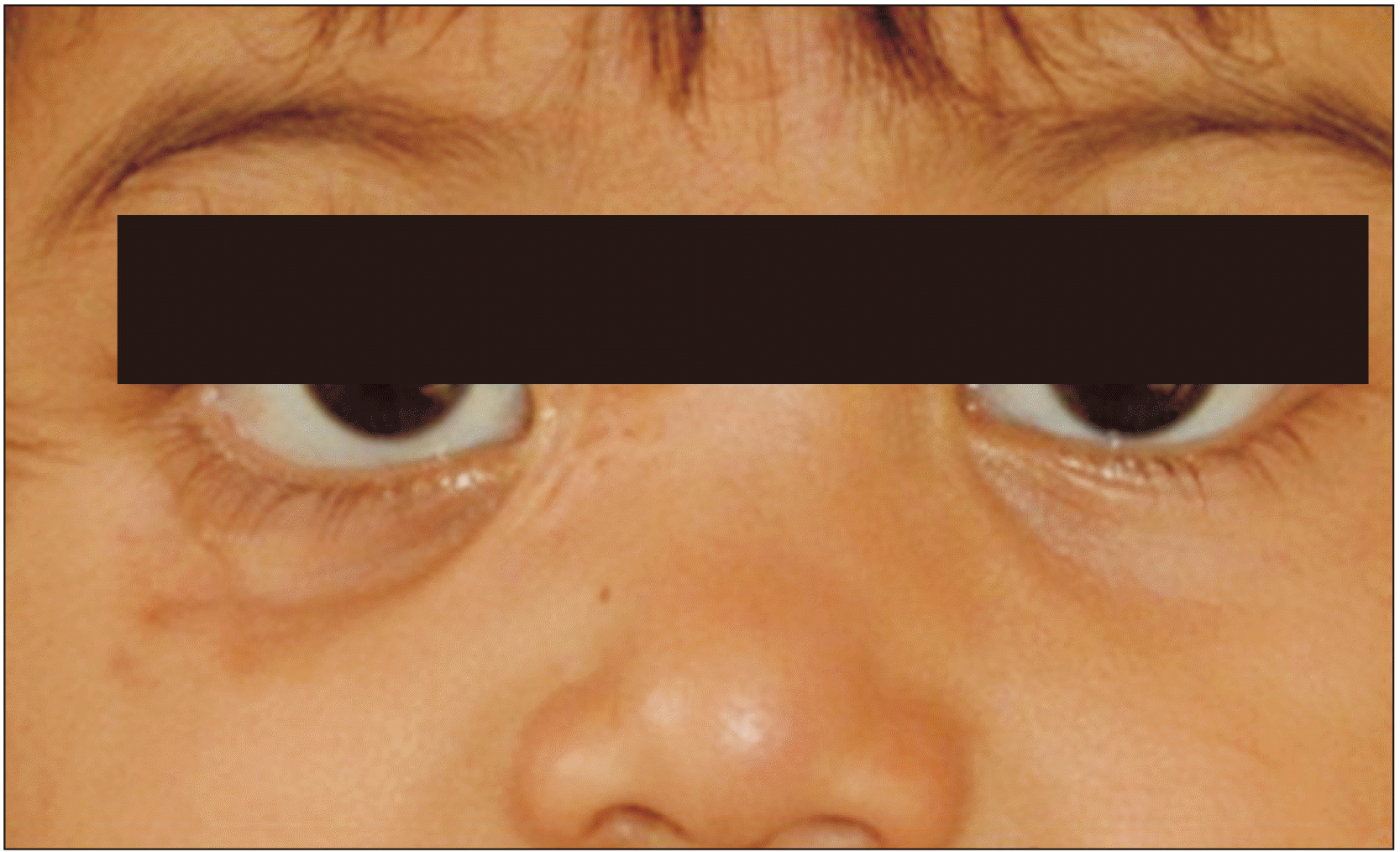
Excessive scleral show was noted on the right side due to eversion of the lower eyelid, which was caused secondary to the scar in the infraorbital region.(Fig. 1)
On eliciting further history, it was discovered that when the child was six months old, she had developed a fluctuant swelling in the infraorbital region that escalated to include the lower eyelid and eventually ruptured to drain pus intraorally and extraorally.
The extraoral site healed leaving a scar but the intraoral draining site persisted as a draining sinus.
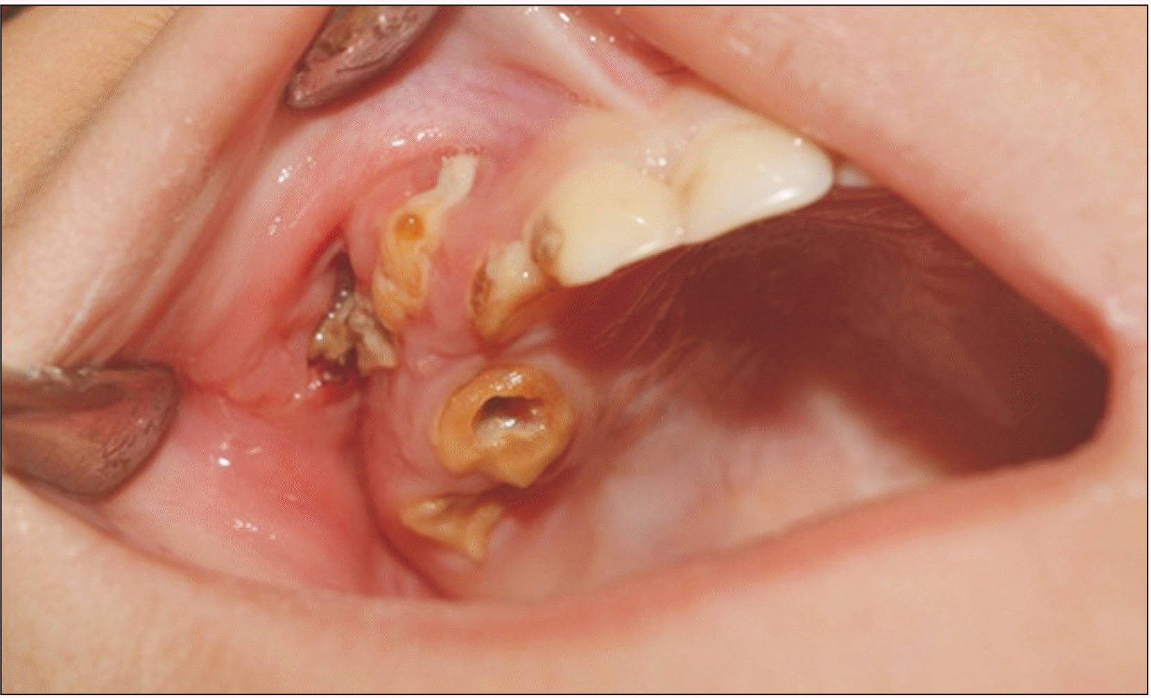
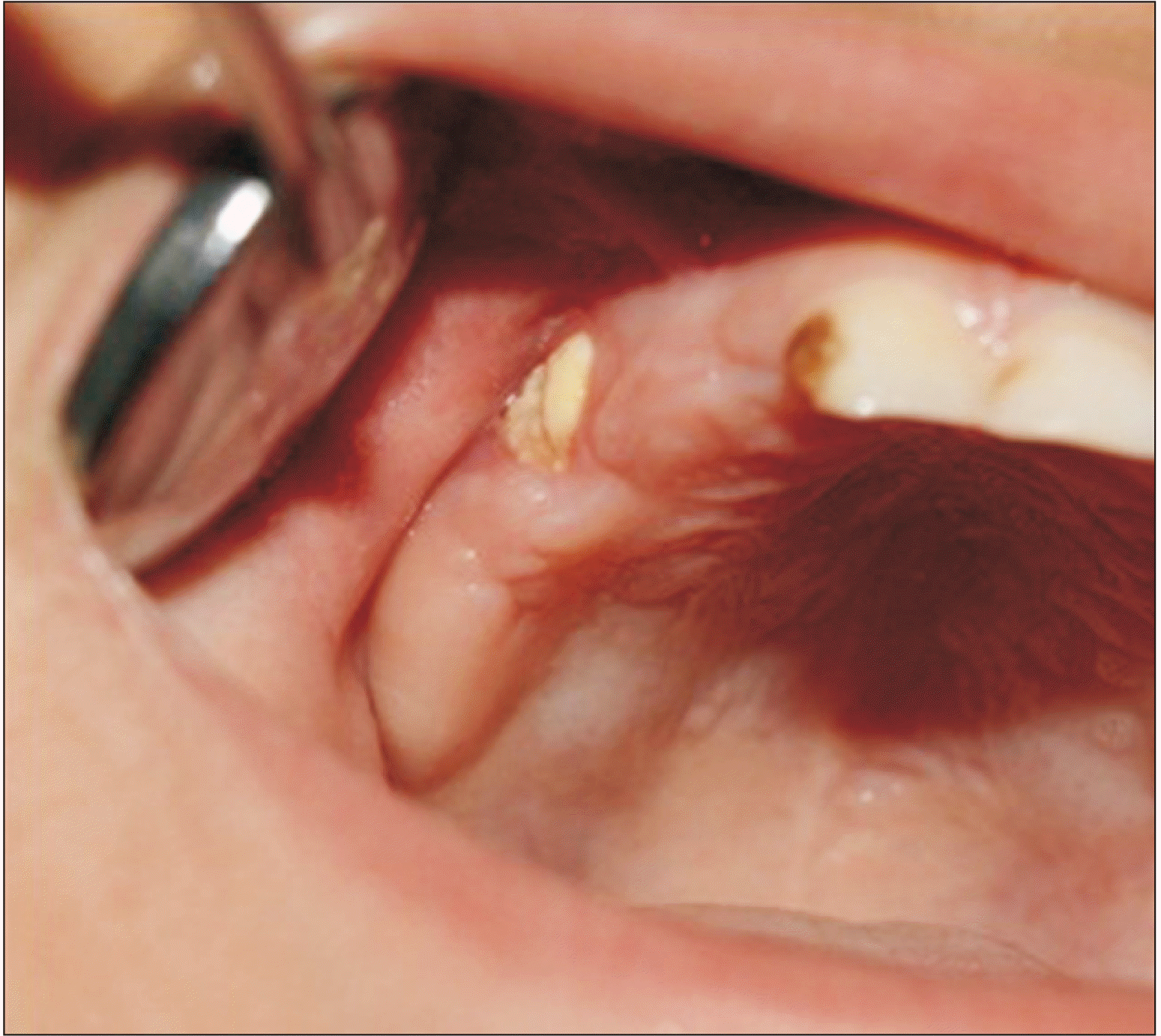
On intraoral examination, an ulcerated lesion was noted along the right buccal sulcus along the carious canine, first and second molars. The underlying bone was exposed and purulent discharge was evident.(Fig. 2)
On admission, the patient’s vitals were as follows: temperature, 100°F; pulse, 97 bpm; respiratory rate, 36 breaths/min. Blood tests revealed a rise in leukocyte count to 21,000 cells/µL and erythrocyte sedimentation rate 90 mm/hr.
She was started on amoxicillin and clavulanic acid 100 mg/kg/day intravenously in two divided doses. Supportive treatment consisted of antipyretics, fluids and a protein rich diet.
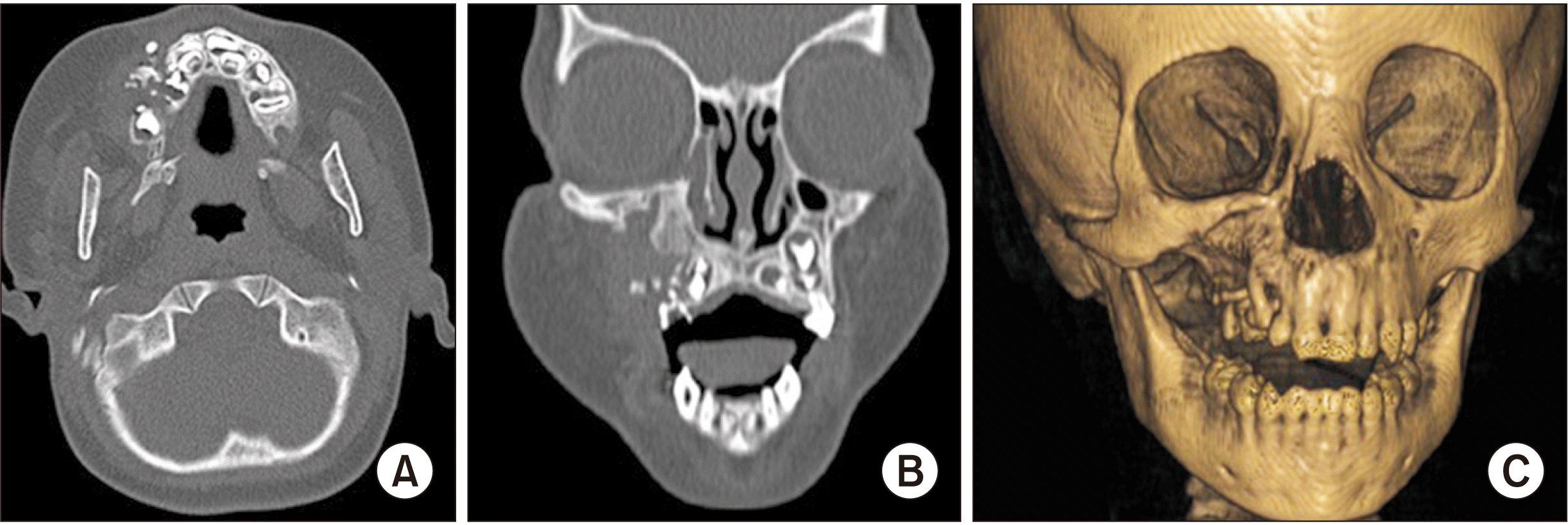
Computed tomography (CT) scan of the head and neck (Siemens Somatom Definition 64 slice CT scanner) showed erosion of the bone and abscess formation along the right maxilla and swelling in the buccal tissues. Thus a provisional diagnosis of osteomyelitis was made.(Fig. 3)
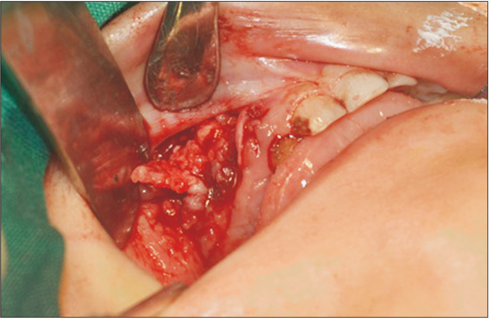
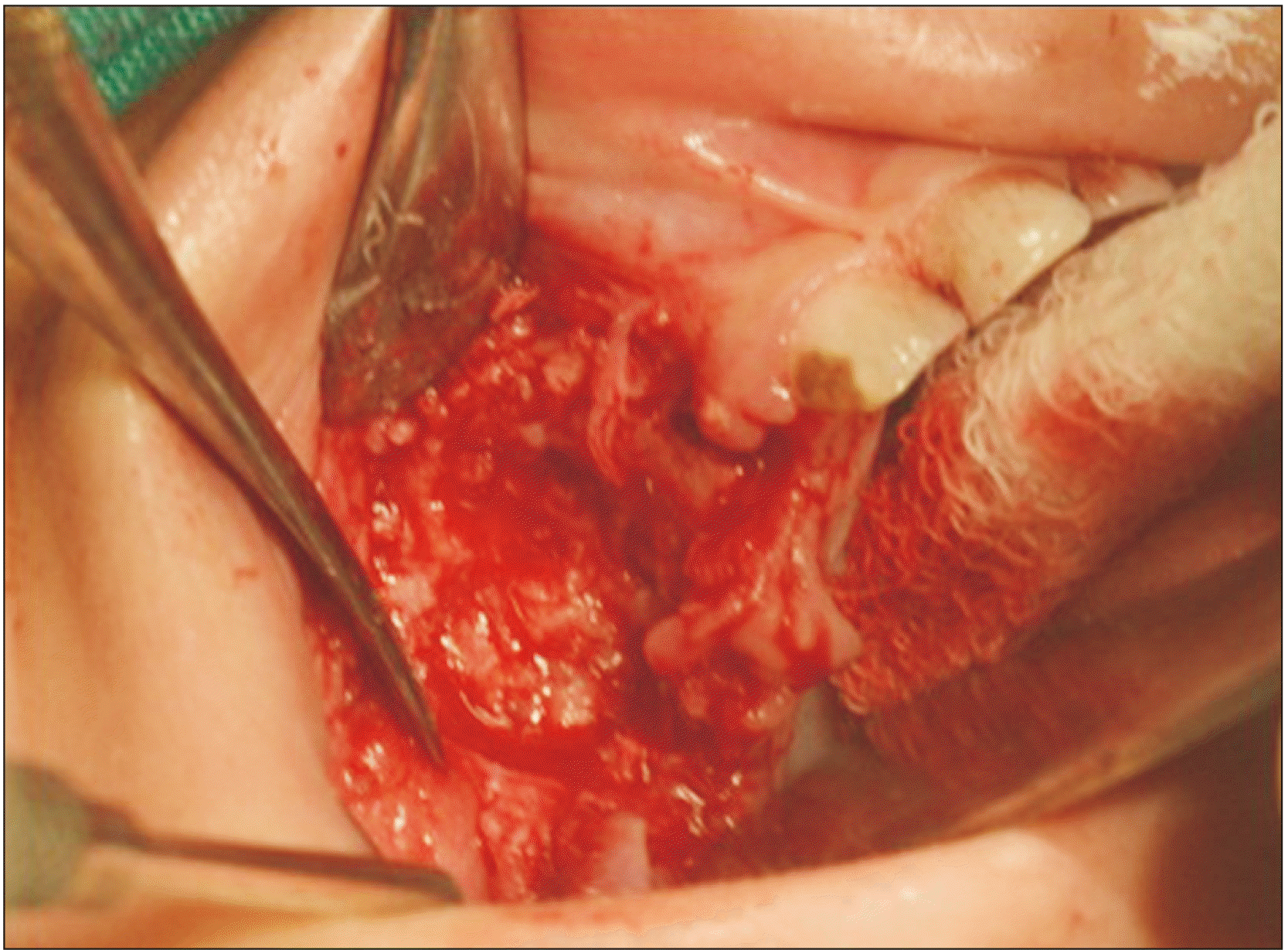
The patient was taken to the operation theatre for debridement under general anesthesia. On exposing the involved area, suppuration and extensive necrosis was noted with sequestration of the maxillary bone.(Fig. 4)
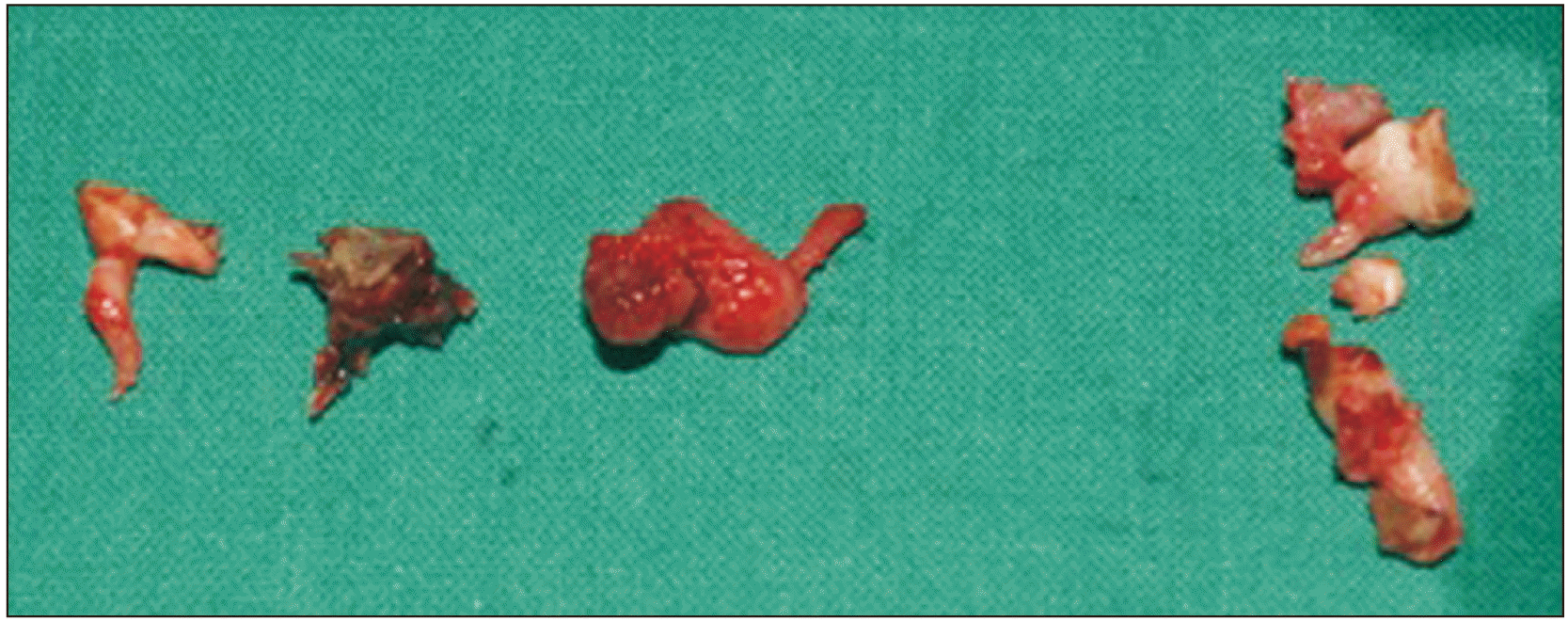
Purulent material was collected and the non-viable soft tissues along with involved deciduous teeth and necrosed bone were removed followed by thorough debridement of the site.(Fig. 5, 6)
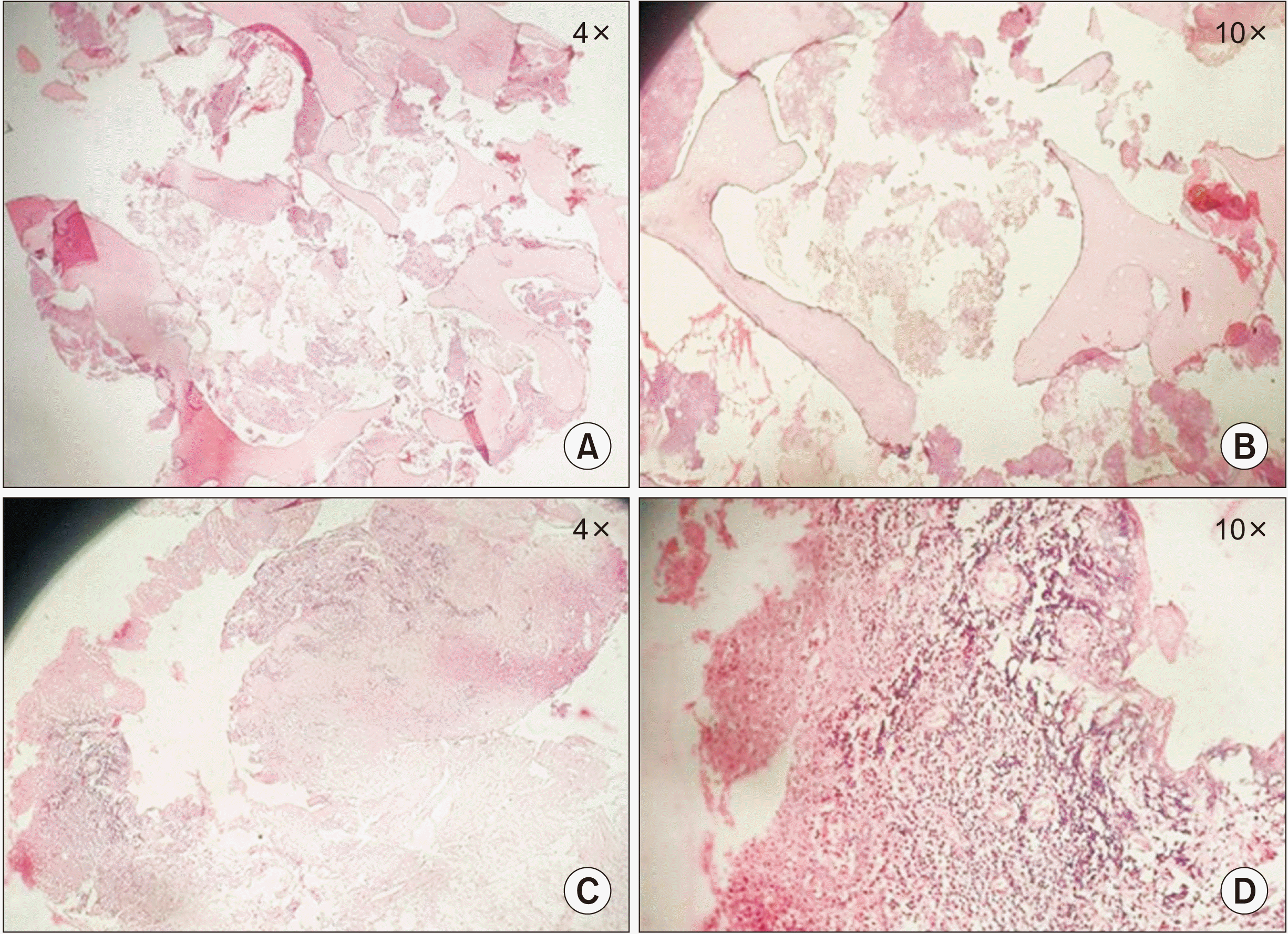
Histopathological examination showed spicules of bone with ragged heamatoxyphilic margins and empty lacunae with reversal lines. Necrotic debris, chronic inflammatory cell infiltrate, and extravasated blood were seen around the bony spicules. Hyperplastic ulcerated stratified squamous parakeratinised epithelium was noted along with an underlying fibrovascular connective tissue. Stroma showed dilated blood vessels along with areas of extravasated erythrocytes and a dense chronic inflammatory cell infiltrate.(Fig. 7) This confirmed the diagnosis of chronic osteomyelitis.
Thus depending on the onset and history of presenting symptoms, clinical findings, and histopathological examination, the clinicopathological diagnosis drawn was infantile osteomyelitis.
The inflammation and discharge of pus reduced the week following the surgery.
After one week, the patient was sent home with adequate recovery. Subsequent follow-up examinations were inadvertent.(Fig. 8)
III. Discussion
Infantile osteomyelitis was never considered as a separate entity until 1932 when Wilensky, after reviewing the literature, suggested that osteomyelitis in infants should be separated from the general subject of osteomyelitis due to its definite clinical features10,11. He reported the first cases of osteomyelitis of jaws in infants most of which were from the pre-antibiotic era. With the advent of antibiotics, this condition has become a rare entity.
Rees in 1847 was the first to describe infantile osteomyelitis of the maxilla12. It is seen to occur mostly during the first few weeks or months of life. The pathogenesis is not clearly understood but development of this condition has been strongly linked to birth injury13.
The most severe injuries in an infant’s mouth are linked to the Mauriceau-Veit-Smellie method of delivery14. There could be injuries varying from mere lacerations of the skin in the corner of the lips to complete avulsion or fracture of the lower jaw, especially if one or two fingers introduced into the mouth are improperly used for strong traction. These may then serve as entry points for microorganisms.
It has also been hypothesized that the infection localizes in the maxillary sinus due to its greater volume. As infants are prone to upper respiratory tract infections that cause nasal discharge or nasal congestion, these symptoms may be the initial signs of infantile osteomyelitis.
Osteomyelitis occurring in the facial bones is mainly due to odontogenic microorganisms, especially Staphylococcus, which result in post-traumatic infection15. The most common pathogen of infantile osteomyelitis is Staphylococcus aureus16. However, hematogenous infantile osteomyelitis caused by streptococci or pneumococci has also been reported8.
In the present case, the causative organism was S. aureus as suggested in the culture sensitivity report. The clinical symptoms subsided after debridement of the intraoral site and administration of intravenous antibiotics for about 2 weeks. Termination of antibiotics was considered after monitoring the white blood cell levels and the general health of the patient.
From the patient’s history, events began with periorbital cellulitis, which could have progressed from sinusitis or some injury during birth. The condition was not diagnosed at this stage either due to misguiding signs or lack of adequate investigations. The periorbital abscess then drained into the oral cavity, eroding the maxillary bone which then advanced to damaging developing tooth buds. With early diagnosis and intervention of the periorbital involvement, sequelae could have been prevented, thereby avoiding unintended damage to the developing tooth buds. In this situation, CT scan helped assess the extent of the lesion and was thus greatly useful in the diagnosis and determining the treatment plan. This further emphasizes the significance of imaging modalities during diagnosis.
Thus, to conclude, dentists and maxillofacial surgeons play an important role in early diagnosis of this condition by identifying oral/facial manifestations during the initial stages of infantile osteomyelitis. A sudden appearance of a peri-orbital swelling needs to be thoroughly investigated, especially in the pediatric age group, in order to eliminate the chances of further complications. Rational use of diagnostic imaging techniques should be adopted to gain control over the disease process and early implementation of the treatment plan.
 XML Download