

 PDF
PDF Citation
 Print
Print


I. Introduction
Injuries to the mandible occur at higher rates than that to other areas of body as it is a prominent bone in the maxillofacial region1. Intraoperative stabilization of mandibular bone fracture segments in an anatomically reduced position before miniplate fixation can be a daunting task2. Bone healing is facilitated by precise anatomic reduction of the fractured bone segments. Fracture healing takes place by contact healing, which leads to primary healing with better stability across the fracture site compared to gap healing of fracture segments, which can increase the risk of malunion and infection3.
II. Technical Note
Various techniques are well documented to obtain anatomic reduction, such as reduction forceps, manual reduction, or a combination of these methods. The use of reduction forceps has not been extensively investigated and is not widespread in oral and maxillofacial surgery despite its promising findings. Forceps were proposed for main use in symphyseal and para-symphyseal regions of the mandible and only a few for the posterior region. However, a complication like lingual flaring due to the design of these forceps is common4. Hence, we proposed a new intra-operative technique for anatomic reduction using screw-wire traction for open reduction and internal fixation of mandibular fractures.
The case was treated under suitable anesthesia with meticulous asepsis.
1) Step 1: Incision is planned as per the anatomical location of the mandibular fracture, and exposure of fracture site can occur through intraoral vestibular/extraoral incision or pre-existing contused lacerated wound. Layer-by-layer dissection is performed to expose the fracture segments to the inferior border of the mandible.
2) Step 2: Intermaxillary fixation is conducted to achieve occlusion.
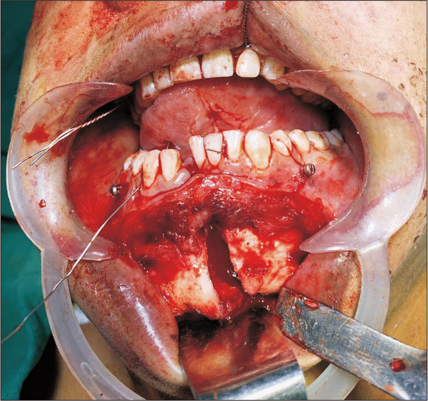
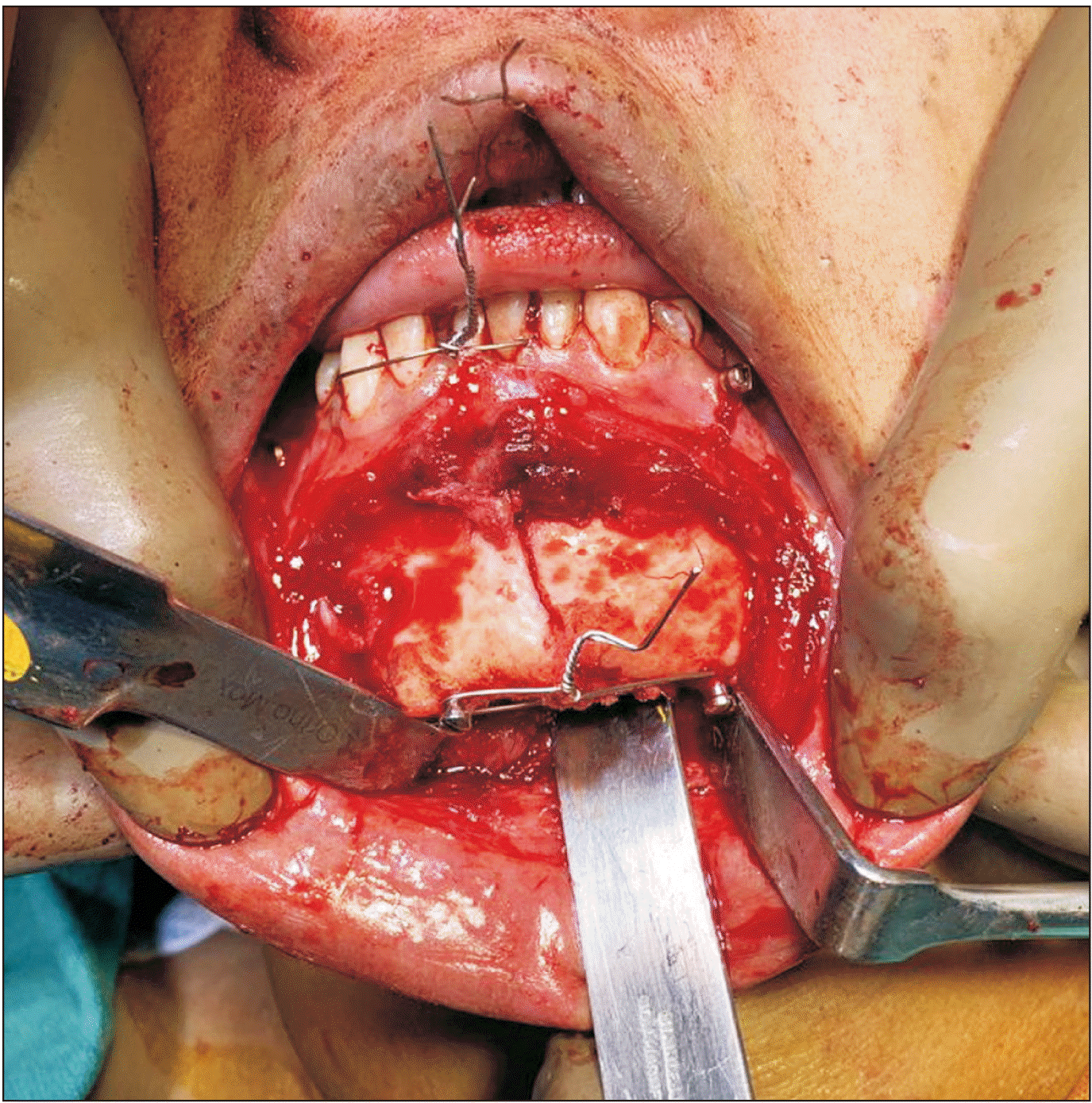
3) Step 3: With help of a 1.5-mm drill bit, an osteotomy hole is drilled at 90° to the inferior border of the mandible (5-10 mm from each fracture segment). Intra-osseous screws 2 mm×8 mm or 2 mm×10 mm (stainless steel or titanium, 2-mm diameter and 8- or 10-mm length) are secured one at each fracture segment in the osteotomy hole. After the screws are anchored well, a 24- or 26-gauge wire is looped around the heads of the screws and tightened to produce traction and achieve maximal contact between the two fracture fragments and anatomical reduction as shown in Fig. 1 and 2.
4) Step 4: Once fracture segments are in maximum contact and anatomically reduced, fixation of the fracture is achieved with mini plates. Configuration of the miniplate will depend upon the type of fracture, and the plate is secured by 2 mm×6 mm or 2 mm×8 mm screws.
5) Step 5: Once the fracture segment is reduced and fixed with miniplates and screws, inferior border screw-wire traction is removed, hemostasis is achieved, copious betadine washes are performed, and layer-by-layer closure is conducted.
III. Discussion
1. Advantages
1) This technique does not require bulky forceps for reduction and manipulation of fracture segments.
2) Risk of accidental injuries to the operating team can be avoided during manual reduction of the fracture.
3) Precise anatomic reduction can be achieved.
4) Ease of fracture reduction and fixation.
5) Easy learning curve.
6) Repeatedly fixing the displaced reduction forceps for holding the fracture segments increases the duration of surgery.
2. Disadvantages
1) Retraction of a soft tissue flap to the lower border of the mandible is performed with conventional forceps.
2) Traction screw can sometimes interfere with a bone plate fixation screw.
3) Two additional holes are drilled for placement of the traction screws.
4) Conventional forceps can be easily and repeatedly guided to the correct position. With the ATOM technique, incorrect fracture reduction might require unwiring and would extend the duration of the surgery.
 XML Download