

 PDF
PDF Citation
 Print
Print


I. Introduction
Sialolithiasis occurs when the salivary gland excretory duct is obstructed by calcareous deposits and commonly found in adults, accounts for 30% of all salivary disorders but rarely in children as only 3% of all sialolithiasis cases1. The submandibular gland is the most frequently involved site due to its long curvy duct and narrow orifice causing viscous mucinous saliva moves against gravity, resulting in saliva retention2,3.
Francis and Larsen2 suggest that sialolithiasis formation involves retrograde migration of any substances or pathogens from oral cavity into the salivary ductal system to act as a nidus which precipitates organic and inorganic materials. Infection also alters the saliva composition, affecting protein precipitation and calcium solubility to result in stone formation2. Though several studies have analyzed the sialolith crystalline structure, studies on pediatric sialoliths remain scarce. Therefore, we report submandibular sialolithiasis management in a 6-year-old child and analyze the sialolith micromorphology to understand the mechanism of mineralization and growth of pediatric sialoliths.
II. Case Report
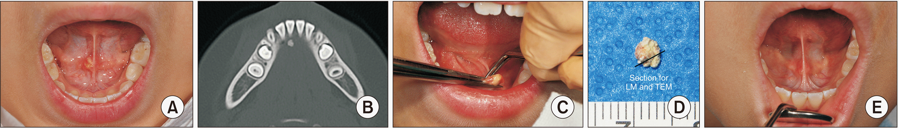
A 6-year-old Korean female presented to the Oral and Maxillofacial Surgery Department, Seoul National University Dental Hospital for swelling under her tongue. This case report was approved by the Institutional Review Board of Seoul National Hospital (S-D20200010) on 3rd March 2021. The intraoral examination revealed a painless yellowish hard mass beneath the tongue near the Wharton’s duct which was suspected as a sialolith.(Fig. 1. A) The patient was informed that the mass would be removed surgically. However, the patient was resistant to the treatment plan and further follow-ups were scheduled to observe the symptom progress. Along the follow-ups, the symptoms progressed to painful, enlarged, festered mass with oral malodor. On the last follow-up, a computed tomography (CT) scan was performed and confirmed the sialolith near the right Wharton’s duct orifice.(Fig. 1. B) After two years and two months from the first visit, the patient finally cooperated with surgery indication.
After receiving the informed consent, the sialolithotomy was performed under local anesthesia using 2% lidocaine with 1:100,000 epinephrine. The sialolith was grasped with dental pincette and removed through blunt dissection using dissecting forceps.(Fig. 1. C) The 3 mm stone was cut transversely into two sections, for light microscope (LM) histopathologic and transmission electron microscope (TEM) slides preparation.(Fig. 1. D) The histologic section was fixed in 10% formalin and the TEM section was fixed in 2.5% glutaraldehyde. The wound was left open without a suture and dressed in the following day. The patient was instructed to maintain oral hygiene and practice tongue movements. At the one-month follow-up, the wound was fully healed without any problems.(Fig. 1. E)
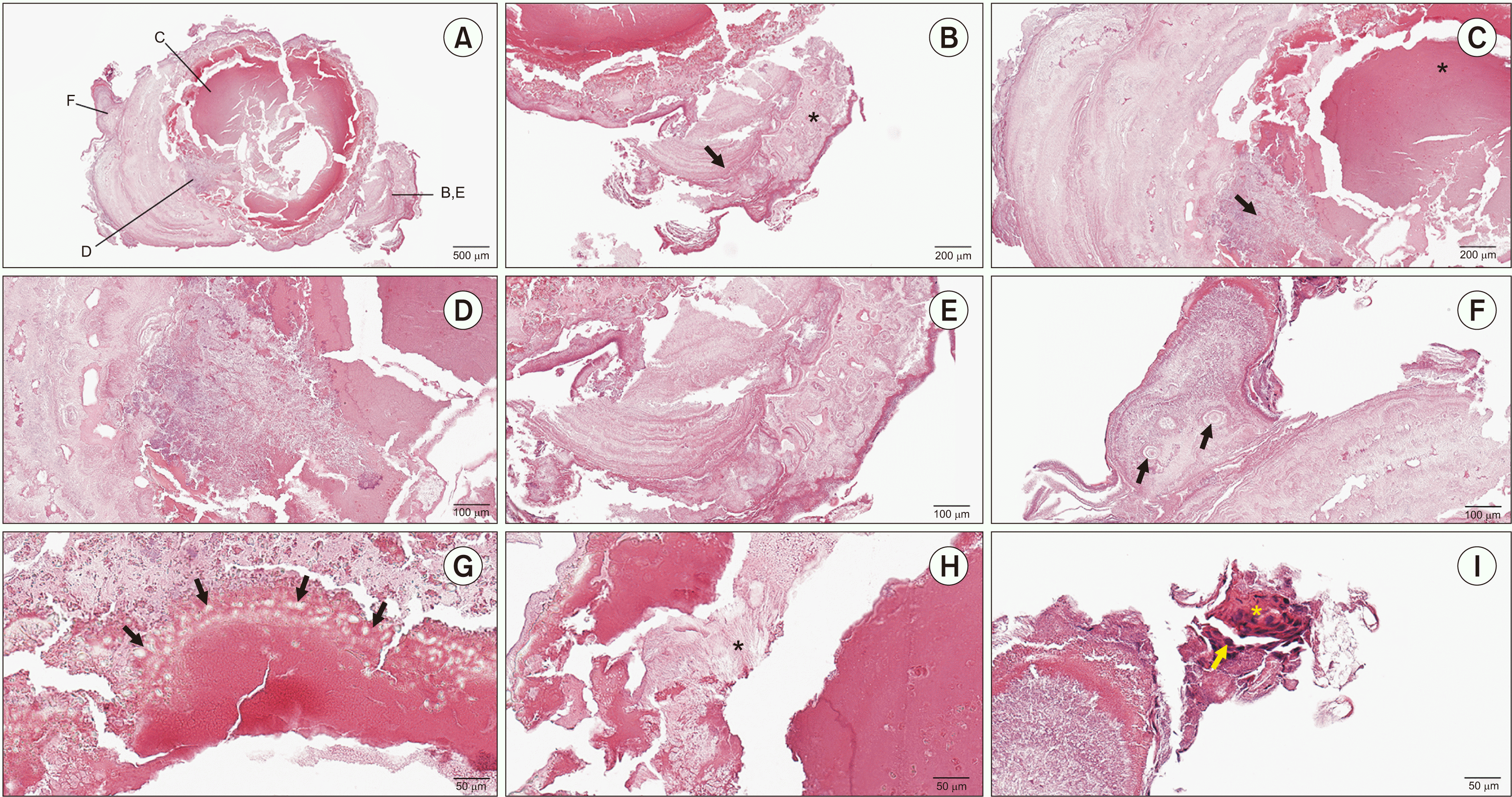
The histologic slide was stained with H&E. Histologically, the stone revealed different degrees of calcification of organic and inorganic layers.(Fig. 2. A-F) Laminated concentric patterns (Fig. 2. B, black arrow [Fig. 2. E]) and mineralized nodules (Fig. 2. B, black asterisk [Fig. 2. E]; Fig. 2. F, black arrows) were observed in the outer layer of sialolith surrounding the eosinophilic core.(Fig. 2. C, black asterisk) Amorphous basophilic materials (Fig. 2. C, black arrow [Fig. 2. D]) were observed in several areas. In the peripheral eosinophilic zone, irregular pattern containing globular structures identified as globuli and tearlike globuli was observed.(Fig. 2. G, black arrows) A needle-like crystallization structure was observed between the peripheral eosinophilic zone and hollow space.(Fig. 2. H, black asterisk) A new branch of stone was observed outside the laminated structure and exhibited mineralized nodules.(Fig. 2. F) Small amounts of bacteria (Fig. 2. I, yellow arrow) were located at the outer shell of the sialolith containing salivary ductal epithelium.(Fig. 2. I, yellow asterisk)
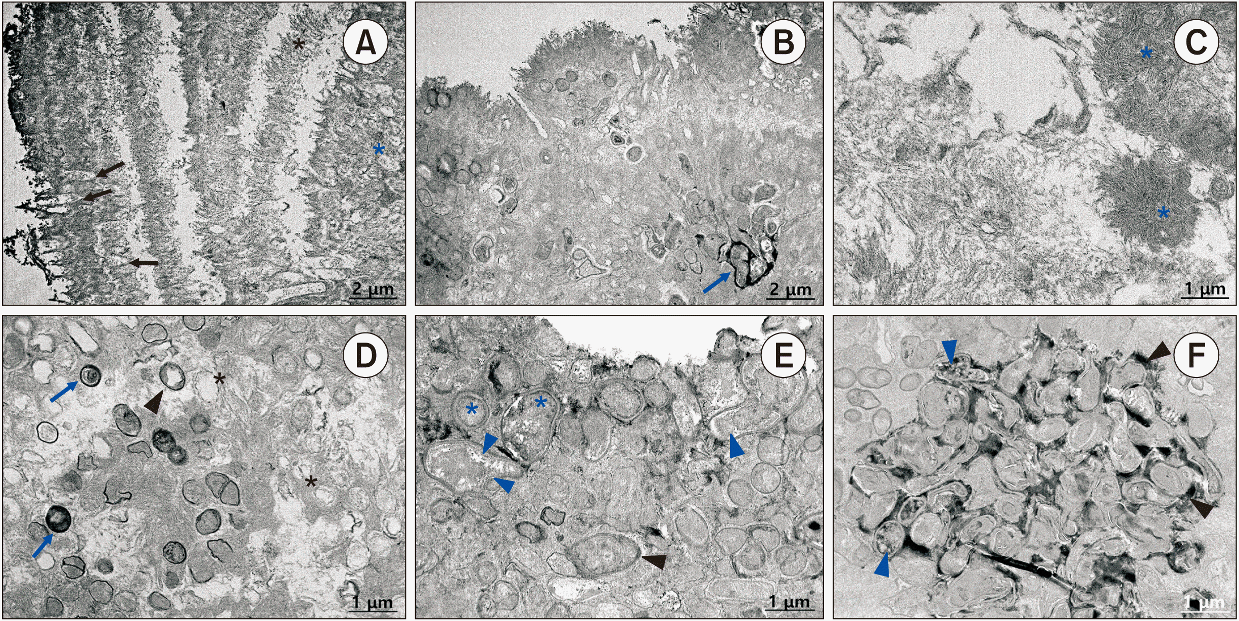
In the TEM examination (JEM-1400 Flash; JEOL, Tokyo, Japan), the stone was stripped into 1 mm×1 mm×1 mm blocks, embedded in epoxy resin, cut into ultrathin sections (70-80 nm) and scanned under 3,000× and 6,000×. The concentric laminar structure is illustrated in Fig. 3. A. In the internal lamellas, the globular structure dominated (Fig. 3. A, blue asterisk), while the crystalline pattern in several outer layers was heterogeneous with some regions of needle-like pattern.(Fig. 3. A, black asterisk) In the most external layer, a finger-like globuli was recorded.(Fig. 3. A, black arrows) Clusters of vesicle structures were observed in the external lamella of several regions, with tear-like structures adjacent to the major vesicles.(Fig. 3. B, blue arrow) Clusters of needle-like crystallites are shown in Fig. 3. C (blue asterisks). Globular exosomal-like structures approximately 0.5 µm in diameter were detected in the internal lamellas.(Fig. 3. D, blue arrows) Membranous bodies were observed adjacent to the vesicles.(Fig. 3. D, black asterisks) Several globules showed opaque internal and surface contractions, indicating deposition of microcrystalline inorganic compounds.(Fig. 3. D, black arrowhead) A detailed image of intra-vesicular (Fig. 3. E, blue arrowheads) and extra-vesicular (Fig. 3. E, black arrowheads) depositions of microcrystalline matter is shown in Fig. 3. E. Interestingly, several vesicle structures exhibited a double membrane.(Fig. 3. E, blue asterisks) Areas with a high density of vesicles are captured in Fig. 3. F. Intra-vesicular (Fig. 3. F, blue arrowheads) and extra-vesicular (Fig. 3. E, black arrowhead) depositions were observed in this cluster.
III. Discussion
Submandibular gland sialolithiasis appears as swelling in the Wharton’s duct area. It can be asymptomatic at first, but discomfort and pain due to duct contractions in an attempt to eliminate saliva will appear gradually4. Symptoms may also be accompanied by the intraoral purulent discharge1. Pediatric sialolithiasis symptoms are short due to patients and parents low tolerance to uncomfortable symptoms5. In our case, the patient initially presented with painless swelling, but pain, oral malodor, pus discharge, and stone enlargement gradually appeared.
Sialolith structure can be classified into concentric and irregular patterns, with high and low mineralization6. In H&E staining, it appears as alternating eosinophilic and basophilic zones or globular calcified zones, formed by organic and inorganic materials7-9. Basophilic zone indicates a highly mineralized area (Fig. 2. D), whereas the eosinophilic zone indicates less mineralization. Frequently, the core is predominated by organic materials while inorganic materials form the concentric pattern. In many studies, the core is globular and highly mineralized or only composed of mineral/inorganic materials6,7,10. The present sialolith core was homogenous eosinophilic without specific structure, indicating organic materials domination.(Fig. 2. A, 2. C) Under high magnification light microscopy, macromolecules were observed, highly suggestive of lipid compounds, which might originate from degenerated salivary gland membrane11. This differs from the five-year-old girl submandibular gland sialolith reported by Lee et al.12, which showed a basophilic core.
In our case, laminated concentric patterns and mineralized nodules in sialolith outer layer and irregular pattern in peripheral eosinophilic zone were observed.(Fig. 2. B, 2. E) The concentric pattern corresponds to steady-state growth, while the irregular pattern indicates sialolith growth perturbations due to chronic inflammation/infection. These structure variations affect the sialolith fracture toughness and mechanical behavior. Meanwhile, mineralized nodules indicate the ongoing growth stages6. The sialolith branch containing mineralized nodules in this case may indicate the ongoing growth and formation of a new layer.(Fig. 2. F) According to Hayashibara et al.13, pediatric sialoliths are more fragile than adult stones during sectioning. In our sialolith, during microscopic slides preparation, the stone was fragile and easily divided which might correlate to the peripheral eosinophilic area irregular growth pattern and core organic composition. Meanwhile, contrary to our core organic composition, Kasaboğlu et al.14 study regarding micromorphology of submandibular sialoliths obtained from six adult patients aged 48 to 62 years revealed no organic material in the cores, and the cores were completely crystallized. However, Hayashibara et al.13, found no significant differences in elements, radiopacities and Ca/P ratios when comparing adult and pediatric sialoliths. The globular structure (Fig. 2. G) was associated with organic components segregation and was considered as lipids accumulation6,15. Furthermore, the tearlike globular structures suggest the globuli as a dynamic structure and indicate the ongoing calcification process with organic materials elimination. Needle-like crystallizations (Fig. 2. H) is thought to be associated with peripheral organic substances resulting from successive precipitation events which follows compositional gradients15.
Infection occurs when intraoral pathogenic bacteria enter the salivary gland or from stagnant saliva in the duct which can cause sialadenitis7. In this case histologic slide, the bacteria appeared only at the outer shell containing salivary gland epithelium indicating bacteria originated from the salivary gland epithelium which was included during sialolithotomy.(Fig. 2. I) This can explain the observed festered swelling.
TEM images of sialolith have revealed intra-vesicular and extra-vesicular calcifications around globular lysosome-like structures.(Fig. 3. A, 3. F) Some vesicles have exhibited mitochondria-like morphology though without the crista structure. The mitochondria and lysosomes seem to be degenerative products from salivary gland and ductal cells, accumulated due to the obstruction and then underwent mineralization within the stone.(Fig. 3. B, 3. D) In the TEM images, the mineralized crystalline regions appeared dark in bright-field imaging because of heavy element diffraction.(Fig. 3. C) Tear-like structures surrounding major vesicles can display organic material leakage from the vesicle structures.(Fig. 3. B) It is suggested that calcification of vesicle structures is accompanied by elimination of organic materials, producing surrounding tear-like structures6.(Fig. 3. E, 3. F)
Treatment modalities for sialolithiasis include a conservative approach through the sialoendoscopy and salivary ductal irrigation, good hydration, analgesic and antibiotic medication, and sialogogues to foster spontaneous stones removal especially stone smaller than 2 mm. However, spontaneous removal in children can be difficult due to the small opening of the duct1,4. Intraoral removal under local anesthesia is a treatment of choice for children with relatively small stones and stones in the distal part of the Wharton’s duct3,5. In our case, we performed intraroral sialolithotomy without suturing the wound to avoid duct obstruction.
In conclusion, the micromorphology and growth processes of pediatric sialolith remain undescribed. We found that the pediatric sialolith core composition is different from the adult sialoliths which may relate to a different growth pattern and mechanical behavior of the stone. However, more comprehensive microscopic studies are needed regarding their distinctive characteristics. By expanding knowledge about sialoliths micromorphology, development of new preventive, diagnostic and patient-tailored treatment methods of pediatric sialolithiasis will be enhanced.
 XML Download