I. Introduction
When a sinus lift is performed on a healthy maxillary sinus, the risk of obstruction of the ostium of the maxillary sinus and subsequent complications is rare. However, a sinus lift performed in the presence of maxillary cysts may significantly reduce the sinus volume, and may increase the risk of obstruction of the ostium of the maxillary sinus and subsequent complications. This phenomenon eventually causes maxillary sinusitis and leads to failure of bone grafting in the maxillary sinus
1,2.
It is important to understand the composition of normal sinus membranes and the pathogenesis of cysts of the maxillary sinus when developing an appropriate treatment plan for a sinus lift in the presence of maxillary cysts
3. The sinus membrane is composed of three layers: the respiratory epithelium (ciliated pseudostratified columnar epithelium) containing goblet cells, the connective tissue layer (termed the lamina propria) containing seromucous glands, and the periosteum. The respiratory epithelium is located at the sinus chamber surface, and in underlain by the lamina propria, and then by the periosteum beneath that overlies the bony walls of the sinus
3-5. The cysts that may be found within the floor of the maxillary sinus include mucoceles, mucous retention cysts, and pseudoantral cysts
3,6. Pseudoantral cysts (also called antral pseudocysts) are classified as nonsecreting cysts because of the lack of epithelial lining. Pseudoantral cysts result from a diffuse accumulation of inflammatory exudate beneath the periosteum. Mucous retention cysts that have epithelial linings are considered true cysts and are classified as secreting cysts. Mucoceles that have epithelial linings are also considered true cysts
3. Pseudoantral cysts are formed as a result of bacterial toxins released from the area of infection increasing local capillary permeability, resulting in accumulation of liquid in the tissues and lifting tissues from the bones. As a result, a characteristic solitary dome-shaped structure is formed within the floor of the sinus
3,7. Mucous retention cysts result from ductal obstructions of the seromucous glands located within the lamina propria
3,8. Blockage of the duct of a seromucous gland by a mucus plug can result in dilatation of the duct. Mucous retention cysts may rupture and continue to produce secretions that leak into the surrounding tissue, thus forming a cavity filled with mucus. Unlike pseudoantral cysts, which are typically singular, mucous retention cysts may be multiple. Large mucous retention cysts may appear similar to pseudoantral cysts and therefore differential diagnosis is not possible through routine radiographic examination alone
7. Mucoceles result from obstructed sinus outflow that leads to an accumulation of fluid in a mucoperiosteal-lined cavity
9. Any condition that obstructs the maxillary sinus ostium may result in a mucocele
10. Mucoceles are destructive, causing bony expansion, bony perforation, and displacement of adjacent structures. In contrast, mucous retention cysts and pseudoantral cysts are noninvasive and asymptomatic
8,11. Mucous retention cysts and pseudoantral cysts can be distinguished from mucoceles on radiographic examination. Cloudy radiopacity, bony expansion, bony perforation, and structural displacement are observed in mucoceles, while, mucous retention cysts and pseudoantral cysts are dome-shaped, nondestructive, and non-expansive and have a smooth margin
12. However, mucous retention cysts and pseudoantral cysts of the maxillary sinus are difficult to distinguish based on radiologic features alone
7,8.
Mucous retention cysts and pseudoantral cysts frequently develop in healthy individuals
6. According to Bhattacharyya
12, the incidence rate of mucous retention cysts of the maxillary sinus was 12.4% among a random selection of 410 patients. Among these, 18% had bilateral cysts. Cysts were mostly located within the sinus floor (50%) and were solitary (88%). And according to Shear and Speight
11, the incidence rate of pseudoantral cysts of the maxillary sinus was reported to be 1.6% to 8.7%, and these were mostly located within the floor of the maxillary sinus.
Various methods to perform a sinus lift in the presence of maxillary sinus cysts have been proposed but no guideline as to the preferred treatment has been established.
In this article, we describe procedures to a perform sinus lift in the presence of mucous retention cysts and pseudoantral cysts and provide a review of the relevant literature.
Go to :

II. Patients and Methods
The case reports of four patients with maxillary cysts were reviewed retrospectively. These patients received a sinus lift between January 2016 and October 2021 at the Wonkwang University Dental Hospital.
1. Case 1
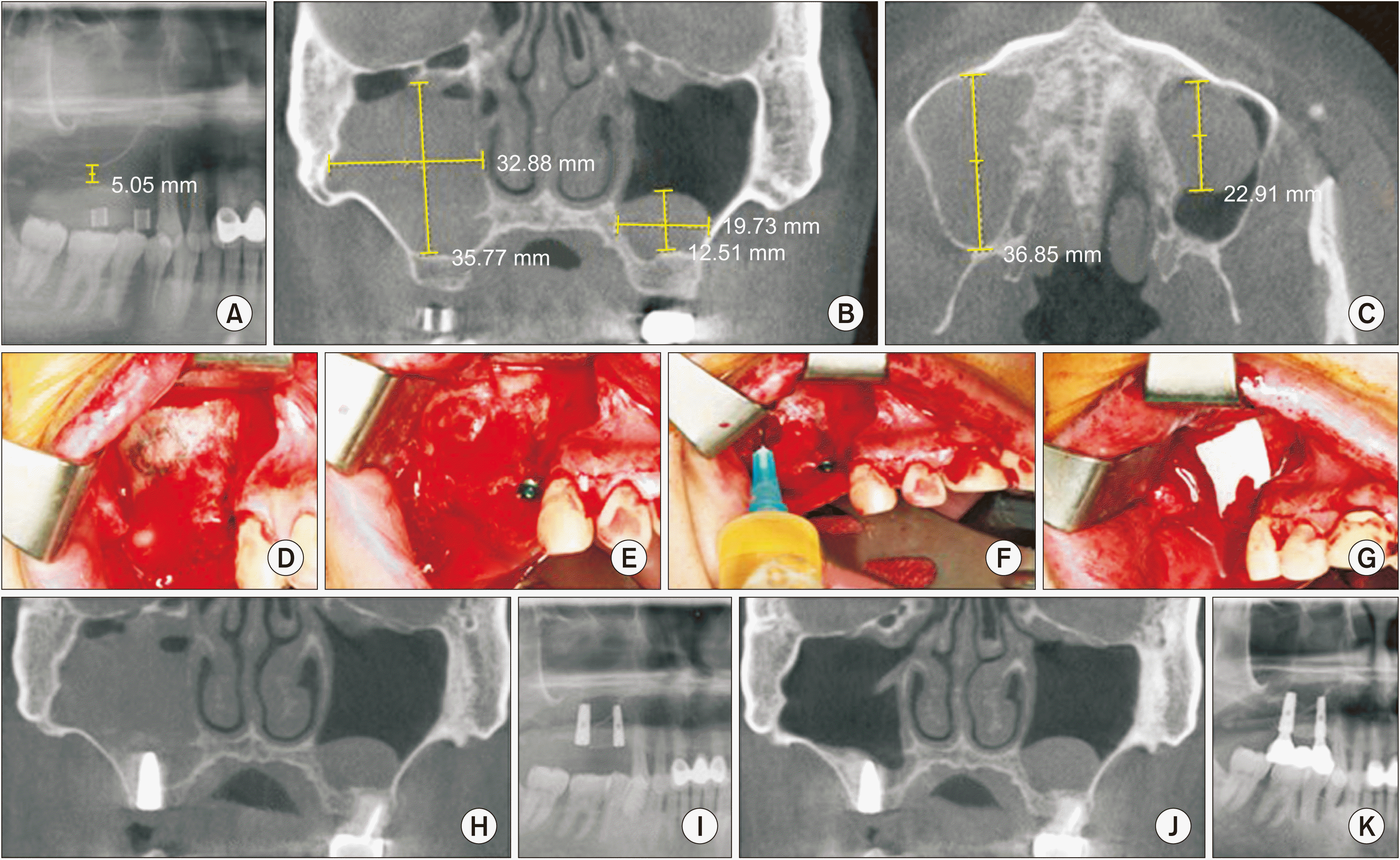
A 45-year-old male patient visited the Department of Oral & Maxillofacial Surgery, Wonkwang University Dental Hospital in November 2016. His chief complaint was masticatory difficulty owing to the loss of his right maxillary posterior tooth. He suffered from diabetes mellitus and hypertension and took medications for these problems, both of which were under control. Two implants were to be placed for the right maxillary first premolar and first molar. A panoramic radiograph that the residual bone height was about 5 mm at the maxillary first molar and there was a sinus radiopacity in the right maxillary sinus. Cone-beam computed tomography (CBCT) showed a 32 mm×35 mm×36 mm, dome-shaped radiopaque lesion on the right maxillary sinus and a 19 mm×12 mm×22 mm, dome-shaped radiopaque lesion on the left maxillary sinus. It was diagnosed as a pseudoantral cyst or mucous retention cyst. It was suspected that the maxillary ostium may become obstructed by edema after a sinus lift procedure and subsequently cause maxillary sinusitis. The treatment plan was therefore to aspirate cystic fluids during the sinus lift and to place an implant on the right maxillary sinus at the same time. The cystic lesion on the left was small, and the plan was to observe it without any treatment. The surgery was performed under general anesthesia. After crestal and lateral oblique releasing incisions, a mucoperiosteal flap was elevated and the lateral wall of the maxillary sinus was exposed. The area that would become the bony window was marked with a pencil. Because the site of the right maxillary first premolar did not require a sinus lift, a 4.0 mm×13 mm-diameter implant (Osstem; Osstem Implant, Seoul, Korea) was first placed directly. Then, a round burr running at a low speed was used to create the bony window. A syringe with a 21-gauge needle was used to aspirate the cystic fluid (total volume 13 mL). The aspirated fluid was light yellow and was free of infection and blood, and the lesion was therefore judged to be a typical pseudoantral cystic fluid. The sinus membrane was then carefully elevated through the bony window area while preventing it from being perforated. The needle puncture site was not provided further treatment. After filling the space created by the elevation of the sinus membrane with 1.0 g of irradiated allogenic cancellous bone & marrow (ICB; Rocky Mountain Tissue Bank, Aurora, CO, USA), a 5.0 mm×13 mm-diameter implant (Osstem) was placed on the right maxillary first molar. After covering the bony window with a collagen membrane (Zimmer Biomet, Warsaw, IN, USA), the epithelium was sutured with 4-0 Vicryl (Johnson & Johnson, New Brunswick, NJ, USA). The patient was prescribed cephalexin (Dong-Wha Pharm, Seoul, Korea) antibiotic, Tylenol (Jassen Korea, Seoul, Korea) analgesic, and Actifed (Samil Pharmaceutical, Seoul, Korea) as a systemic nasal decongestant for 10 days. The patient was instructed to gargle with 0.1% chlorhexine solution (Hexamedine; Bukwang Pharm, Ansan, Korea).
The sutures were removed after 10 days. A postoperative radiograph one day after the procedure showed that the implants were placed well and there was fluid retention. Six months later, a three-bridge type dental prosthesis was completed. A postoperative CBCT 4 years after the procedure showed that the cystic lesion on the right maxillary sinus had disappeared completely, but the cystic lesion on the left maxillary sinus remained unchanged. At the time of records review, 5 years and 3 months had elapsed since the operation. There were no complications such as sinusitis or peri-implantitis.(
Fig. 1)
 | Fig. 1A. Preoperative panoramic radiograph. Residual bone height was about 5 mm at the maxillary first molar and sinus radiopacity was observed in the right maxillary sinus. B. Preoperative cone-beam computed tomography (CBCT) showed a 32 mm×35 mm radiopaque, dome-shaped lesion on the right maxillary sinus and a 19 mm×12 mm radiopaque, dome-shaped lesion on the left maxillary sinus in a sagittal plane. C. Preoperative CBCT showed a 36 mm radiopaque, dome-shaped lesion on the right maxillary sinus and 22 mm radiopaque, dome-shaped lesion on the left maxillary sinus in a coronal plane. D. Intraoperative view. After elevation of the mucoperiosteal membrane, the bony window was marked with pencil on the right maxillary wall. E. Intraoperative view. Because the right maxillary first premolar did not require a sinus lift, the implant was placed first without a sinus lift, and then a round burr at a low speed was used for the creation of the bony window. F. Intraoperative view. After aspiration of the cystic fluid with a syringe a 21-gauge needle, the sinus membrane was carefully elevated. 1.0 g of ICB (Rocky Mountain Tissue Bank, USA) was used to fill the space created by elevation of the sinus membrane. And then, the implant was placed on the right maxillary first molar. G. Intraoperative view. The bony window was covered with a collagen membrane. H. Postoperative CBCT 1 day after operation showed fluid rertention on the right maxillary sinus. I. Postoperative panoramic view 1 day after operation showed two well-placed implants. J. Postoperative CBCT 4 years after operation. The cystic lesion on the right maxillary sinus disappeared completely, but the cystic lesion on the left maxillary sinus remained without any change. K. Postoperative panoramic view 5 years and 3 months after operation. The 3-bridge type dental prosthesis was well maintained. 
|
2. Case 2
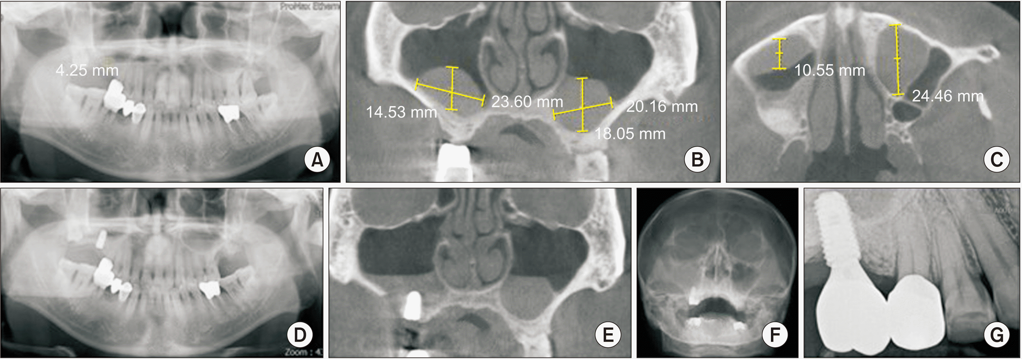
A 62-year-old male patient visited in April 2021. He was a heavy smoker and drinker. He had no other medical problems. One implant was to be placed on the right maxillary first molar. A panoramic radiograph showed the residual bone height was about 4 mm at the maxillary first molar. A CBCT scan showed a 23 mm×14 mm×10 mm dome-shaped radiopaque lesion on the right maxillary sinus and a 20 mm×18 mm×24 mm dome-shaped radiopaque lesion on the left maxillary sinus. The lesions were diagnosed as pseudoantral cysts or mucous retention cysts. The treatment plan was to aspirate the cystic fluids during the sinus lift and to place an implant on the right maxillary sinus at the same time. The cystic lesion on the left was observed without any treatment. The surgery was performed under local anesthesia (2% lidocaine hydrochloride with 1:100,000 epinephrine). A syringe with a 21-gauge needle was used to aspirate the cystic fluid (total volume 2 mL). The aspirated fluid was light yellow, and the cyst lesion was therefore judged to be a typical pseudoantral cyst. Then, the sinus membrane was carefully elevated through the bony window area while preventing it being perforated. After filling the space created by the elevation of the sinus membrane with 2.0 g of ICB, a 5.0 mm×10 mm-diameter implant (Osstem) implant was placed. After covering the bony window with a collagen membrane, the epithelium was sutured with 4-0 Vicryl. The patient was prescribed the same drugs as in Case 1. The sutures were removed after 10 days. A postoperative radiograph one day after operation showed that the implants were well placed, although fluid retention was observed. A Water’s view radiograph 4 months after the operation showed haziness on the right maxillary sinus. Six months later, a dental prosthesis was fitted. At the time of records review, eight months had passed since the operation. Even though a problem was noted on radiography, the prosthesis was being used regularly and there were no specific clinical symptoms.(
Fig. 2)
 | Fig. 2A. Preoperative panoramic radiograph. The residual bone height was about 4 mm at the maxillary first molar. B. Preoperative cone-beam computed tomography (CBCT) showed a 23 mm×14 mm radiopaque, dome-shaped lesion on the right maxillary sinus and a 20 mm×18 mm radiopaque, dome-shaped lesion on the left maxillary sinus in a sagittal plane. C. The preoperative CBCT showed a 10 mm radiopaque, dome-shaped lesion on the right maxillary sinus and a 24 mm radiopaque, dome-shaped lesion on the left maxillary sinus in a coronal plane. D. A panoramic view 1 day after operation showed a well-placed implant. E. A CBCT 1 day after operation showed fluid retention on the right maxillary sinus. F. Water’s view 4 months after operation showed haziness on the right maxillary sinus. G. Periapical view 6 months after operation showed a well-maintained dental prosthesis. 
|
3. Case 3
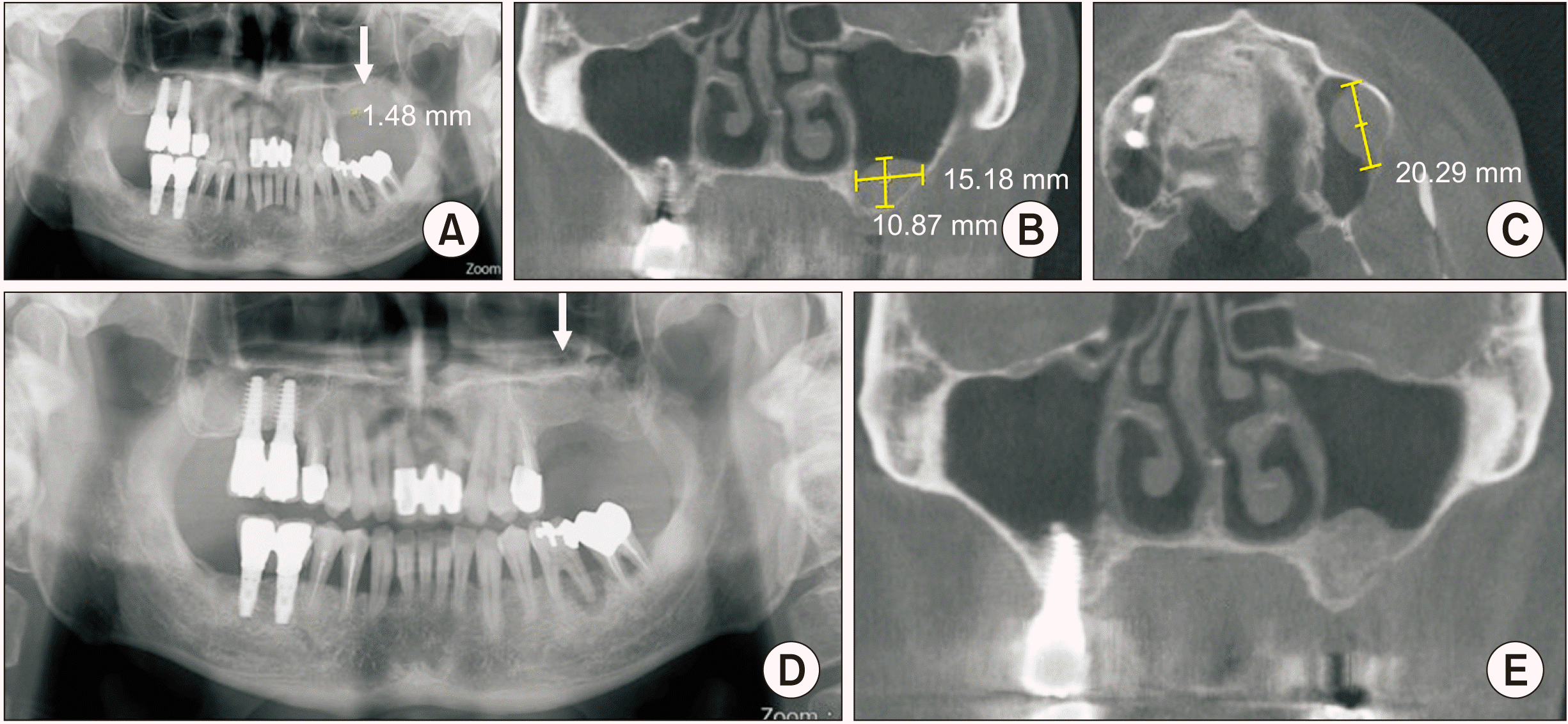
A 54-year-old male patient visited in June 2020. A private clinic had requested a sinus lift. The left maxillary molars were missing. He had no special medical problems. A panoramic radiograph showed residual bone height was about 1.4 mm at the maxillary molar area and there was a radiopacity on the left maxillary sinus. A CBCT showed a 15 mm×10 mm, dome-shaped radiopaque lesion in the sagittal plane and a 20 mm dome-shaped radiopaque lesion in the coronal plane. The lesion was diagnosed as a pseudoantral cyst based on clinical judgment. The surgery was performed under local anesthesia. The sinus lift was completed at the same time as the cystic fluid was aspirated. There was little aspiration during the operation. Then, 2.5 g of ICB was used to fill the space created by the elevation of the sinus membrane. The prescribed drugs were the same as in Cases 1 and 2. The sutures were removed after 10 days. A panoramic radiograph 1 day after the operation showed a well-augmented maxillary sinus. A CBCT 4 months after the operation showed an augmented maxillary sinus without any problems. At the time of records review, 2 years had passed, and there have been no clinical problems.(
Fig. 3)
 | Fig. 3A. Preoperative panoramic radiograph. The residual bone height was about 1.4 mm at the left maxillary molars are and sinus radiopacity (arrow) was observed on the left maxillary sinus. B. A preoperative cone-beam computed tomography (CBCT) showed a 15 mm×10 mm radiopaque, dome-shaped lesion in a sagittal plane on the left maxillary sinus. C. A preoperative CBCT showed a 20 mm radiopaque, dome-shaped lesion in a coronal plane on the left maxillary sinus. D. A panoramic view 1 day after operation showed an augmented left maxillary sinus (arrow). E. A CBCT 4 months after operation showed an augmented maxillary sinus without any problems. 
|
4. Case 4
A 39-year-old male patient visited in May 2020. He had no special medical problems. A private clinic had requested bilateral sinus lifts. The right maxillary molars remained, but the left maxillary molars were missing. The right maxilla was scheduled for implant surgery after extraction of the molars and the patient wanted a sinus lift in advance. A panoramic radiograph showed the residual bone height was about 3 mm at the left maxillary molar area and there was a sinus radiopacity on the left maxillary sinus. A CBCT showed multiple, dome-shaped radiopaque lesions. The lesions were diagnosed as pseudoantral or mucous retention cysts. The surgery was performed under general anesthesia. A sinus lift was performed in the right maxillary sinus using a conventional lateral approach and the left maxillary sinus lift was performed in the same way as for Case 1. During aspiration, the cyst wall on the left maxillary sinus was torn due to careless aspiration and suction. Unlike pseudoantral cysts, this cyst had an epithelial lining, so the cyst was removed conservatively. After removing the cyst, a large sinus perforation developed. The perforation was covered with a collagen membrane. Tibial bone was harvested. The harvested tibial bone was used to augment both maxillary sinuses. A CBCT 1 day after the operation showed fluid retention in the left maxillary sinus due to sinus perforation and no problems in the right maxillary sinus. Examination of a biopsy sampled indicated a mucous retention cyst. A Water’s view radiograph 4 months after the operation showed decreased mucosal swelling of the left sinus membrane. At the time of the record review, 7 months had passed, and there had been no clinical problems.(
Fig. 4)
 | Fig. 4A. Preoperative panoramic radiograph. The residual bone height was about 3mm at the maxillary first molar and a sinus radiopacity (arrow) was observed on the maxillary sinus. B, C. Preoperative cone-beam computed tomography (CBCT). A CBCT showed multiple, dome-shaped, radiopaque lesion on the left maxillary sinus (arrow). D. Intraoperative view. After elevation of the mucoperiosteal membrane, a bony window was created. E. Intraoperative view. The cyst wall on the left maxillary sinus was torn a lot due to careless aspiration and suction. Unlike in pseudoantral cysts, an epitheilial lining existed. F. Intraoperative view. The cyst was removed conservatively. G. A CBCT 1 day after operation showed fluid retention due to sinus perforation and no problems on the right maxillary sinus. H. Histologic specimen. It was lined with a pseudostratified ciliated columnar epithelium (bottom; arrow) and was filled with mucins (top; asterisk) (Periodic-acid Schiff staining, ×200). I. Water’s view 4 months after operation showed decreased mucosal swelling on the left sinus membrane. 
|
Go to :

IV. Discussion
It has long been debated whether a maxillary cyst must be removed before a sinus lift procedure is performed. The presence of a maxillary sinus cyst is typically considered an absolute contraindication for sinus grafting
6,13,14. The sinus membrane tends to swell more in the presence of maxillary cysts, and mucosal swelling may be a contributing factor in sinusitis
15. Maxillary cysts sometimes remain infected, and membrane perforation during a sinus lift could contaminate the graft materials due to leakage of cystic fluids, which may result in uncontrolled graft infection
15,16. The presence of a maxillary cyst may change the normal drainage of the maxillary sinus and the ventilatory problem may be worsen by the surgery, which leads to inflammation of the maxillary sinus
17. The presence of maxillary cysts reduces the volume of the maxillary sinus, and lifting the sinus membrane in the presence of a cyst may further reduce the volume of the maxillary sinus. In this situation, consequent edema of the sinus membrane may block the ostium of the maxillary sinus, leading to maxillary sinusitis
6,15,17. Removal of the maxillary mucosal cyst is therefore recommended prior to performing sinus lift
6,14. However, it has been proposed that the presence of cysts is not an contraindication for a sinus lift
1,18 if there were no clinical symptoms and the cyst was not large enough to block the maxillasry ostium
15. Mardinger et al.
1 reported that benign mucosal cysts in the maxillary sinus, such as pseudoantral cysts and mucous retention cysts, were not symptomatic and did not cause obstruction of the ostium of maxillary sinus and that the presence of maxillary cysts did not affect the prognosis of a sinus lift. Because there is a low incidence of perforation of the sinus membrane and consequent maxillary sinusitis after a sinus lift despite the presence of maxillary cysts, it is generally safe to perform sinus lift procedures in the presence of maxillary cysts
19. However, opinions are divided with respect to mucoceles. A sinus lift may be performed safely in the presence of small mucoceles: Perfetti et al.
20 reported that a sinus lift could be carried out without complications on the condition that the distance between the top of the mucocele and the maxillary ostium was not less than 22 mm and the volume of the mucocele was less than 18 mL. If there is sufficient distance between the top of the mucocele and the maxillary ostium, lifting of the sinus membrane will not alter the antral drainage and the maxillary ostium will not be obstructed even if the mucocele is not removed preoperatively
19. The numbers mentioned above are not absolute criteria; however, it is imperative that there is sufficient space between the two structures, such that a lifted membrane will not interfere with antral drainage. Although small maxillary sinus mucoceles are not considered an absolute contraindication of sinus lift, the procedure should be avoided when large mucoceles are present because most of the maxillary sinus is occupied by such mucoceles, and maxillary sinusitis is likely to occur due to postoperative swelling of the mucous membrane
9. Further evaluation of large lesions such as mucoceles and postoperative maxillary cysts is needed before a sinus lift and the procedure should be postponed until an accurate diagnosis is made
1.
Several surgical approaches for a sinus lift in the presence of benign maxillary cysts have been proposed. Since most of the recommended approaches are based on case reports, there is no research-based consensus. Anitua et al.
8 reported a systemic review examining whether cysts should be removed before or during a sinus lift. This study included 182 people with previous mucous retention cysts or pseudoantral cysts in 195 maxillary sinuses where 233 implants were installed. In 6% of cases, cysts were removed before completing a sinus lift and implant placement. In 54%, aspiration or cyst removal was completed simultaneously. In 31.5%, there was no treatment. In 9%, the treatment was not clearly stated. The follow-up period ranged from 4 to 90 months. Of the 233 implants, only two implants were lost, indicating 99% of the implants were successful regardless of the type of treatment provided. No statistical differences in implant survival were observed in relation to age, sex, surgical approach, implant surgery, or grafting material. They concluded that the presence of mucous retention cysts or pseudoantral cysts had not been considered a contraindication for sinus lift and implant placement and that removal of the cysts did not affect implant survival
8.
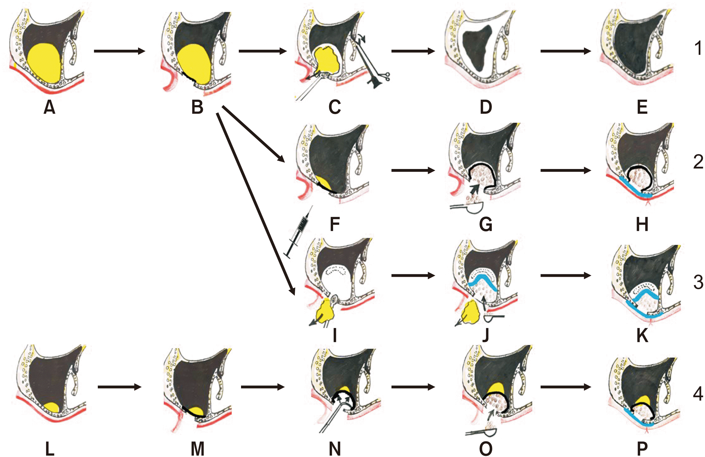
Various methods to perform sinus lift in the presence of benign maxillary sinus cysts have been advocated, and can be roughly categorized into five types
5,21.(
Fig. 5)
 | Fig. 5Various treatment methods are illustrated. 1. Before a sinus lift, a maxillary cyst is removed. A. A large maxillary cyst (yellow color). B. Mucoperiosteal elevation. C. Removal of maxillary cyst is performed through a Caldwell-Luc operation or endoscopic sinus surgery. D. Postoperative swelling of the sinus membrane. E. After decrease of the swelling of sinus membrane, a sinus lift is later performed. 2. Doing the intraoperative management of the maxillary sinus cyst, where maxillary sinus elevation is done after cystic fluid aspiration without removal of the cyst. F. Cystic fluid is aspirated with a syringe. G. Inserting grafting material into the maxillary sinus. H. the bony window is covered with a collagen membrane. 3. Simultaneous sinus lift with the removal of cyst. I. Removal of a cyst was achieved while preserving the integrity of the periosteal layer of the Schneiderian membrane. Perforation of the periosteum (dotted part) may develop. J. When perforation of the periosteum develops, it is covered with a collagen membrane (sky blue color). Grafting material is inserted into the maxillary sinus. K. The bony window is covered with a collagen membrane. 4. Direct maxillary sinus surgery. L. A small maxillary cyst. M. Mucoperiosteal elevation. N. Elevation of the sinus membrane. O. Inserting graft material into the maxillary sinus. P. the bony window is covered with a collagen membrane. 
|
(1) Before performing a sinus lift, maxillary cysts are removed. Maxillary cyst removal is performed using a Caldwell-Luc operation or endoscopic sinus surgery and at least 3-6 month’s healing is suggested before a sinus lift
22,23. The Caldwell-Luc approach tends to destroy the normal sinus membrane, making it impossible to complete a sinus lift at the same time. Endoscopic sinus surgery is less invasive than the Caldwell-Luc approach, but dentists are typically not familiar with endoscopic sinus surgery, so the patient must referred to an otolaryngologist. After reduction of the swelling of sinus membrane is confirmed by CBCT, sinus lift is performed. This treatment option requires a longer healing time and has the disadvantage of unnecessary maxillary sinus surgery
5,23. When this approach is applied, it should be remembered that conservative surgery should be perforrmed
18. According to Lin et al.
23, if conservative surgery is performed, the treatment period can be shortened to 3 months by preserving the normal sinus epithelium; this shorter healing period is sufficient to allow for closure of the sinus membrane perforation.
(2) The maxillary sinus cyst may also be managed intraoperatively, such that maxillary sinus elevation is performed after cystic fluid aspiration without removal of the cysts
21. In the process of aspirating the cystic fluids, there is little damage to the maxillary sinus mucosa and reduction of the volume of cystic fluids can reduce infection of transplanted bone caused by cystic fluid
24. The treatment period can be shortened without waiting for healing of the sinus mucosa. The clinicians should keep in mind that some cysts cannot be aspirated. According to Nosaka et al.
5, pseudoantral cysts can be classified into 4 categories according to color and transparency: whitish transparent cysts, yellowish transparent cysts, dark purple transparent cysts, and milky-white opaque cysts. Of these, milky-white opaque cysts accounted for 30% of the cysts identified. Milky-white cysts are composed of viscous or elastic soft tissues, and attempts to aspirate the contents are generally unsuccessful; it is impossible to specify the character of the contents of pseudoantral cysts by preoperative radiography. Instead of using the traditional lateral approach, both sinus membrane elevation and cyst removal through a crestal approach can be another treatment method
25. This technique uses aspiration of the cysts through a drilled hole and a hydraulic sinus elevation system. It allows for minimally invasive removal of cysts at the time of sinus grafting and simultaneous implant placement, preserving the integrity of the sinus membrane. This technique may be helpful if the cysts are located within the floor of the maxillary sinus but may not be helpful if they are located within the side wall of the maxillary sinus.
(3) Direct maxillary sinus surgery is performed when the cysts are small in size
1,9,15. Direct maxillary sinus surgery means that direct sinus lifting is performed without any treatment of maxillary cyst. If the cyst is small and far from the osteomeatal complex, a sinus lift is possible without any treatment. Direct maxillary sinus surgery is possible when there is no significant expansion through the sinus with clinical symptoms or maxillary sinus infection.
(4) Simultaneous sinus lift with removal of cysts
2 involves simultaneous removal of cysts through an additional small bony access or bony window, while preserving the integrity of the periosteal layer of the Schneiderian membrane. This method makes it possible to shorten the period of implant treatment and reduce the number of surgeries
19. There is a possibility of perforation of the sinus membrane during cyst removal. If a small perforation of the sinus membrane occurs during cyst removal, covering the perforation with a barrier membrane will often produce good results. If a large perforation occurs, the sinus lift cannot be performed.
(5) Periodic observation can also be a treatment method
26 because maxillary cysts have a cycle of expansion and reduction. Cysts can even spontaneously disappear. Spontaneous rupture of the cyst wall and drainage through the nose may also occur. Therefore, periodic observation is recommended if the patient can endure a certain period of follow-up. When cysts disappear naturally, there is no need for additional surgery. However, a disadvantage of this option is that it not clear how long observation should be continued.
When maxillary cysts are identified, the mucins or inflammatory exudates can be aspirated prior to sinus membrane elevation. Aspiration can decompress internal pressure, reduce the size of the cysts, and decrease the possibility of laceration of the sinus membrane during elevation of the sinus membrane
1. During aspiration, the first layer that is perforated is not a sinus membrane that underlies the wall of the maxillary sinus. In pseudoantral cysts, connective tissue is present between the sinus membrane and the cyst, while in mucous retention cysts, the sinus membrane is located above the lining of the retention cyst
3. Because the sinus membrane does not directly overlie the bony wall of the maxillary sinus, sinus perforation occurs very infrequently during the aspiration procedure if the surgeon manipulates only connective tissue and epithelial linings. Reducing the size of cysts makes sinus lifts easier. If sinus perforation happens, it can be managed intraoperatively and does not prevent sinus grafting, nor result in any postoperative complications of great importance. The position of the cystic fluid changes depending on the patient’s head position, and the cystic fluids should be aspirated several times by changing the direction of the needle placed through a single puncture site. Maiorana et al.
27 noted that when sinus lift and implant placement procedures are performed simultaneously in the presence of a maxillary mucosal cyst, a 2-mm hole is made at the upper margin of the bony window, and then the cystic fluids are aspirated through this hole using a needle and syringe; the resulting 2 mm bone hole is left untreated. However, the authors of the current paper and various literature reviews indicate aspiration of cystic fluids and sinus mucosal elevation is possible through a single bony window opening.
The occurrence of postoperative sinusitis appears to be limited to patients with a history of preoperative sinusitis and thick mucosa even when control of these conditions has been achieved prior to sinus lift
17. Intraoperative surgical complications such as membrane perforation are unlikely to have a negative effect. If the sinus membrane is torn during a sinus lift procedure and is then sealed with appropriate materials, maxillary cysts are not considered a contraindication of a sinus lift
17. Even if a large perforation occurs during a sinus lift procedure when benign maxillary cysts are present, it can be repaired safely in various ways
28. A perforation due to needle puncture could also be covered by a collagen membrane to prevent residual cystic leakage.
It is very important to develop an appropriate treatment plan
15 that addresses the type, size and location of the cyst, and the presence of symptoms of sinus pathology. In addition, CT scans are essential to accurately establish the cyst location for treatment. Compared with conventional CT, CBCT is widely used in dental clinics due to the low radiation dose, short scanning time and good image quality
15.
Go to :


 PDF
PDF Citation
 Print
Print


 XML Download