Abstract
Retropharyngeal schwannoma is rare. To the best of our knowledge, only 18 cases have been published in the English literature. Complete surgical excision is the treatment of choice for schwannomas. Transoral approaches have been applied for smaller lesions, and external cervical approaches are preferred for larger and more complex lesions. In this report, we present a case of large retropharyngeal schwannoma that was excised using an endoscopic-assisted transoral approach with palatal splitting. Postoperative functional and oncologic outcomes were satisfactory with no reported intraoperative/postoperative complications.
Approximately 25%-45% of schwannomas are found in the head and neck1. Most of these are located in the parapharyngeal space, including those associated with neurofibromatosis, but they rarely involve the retropharyngeal space since this space contains few anatomical structures1,2.
The retropharyngeal space only contains fat and lymph nodes. Primary tumors arising from this space are extremely rare3. The most common tumors located here are metastasis from primary head and neck tumors, lipoma, neuroblastoma, and pleomorphic adenoma4. To the best of our knowledge, only 18 cases of retropharyngeal schwannomas have been published in the English literature.
The standard treatment of schwannoma is surgical excision5, and several approaches have been reported for resection of tumors in the retropharyngeal space. Transoral approaches have been applied for localized small lesions. On the other hand, external cervical approaches are usually preferred for complete resection of large and complex lesions6. In this study, we report a case of a giant retropharyngeal schwannoma that was completely excised through a transoral approach, endoscopically assisted, with successful functional and oncologic outcomes.
A 31-year-old male patient presented to the Otorhinolaryngology Emergency Department, Mansoura University Hospitals, Egypt, with severe respiratory distress, stertor, muffled voice, bilateral nasal obstruction, snoring, and dysphagia. The patient declared that his symptoms had gradually progressed over the last six months.
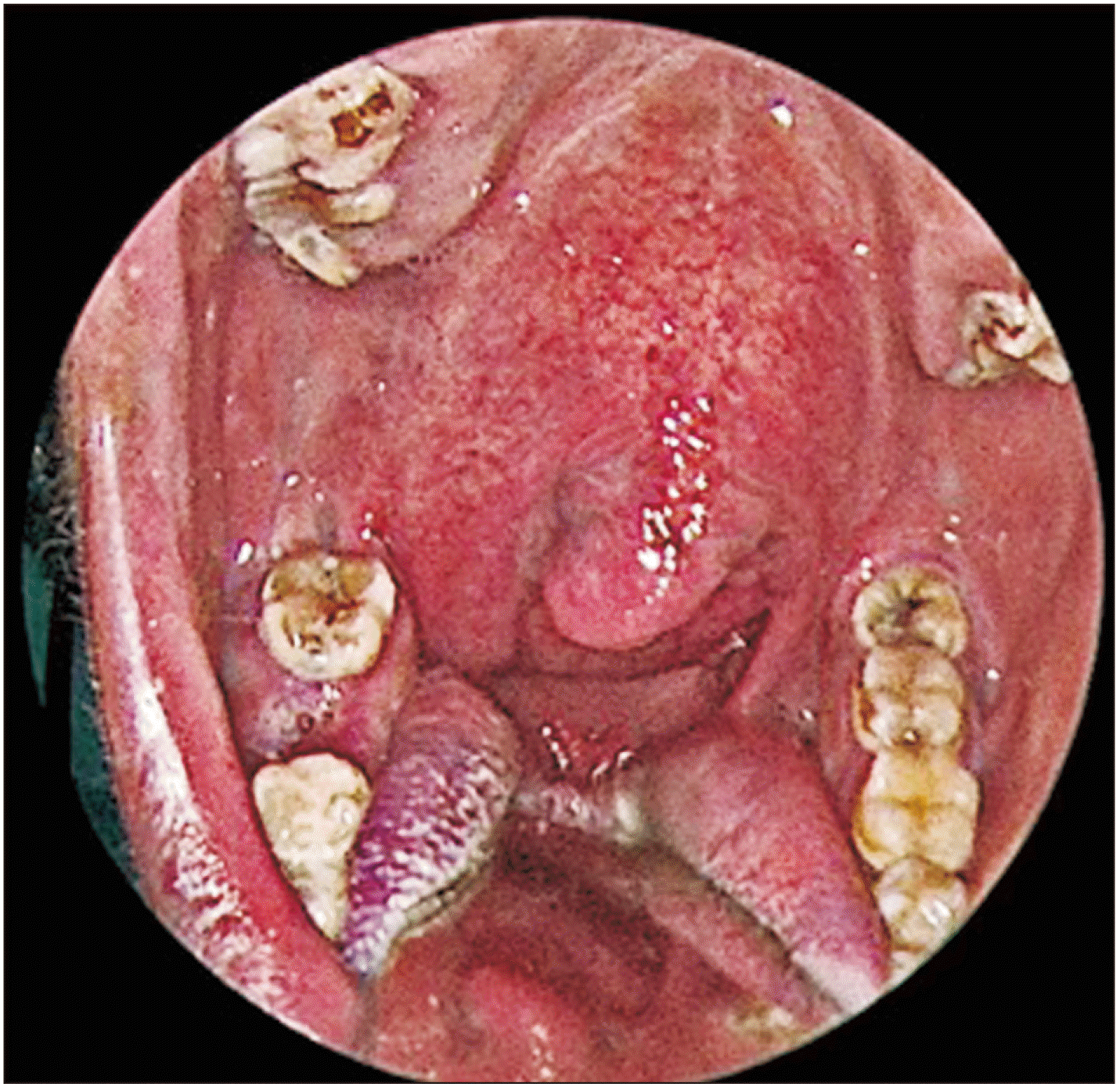
Clinical oral/oropharyngeal examination showed anterior bulging of the posterior nasopharyngeal and oropharyngeal walls, with smooth and intact mucosa. This was associated with significant airway compromise as the swelling was in contact with the soft palate and pushing it anteriorly.(Fig. 1) Nasal endoscopic examination showed non-patent choanae due to the bulging of the posterior pharyngeal wall. Laryngeal examination was not possible in this emergency situation. Examination of the neck was irrelevant and showed no external swelling.
The provisional diagnosis based on the clinical findings was a retropharyngeal retention cyst/tumor. The decision to perform an urgent tracheostomy under local anesthesia was made by the head and neck surgery team in order to secure the airway. This was followed by imaging studies for assessment of the retropharyngeal pathology.
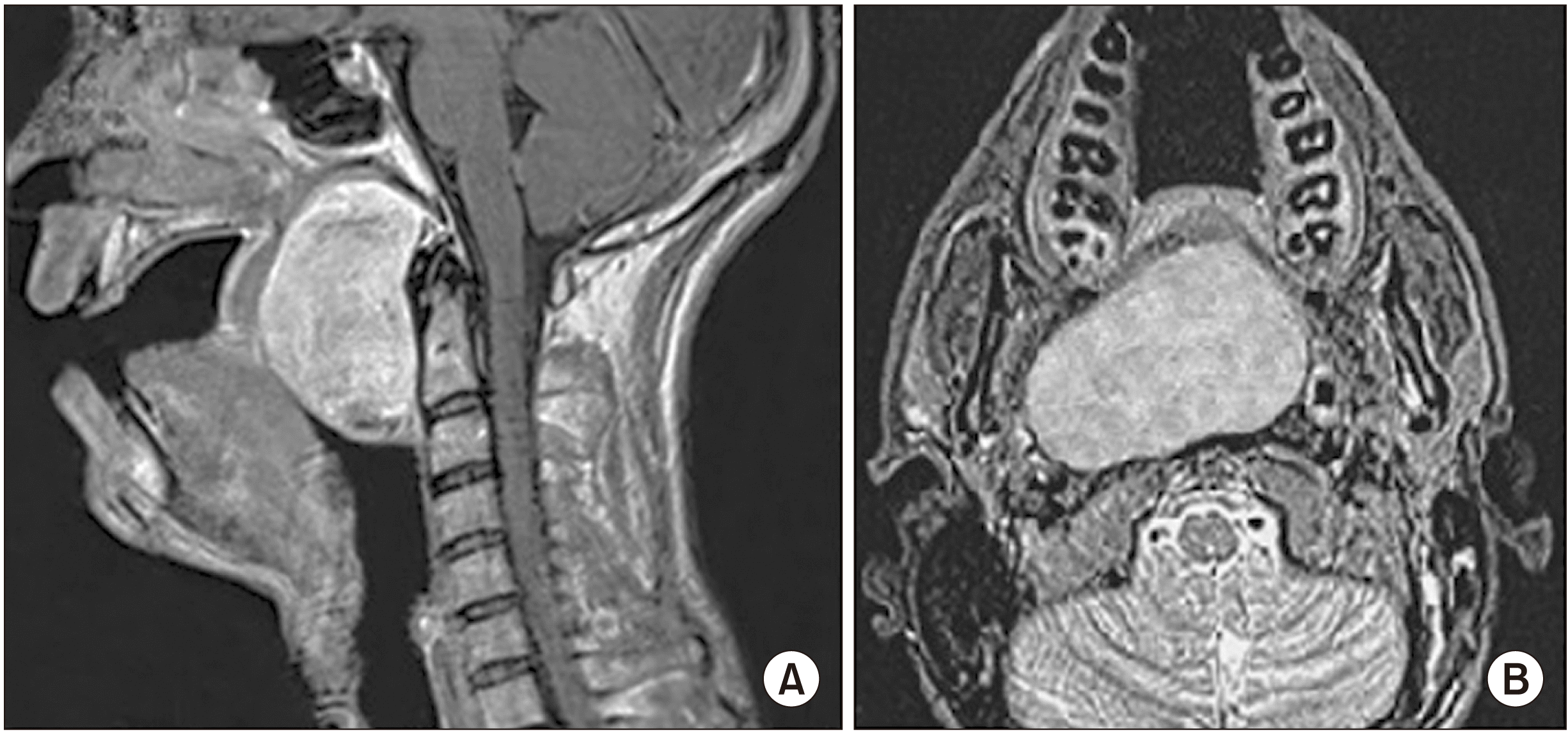
Magnetic resonance imaging showed a well-defined encapsulated tumor in the retropharyngeal space with high signal intensity on T2-weighted images and low signal intensity on T1-weighted images. Gadolinium contrast administration showed significant enhancement of the lesion on T2-weighted images. The tumor extended from the roof of the nasopharynx (the skull base) superiorly down to the level of the third cervical vertebra inferiorly. It measured about 9 cm×8 cm in the transverse and cranio-caudal dimensions, respectively, with almost complete obliteration of the airway.(Fig. 2) The tumor extended to the right parapharyngeal space, resulting in lateral displacement of the great vessels with an apparently preserved dissection plane.
A surgical plan was prepared by the head and neck team to remove the tumor using an endoscopic-assisted transoral approach with palatal splitting. The main purpose of palatal splitting was to allow wider access through the oral cavity for complete en bloc tumor excision. The use of endoscopes with different angles (30°, 45°, and 70°) offered wider panoramic visualization and allowed meticulous dissection, especially in the less accessible lateral and lower extensions.
General anesthesia was administered via the tracheostomy tube. The patient was placed in the supine position with the head in a neutral position, and the neck slightly extended. A low-profile self-retaining transoral retractor system (Spetzler-Sonntag; Aesculap, San Francisco, CA, USA) was used to achieve wide exposure of the posterior oropharynx.
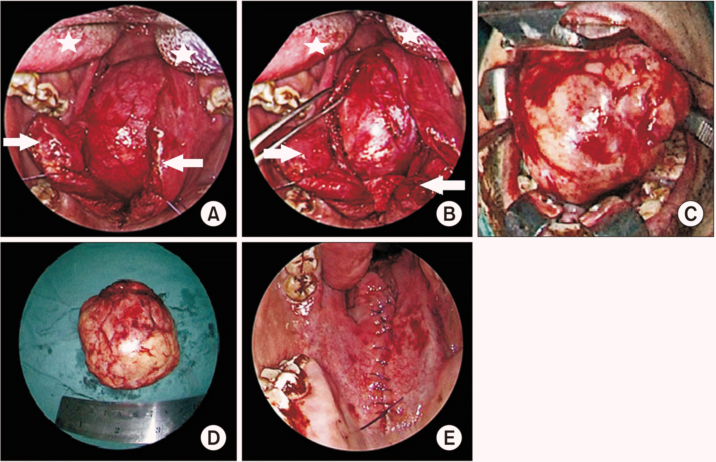
First, palatal splitting was performed using electrocautery with a Colorado needle tip dividing all three layers of the soft palate (oral mucosa, muscle layer, and nasal mucosa). Each half of the palate was then retracted using a Vicryl suture loop fixed to the retractor system. This was followed by a straight midline incision of the posterior pharyngeal wall where the well encapsulated tumor was exposed.(Fig. 3. A, 3. B)
Dissection was then performed at the left and lower borders of the mass as they were more easily accessible. Those limits of the mass were freed from their attachments with the aid of the angled endoscopes. After releasing the left and lower poles, successful dissection and delivery of the upper poles of the swelling were completed.
Due to lateral extension of the mass into the right parapharyngeal space, the right border of the tumor was the most challenging area to dissect. Again, the use of endoscopes was helpful to free the tumor and to deliver it en bloc with an intact capsule, without jeopardizing any of the surrounding great vessels or neural structures.(Fig. 3. C, 3. D)
Operative blood loss was unremarkable. Hemostasis of the surgical bed was secured by application of Surgiflo (Ethicon, Raritan, NJ, USA) over the prevertebral fascia and muscles. Primary repair of the posterior pharyngeal wall was conducted using interrupted Vicryl 3-0 sutures. Repair of soft palate was completed in three separate layers to avoid postoperative wound dehiscence. A nasogastric tube was inserted for early postoperative feeding.(Fig. 3. E)
The specimen was sent for histopathological examination. The microscopic findings showed the tumor surrounded by a fibrous capsule, composed of spindle cells with focal palisading of the nuclei arranged in short bundles and fascicles. The tumor was diagnosed as a schwannoma (Antoni A type).
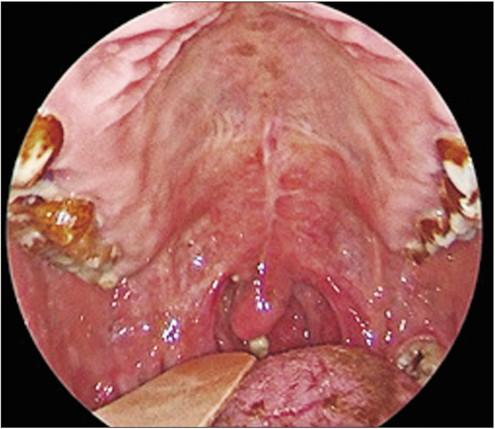
Removal of the nasogastric tube as well as tracheostomy decannulation were performed on the third postoperative day. The patient was discharged on the next day. The postoperative course was uneventful. Remarkable healing of the palate and retropharyngeal wall was noted during the follow-up.(Fig. 4) The patient regained normal swallowing and voice functions with the help of phoniatric and swallowing therapists. The patient returned to routine daily activities after three weeks. The patient was symptom free, and no cranial nerve or sympathetic chain dysfunction were reported during the one-year follow-up period.
Retropharyngeal schwannomas are exceptionally rare1. A systematic search of the English literature in the Medline, PubMed, and Google Scholar databases revealed only 18 published cases of retropharyngeal schwannoma1,3,4,6,7-20.(Table 1)
Retropharyngeal schwannoma has been observed at all ages (16 to 72 years), and the male to female ratio is 3:1 (12 males and 4 females). The most common site for schwannoma in the head and neck area is the parapharyngeal space, and the nerve of origin can be the IXth, Xth, XIth, or XIIth cranial nerves or the cervical sympathetic chain8. In the retropharyngeal space, the tumor can originate from the peripheral plexus in the retropharyngeal space, as suggested in the present case, as well as in all previously reported cases. Those patients did not manifest paresthesia, anesthesia, or any neurological sequelae after tumor excision.
In the current case, the size of the schwannoma was about 9 cm×8 cm. This is the second largest reported retropharyngeal schwannoma, after the case reported by Abou-Elfadl et al.15. The size of all reported cases ranged from 2 to 13.7 cm in the largest dimension (mean, 5.4 cm).
Complete surgical excision is the treatment of choice for schwannomas. Of the 18 previously reported cases, a transoral approach was used in 72.2% (n=13). However, it was usually considered for small tumors confined to the retropharyngeal space. On the other hand, an external approach was preferred for larger tumors or those with parapharyngeal space extension with/without displacement of the neck vessels (27.8%; n=5). In the present case, despite the large size of the tumor and its extension into the right parapharyngeal space, it was possible to dissect and remove the whole tumor trans-orally. This was aided by palatal splitting and the good visualization offered by the angled endoscopes.
Gungadeen et al.6 reported transoral robotic resection of a large schwannoma (6.7 cm×3 cm). The authors concluded that transoral excision of the large schwannoma was possible due to the advantages of the robotic system, such as the three-dimensional high-definition visualization and the improved dexterity with the use of wristed surgical instruments. In the present case, the use of angled endoscopes and of palatal splitting offered a secure panoramic visualization and accessibility that allowed precise dissection and excision of the large mass.
The transoral approach is a less invasive technique that promotes rapid healing, early postoperative oral feeding, and a shorter hospital stay. In the present case, oral feeding was permitted on the third postoperative day. This result was similar to results reported by Righini and Atallah17 and Hsieh et al.4, who reported oral feeding on the first and second postoperative days, respectively, after transoral excision of the retropharyngeal schwannoma. On the other hand, Escamilla Carpintero et al.20 inserted a nasogastric tube for one month until safe swallowing was regained after the external approach.
Regarding hospital stay, early discharge of the patient was reported with transoral approaches. Gungadeen et al.6 and Righini and Atallah17 discharged the patient on the second postoperative day. In the present case, discharge of the patient was allowed on the fourth day. A longer hospital stay was reported by Gallo et al.18 (12 days) after a combined transoral and external trans-mandibular approach.
In this reported patient, transoral resection of a huge retropharyngeal schwannoma was possible, aided by palatal splitting and angled endoscopes. This proposed less invasive approach avoided the need for an external incision and its associated morbidity, which includes a visible external scar, a longer hospital stay, higher costs, and more severe associated pain. Rapid resumption of swallowing and normal daily activities was also achieved. Moreover, the transcervical technique poses the risk of damaging cranial nerves IX, X, and XII, and major vascular structures12.
In the present case, the histopathology confirmed the diagnosis of a benign schwannoma. This was similar to all previously reported cases except the one reported by Supiyaphun et al.11, which was a malignant retropharyngeal schwannoma that was treated by local excision and postoperative radiotherapy.
The prognosis of a retropharyngeal schwannoma is usually good. In the present case, the patient was symptom free with no evidence of tumor recurrence during the one-year postoperative follow-up period. To the best of our knowledge, this is the largest retropharyngeal schwannoma to be excised via a transoral approach using palatal splitting and the endoscopic-assisted technique.
In conclusion, a transoral approach for excision of a giant retropharyngeal schwannoma was possible, assisted by the panoramic three-dimensional visualization offered by angled endoscopes, which allowed dissection of the difficult to access extensions. Palatal splitting also allowed wider access for meticulous dissection and complete en bloc tumor resection. Primary healing of the posterior pharyngeal wall as well as the palate was successful with achievement of a satisfactory outcome regarding voice and swallowing functions.
Notes
Authors’ Contributions
A.M.A.E.F. participated in conceptualization and study design. M.A. participated in data collection. H.A.E. participated in data collection and wrote the manuscript. All authors read and approved the final manuscript.
References
1. Sakhrekar R, Peshattiwar V, Jadhav R, Kulkarni B, Badhwar S, Kale H, et al. 2020; Extremely rare case of retropharyngeal space benign plexiform schwannoma - excised through Smith- Robinson approach. Surg Neurol Int. 11:182. https://doi.org/10.25259/SNI_317_2020. DOI: 10.25259/SNI_317_2020. PMID: 35079447. PMCID: PMC8781240.

2. Giraddi G, Vanaki SS, Puranik RS. 2010; Schwannoma of parapharyngeal space. J Maxillofac Oral Surg. 9:182–5. https://doi.org/10.1007/s12663-010-0059-y. DOI: 10.1007/s12663-010-0059-y. PMID: 22190783. PMCID: PMC3244085.

3. Kumagai M, Endo S, Shiba K, Masaki T, Kida A, Yamamoto M, et al. 2006; Schwannoma of the retropharyngeal space. Tohoku J Exp Med. 210:161–4. https://doi.org/10.1620/tjem.210.161. DOI: 10.1620/tjem.210.161. PMID: 17023770.

4. Hsieh CY, Hsiao JK, Wang CP. 2006; Retropharyngeal schwannoma excised through a transoral approach: a case report. Kaohsiung J Med Sci. 22:465–9. https://doi.org/10.1016/S1607-551X(09)70340-7. DOI: 10.1016/S1607-551X(09)70340-7. PMID: 17000449.

5. Saito DM, Glastonbury CM, El-Sayed IH, Eisele DW. 2007; Parapharyngeal space schwannomas: preoperative imaging determination of the nerve of origin. Arch Otolaryngol Head Neck Surg. 133:662–7. https://doi.org/10.1001/archotol.133.7.662. DOI: 10.1001/archotol.133.7.662. PMID: 17638778.

6. Gungadeen A, Lisseter R, Manickavasagam J, Paleri V. 2016; Transoral robotic resection of a large schwannoma in the retropharyngeal space. J Laryngol Otol. 130:401–3. https://doi.org/10.1017/S0022215116000190. DOI: 10.1017/S0022215116000190. PMID: 26847473.

7. Triaridis C, Tsalighopoulos MG, Kouloulas A, Vartholomeos A. 1987; Posterior pharyngeal wall schwannoma (case report). J Laryngol Otol. 101:749–52. https://doi.org/10.1017/s002221510010266x. DOI: 10.1017/S002221510010266X. PMID: 3625033.

8. Singh B, Ramjettan S, Maharaj TP, Ramsaroop R. 1995; Schwannoma of the posterior pharyngeal wall. J Laryngol Otol. 109:883–5. https://doi.org/10.1017/s0022215100131585. DOI: 10.1017/S0022215100131585. PMID: 7494128.

9. Haraguchi H, Ohgaki T, Hentona H, Komatsuzaki A. 1996; Schwannoma of the posterior pharyngeal wall: a case report. J Laryngol Otol. 110:170–1. https://doi.org/10.1017/s0022215100133079. DOI: 10.1017/S0022215100133079. PMID: 8729506.

10. Moore CE, Putzi M, McClatchey KD, Terrell J. 1997; Ancient schwannoma of the posterolateral pharynx: a benign lesion commonly mistaken for sarcoma. Otolaryngol Head Neck Surg. 117:S125–8. https://doi.org/10.1016/s0194-5998(97)70078-0. DOI: 10.1016/S0194-5998(97)70078-0. PMID: 9419124.

11. Supiyaphun P, Snidvongs K, Shuangshoti S, Khowprasert C. 1997; Malignant transformation in a benign encapsulated schwannoma of retropharyngeal space: a case report. J Med Assoc Thai. 80:540–6. PMID: 9277088.
12. Huang CM, Leu YS. 2002; Schwannoma of the posterior pharyngeal wall. J Laryngol Otol. 116:740–1. https://doi.org/10.1258/002221502760238091. DOI: 10.1258/002221502760238091. PMID: 12437815.

13. Gaball CW, Meyer T, William P, Magdycz WP. 2004; Transoral approach to a retropharyngeal schwanoma: a case report and literature review. Otolaryngol Head Neck Surg. 131:304. https://doi.org/10.1016/j.otohns.2004.06.674. DOI: 10.1016/j.otohns.2004.06.674.

14. Jovanovic MB, Milutinovic Z, Milenkovic S. 2008; Posterior pharyngeal wall schwannoma - an unusual case. J BUON. 13:577–9. PMID: 19145685.
15. Abou-Elfadl M, Lrhazi S, Mahtar M, Roubal M. 2015; Unusual retropharyngeal mass. Eur Ann Otorhinolaryngol Head Neck Dis. 132:115–6. https://doi.org/10.1016/j.anorl.2015.01.002. DOI: 10.1016/j.anorl.2015.01.002. PMID: 25703405.

16. Raimondo L, Garzaro M, Mazibrada J, Pecorari G, Giordano C. 2015; Plexiform schwannoma of the posterior pharyngeal wall in a patient with neurofibromatosis 2. Ear Nose Throat J. 94:E17–9. PMID: 25738721.
17. Righini CA, Atallah I. 2015; A retropharyngeal mass. Diagnosis: Antoni A type schwannoma. Eur Ann Otorhinolaryngol Head Neck Dis. 132:57–8. https://doi.org/10.1016/j.anorl.2014.01.008. DOI: 10.1016/j.anorl.2014.01.008. PMID: 25439624.

18. Gallo S, Bandi F, Maffioli MP, Giudice M, Castelnuovo P, Fazio E, et al. 2017; Retropharyngeal space schwannoma: a rare entity. Iran J Otorhinolaryngol. 29:353–7. PMID: 29383317. PMCID: PMC5785115.
19. Ngu CYV, Gan CC, Tang IP. 2017; Transoral excision of retropharyngeal schwannoma: case report. Acta Otolaryngol Case Rep. 2:64–7. https://doi.org/10.1080/23772484.2017.1303773. DOI: 10.1080/23772484.2017.1303773.

20. Escamilla Carpintero Y, Aguilà Artal AF, Prenafeta Moreno M. 2020; Retropharyngeal schwannoma. An uncommon location. Acta Otorrinolaringol Esp (Engl Ed). 71:193–4. https://doi.org/10.1016/j.otorri.2019.02.002. DOI: 10.1016/j.otorri.2019.02.002. PMID: 31113533.

Fig. 1
Oral examination showing marked anterior bulging of the posterior pharyngeal wall, pushing the soft palate.

Fig. 2
Magnetic resonance imaging scan showing the retropharyngeal tumor. A. Sagittal view. B. Axial view.

Fig. 3
The surgical technique. A. Palatal splitting. The arrows point to the edges of the palate after splitting, retracted with a suture loop. The asterisks point to the tongue, retracted by the retractor system. The posterior pharyngeal wall is bulging with intact mucosa. B. Posterior pharyngeal wall incision. The edge is grasped with Allis forceps. The tumor capsule is exposed. C, D. En bloc delivery of the mass. E. Palatal repair.

Table 1
Data from previously published cases
| Study | Age (yr) | Sex | Site | Size (cm) | Symptoms | Approach | Pathology |
|---|---|---|---|---|---|---|---|
| Triaridis et al.7 (1987) | 21 | Male | Right posterior wall of the pharynx, protruding behind the ipsilateral tonsillar pillar | 3×2 | Malodorous breath, dysphagia, sore throat, mild fever | Trans-oral using a strong tonsillar snare | Schwannoma (Antoni A) |
| Singh et al.8 (1995) | 36 | Male | Retropharyngeal from the second to the fourth cervical vertebrae | 6×5 | Snoring, stridor | Trans-oral | Benign schwannoma |
| Haraguchi et al.9 (1996) | 60 | Male | Right posterior wall of the pharynx | 2×1 | Discomfort in pharynx | Trans-oral | Schwannoma |
| Moore et al.10 (1997) | 39 | Male | Right posterolateral pharyngeal wall | 4.5×4 | Chronic throat clearing, dysphagia | Trans-oral | Schwannoma (mixed Antoni A and Antoni B) |
| Supiyaphun et al.11 (1997) | - | - | Retropharyngeal | - | - | Trans-oral | Malignant schwannoma |
| Huang and Leu12 (2002) | 24 | Female | Posterior pharyngeal wall just above the larynx | 3×2 | Sensation of a lump in the throat, difficulty swallowing, stridor | Trans-oral | Schwannoma (mixed Antoni A and Antoni B) |
| Gaball et al.13 (2004) | 26 | Female | Retropharyngeal | - | Postnasal drip | Trans-oral | Schwannoma |
| Hsieh et al.4 (2006) | 44 | Female | Posterior oropharyngeal wall behind the left posterior pillar | 3.5×3 | Snoring, sensation of a lump in the throat | Trans-oral | Schwannoma |
| Kumagai et al.3 (2006) | 24 | Male | Retropharyngeal, extending to the right parapharyngeal space | 4×2 | Sensation of a foreign body in the pharynx, dysphagia | Trans-oral | Schwannoma (Antoni A) |
| Jovanovic et al.14 (2008) | 59 | Male | Posterior pharyngeal wall | - | Dysphagia | External lateral pharyngotomy approach | Schwannoma |
| Abou-Elfadl et al.15 (2015) | 16 | Male | Retropharyngeal, causing lateral displacement of neck vessels | 13.7×8.7 | Neck swelling, respiratory discomfort, dysphagia | External lateral neck incision | Benign schwannoma |
| Raimondo et al.16 (2015) | 37 | Male | Posterior pharyngeal wall | - | - | Trans-oral laser excision | Plexiform schwannoma |
| Righini and Atallah17 (2015) | 32 | Male | Right posterior pharyngeal wall | 3.5×2.7 | Sensation of a pharyngeal foreign body | Trans-oral | Schwannoma (Antoni A) |
| Gungadeen et al.6 (2016) | 48 | Male | Retropharyngeal, extending from the level of the post-nasal space to the level of the arytenoids | 6.7×3 | Snoring, sensation of a lump, dysphagia | Robot-assisted transoral excision | Schwannoma |
| Gallo et al.18 (2017) | 36 | Female | Retropharyngeal, from the nasopharynx to the hypopharynx | 6×5.5 | Perception of a foreign body in the throat, dysphagia, odynophagia, significant weight loss | Combined trans-oral and external trans-mandibular approach | Schwannoma |
| Ngu et al.19 (2017) | 27 | Male |
Retropharyngeal, extending to the left pyriform fossa |
5.5×3.6 | Asymptomatic | Trans-oral | Schwannoma (mixed Antoni A and Antoni B) |
| Escamilla Carpintero et al.20 (2020) | 72 | - | Posterior wall of the hypopharynx | 6.4×3.4 | Dysphagia, left neck swelling | Bilateral external cervical approach | Schwannoma |
| Sakhrekar et al.1 (2020) | 30 | Male | Retropharyngeal, from C2 to C5 | 7.5×4.8 | Feeling of a lump in the throat, impaired phonation, snoring dysphagia | External cervical approach (right-sided Smith-Robinson approach) | Benign plexiform schwannoma |


 PDF
PDF Citation
 Print
Print


 XML Download