Journal List > Blood Res > v.57(S1) > 1516078611

Park: Diagnosis and management of thrombocytopenia in pregnancy

Abstract

Thrombocytopenia, defined as platelet count <150×109/L, is frequently observed by physicians during pregnancy, with an incidence of approximately 10% of all pregnancies. Most of the cases of thrombocytopenia in pregnancy are due to gestational thrombocytopenia, which does not confer an increased risk of maternal bleeding. However, because other causes can be associated with life-threatening events, such as severe bleeding, that can affect to maternal and fetal outcomes, differentiating other cause of thrombocytopenia, which includes preeclampsia, HELLP (hemolysis, elevated liver enzymes, low platelets) syndrome, acute fatty liver of pregnancy, immune thrombocytopenia, hereditary thrombocytopenia, antiphospholipid syndrome, thrombotic thrombocytopenic purpura, and atypical hemolytic uremic syndrome, is important. Understanding the mechanisms and recognition of symptoms and signs are important to decide an adequate line of investigation. In this review, the approach to diagnosis and the management of the thrombocytopenia commonly observed in pregnancy are presented.

INTRODUCTION

Thrombocytopenia, defined as platelet count of <150×109/L, is frequently observed during pregnancy, with an incidence of approximately 10% of all pregnancies [1]. It may be related to physiologic changes or pathologic conditions, some of which are unique to pregnancy and may pose significant risk to both mother and child [1]. Thrombocytopenia usually results in mucosal bleeding consequent to primary hemostasis defect. Clinical presentation includes epistaxis, gingival bleeding, or abnormal uterine bleeding [2]. Life-threatening bleeding is infrequent and is limited to patients with extreme thrombocytopenia, presenting as hematuria, gastrointestinal bleeding, and rarely, intracranial bleeding. In pregnancy, most cases of thrombocytopenia are resulted from hemodilution and increased platelet destruction [1, 3].

GENERAL APPROACH TO DIAGNOSIS

A complete blood count assessment and review of the peripheral blood smear are the important first step in evaluating thrombocytopenia in pregnancy. In addition, a careful family and personal medical history will permit categorizing of thrombocytopenia. The diagnostic evaluation for thrombocytopenia in pregnancy includes test for markers of hemolysis, liver function test, or testing for infections (hepatitis B virus, hepatitis C virus, human immune deficiency virus, Helicobacter pylori, cytomegalovirus) [1]. Depending on the specific medical history and clinical suspicion, further diagnostic tests may include tests for antiphospholipid antibodies, antinuclear antibodies, or von Willebrand syndrome type 2B. Prevalence of laboratory abnormalities by cause of thrombocytopenia in pregnancy is presented in Table 1. The trimester when thrombocytopenia develops can provide an important clue to etiology. A gradual decline in platelet count occur in the middle of the second trimester, i.e. gestational thrombocytopenia [1]. A decline before second trimester suggests a cause of thrombocytopenia other than gestational thrombocytopenia [1].

PHYSIOLOGIC CHANGES IN PREGNANCY

Thrombocytopenia could be often observed in pregnant women, beginning in the first trimester, and gradually decreasing through gestation, with nadir at delivery [4]. This condition is resulted from physiologic hemodilution due to large plasma volume, increased platelet activation and clearance [3].

GESTATIONAL THROMBOCYTOPENIA

Gestational thrombocytopenia (GT) is the most common cause of thrombocytopenia during pregnancy, with incidence of 5% to 11% of all pregnancies [1]. It may be resulted from hemodilution caused by the increased plasma volume and accelerating platelet clearance [4]. GT is observed frequently during mid- to late-second or third trimester [1, 4]. Because no specific laboratory tests to diagnose GT exists, diagnosis of exclusion is needed according to onset time, severity of thrombocytopenia, and variable clinical features that can be presented in other causes of thrombocytopenia in pregnancy. Early onset of thrombocytopenia in pregnancy, platelet count <80×109/L, and precipitous decline in platelet count should be considered to alternative diagnosis [1, 5]. Stable platelet count (>100×109/L) in asymptomatic women do not require further investigation nor specific intervention other than regular platelet count monitoring [1]. Interventions as elective cesarean delivery are not indicated, and GT does not confer an increased risk of maternal bleeding complications nor fetal bleeding [6]. Recovery to normal platelet count occurs within 1 and 2 months of delivery [6].

PREECLAMPSIA

Preeclampsia (PEC) is characterized by the new onset of hypertension after 20 weeks of gestation and proteinuria [7]. In severe PEC, there is significant end-organ damage, including thrombocytopenia, impaired liver function, renal insufficiency, pulmonary edema, cerebral or visual disturbance, or high blood pressure [7]. The pathogenesis of PEC may be related with abnormal placental angiogenesis and vascular function [8]. PEC should be suspected if high blood pressure and/or proteinuria is observed after 20 weeks of gestation [9]. Platelet counts are usually above 100×109/L and rarely below 50×109/L [9]. Thrombocytopenia (<100×109/L) is one of the criteria for severe PEC [9]. Overt hemolysis may be a feature of hemolysis, elevated liver enzyme and low platelet count (HELLP) syndrome or thrombotic thrombocytopenic purpura (TTP) than that of PEC [8]. Mild elevation of liver enzymes may be present in PEC, however, high elevation of liver enzymes is more indicative of HELLP syndrome [8]. Disseminated intravascular coagulation (DIC) may be develop, but overt bleeding or thrombosis is rare [8]. Delivery is definite management, but inpatient expectant management until 34 weeks can be considered for some patients who are <34 weeks of gestation [10]. Supportive cares include control of severe hypertension with anti-hypertensive agents and administration of magnesium sulfate to prevent seizures [10].

HELLP SYNDROME

The HELLP syndrome is characterized by platelet count <100×109/L, elevated liver enzyme, and microangiopathic hemolytic anemia, occurring in approximately 0.2–0.8% of all pregnancies [7, 11]. It may be a severe form of PEC with high morbidity and mortality, being more frequent in the third trimester [7]. Up to 8–24% of patients with severe PEC or eclampsia will develop HELLP syndrome in some cases [12]. Clinical manifestations include upper abdominal pain, nausea, vomiting, headache, and, rarely, jaundice. DIC occurs in 20% of cases, which can result in massive bleeding, placental abruption, and hepatic rupture [1, 7, 12]. The diagnosis should be considered when progressive thrombocytopenia, overt hemolysis, and worsening hepatic function develop in the setting of PEC [1, 7, 12]. Urgent delivery is the cornerstone of management to prevent rapid deterioration, regardless of gestational age [7, 12]. Management of maternal hypertension and magnesium to prevent seizures may be required [12]. In case of DIC, fresh frozen plasma with or without cryoprecipitate may be required. In a meta-analysis, it was reported that 7.2% of patients with HELLP syndrome had a recurrence during subsequent pregnancies and 36.3% developed either another hypertensive syndromes or fetal growth restriction [13].

ACUTE FATTY LIVER OF PREGNANCY

Acute fatty liver of pregnancy (AFLP) is rare life-threatening condition (1/70,000–1/15,000 pregnancies) characterized by the rapid onset of hepatic failure, typically occurring in the third trimester [1]. Although the pathogenesis is incompletely understood, it seems to be related to estrogen elevation in pregnancy, defective fatty acid metabolism, and mitochondrial dysfunction [14]. Clinical manifestations include abdominal pain, anorexia, nausea, vomiting, if AFLP progresses, liver failure and encephalopathy. Hypoglycemia and coagulopathy are key features of severe disease that helps to distinguish AFLP from other causes of thrombocytopenia in pregnancy [1, 15]. The Swansea criteria is commonly used to diagnose AFLP, including vomiting, elevated uric acid (>5.7 mg/dL), abdominal pain, polydipsia/polyuria, encephalopathy, bilirubin >0.8 mg/dL, hypoglycemia (<72 mg/dL), coagulopathy (prothrombin time >14 s or activated partial thromboplastin time >34 s), creatinine >1.7 mg/dL, ammonia >27.5 mg/dL, alanine aminotransferase >42 U/L, white blood cell count >11×109/L, ascites or bright liver on ultrasound, and microvascular steatosis on liver biopsy [16]. Six or more of these terms are required to diagnose AFLP. Early delivery is the cornerstone of management to prevent rapid deterioration, regardless of gestational age [1, 16]. Supportive care may be required for several days, including red blood cell transfusion, dialysis, plasmapheresis, and correction of coagulopathy (fibrinogen ≥200 mg/dL) [1, 14, 15].

IMMUNE THROMBOCYTOPENIA

Immune thrombocytopenia (ITP) is an acquired autoimmune disorder characterized by accelerate platelet destruction and impaired platelet production, accounting for 1–4% of pregnancy thrombocytopenia [1, 4]. It is the most common cause of a platelet count <50×109/L in the first or second trimester [1, 17]. Patients with a history suggestive of ITP or those with a platelet count <80×109/L should be investigated for possible ITP [1]. Because no definite diagnostic tests are lacking, diagnosis of ITP is based mainly on the exclusion of other cause of thrombocytopenia in pregnancy. Although most cases present with platelet count of >70×109/L, approximately 20% of cases may require treatment before labor [1]. According to the International Consensus Report (ICR) guidelines for ITP, a platelet count between 20 and 30×109/L in a nonbleeding patient is safe for most pregnancy and a platelet count ≥50×109/L is preferred for delivery [3]. Corticosteroids and intravenous immunoglobulin (IVIG) are first-line treatment options [3, 17]. Although corticosteroids are generally safe in pregnancy, there are potentially serious side effects, especially when high-dose corticosteroids are used, including an increased risk of gestational diabetes, hypertension, emotional lability, and an increased risk of cleft palate in the fetus when given in the first trimester [17-19]. If a rapid response is needed or intolerance to corticosteroids occurs, IVIG may be preferable [17]. There is no consensus on optimal second-line treatment for ITP in pregnancy. Immunosuppressant, such as azathioprine and cyclosporine, have been used in pregnant women with acceptable adverse effects [20]. Rituximab, CD20 monoclonal antibody, can be considered in refractory cases, but can cause prolonged lymphopenia in newborn, resulting from suppression of neonatal B lymphocyte development [20]. Splenectomy may be performed for refractory ITP, preferably in the second trimester [20]. As safety data for thrombopoietin receptor agonists (eltrombopag and romiplostim) is limited in pregnancy, thus routine use of these agents during pregnancy is not recommended [20]. Platelet transfusions are not routinely recommended except in the setting of significant bleeding or platelet count of <50×109/L near delivery. Given that the method of delivery is determined by obstetric indications, platelet count >20×109/L for vaginal delivery and >50×109/L for cesarean delivery are recommended to prevent excessive blood loss [3, 20]. Although 10–15% of neonates are born with thrombocytopenia, severe thrombocytopenia is uncommon and intracranial hemorrhage is rare [21].

HEREDITARY THROMBOCYTOPENIA

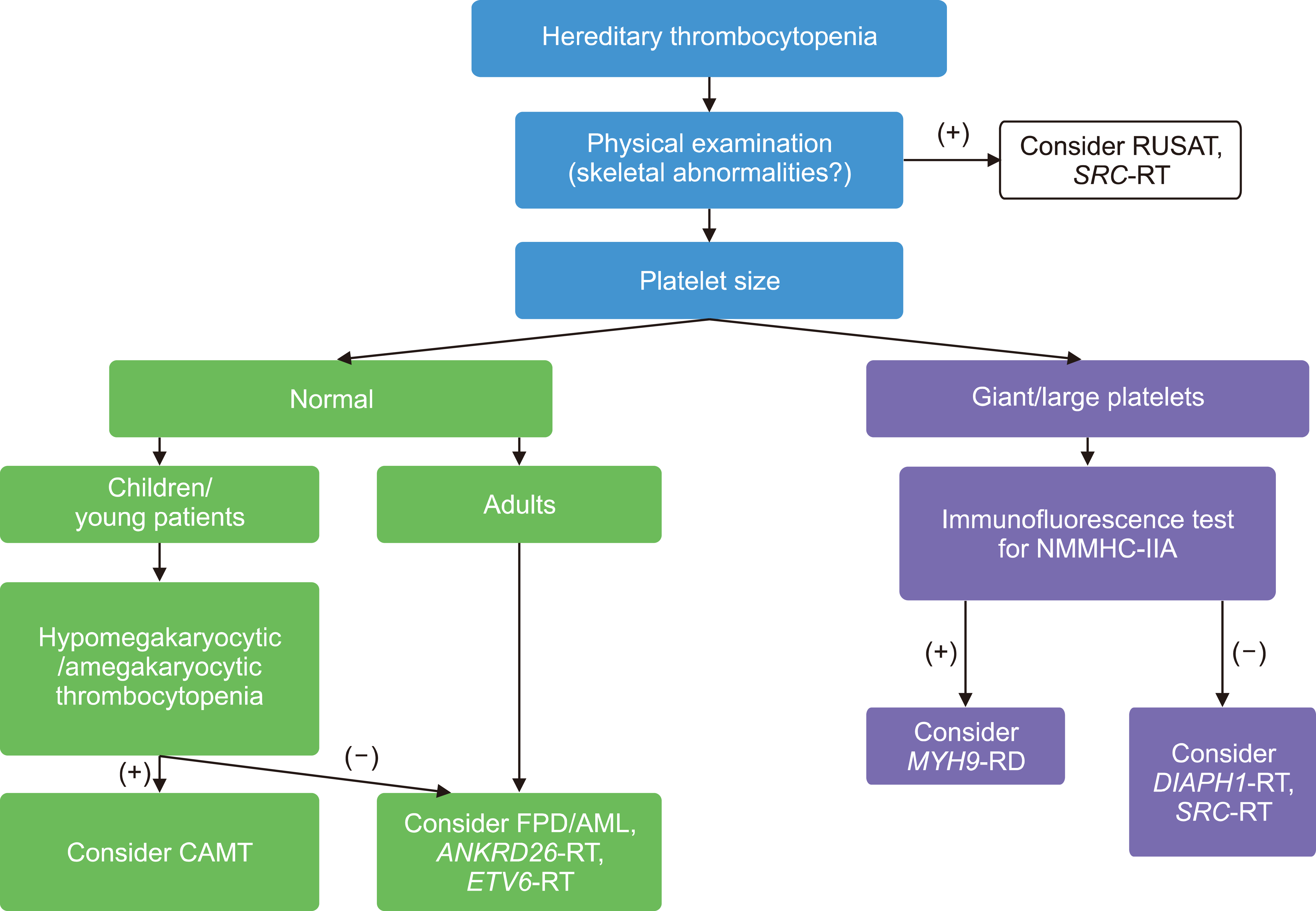
The hereditary thrombocytopenias comprise a diverse group of disorders that vary in their phenotype based on the specific gene defects in one of the steps in megakaryocyte and platelet development [1, 22]. However, it is often difficult for patients with hereditary thrombocytopenia syndromes to be diagnosed with or suspected of hereditary thrombocytopenia only by family history because many of them are autosomal recessive transmission or several patients have de novo mutations [22]. A diagnostic algorithm for hereditary thrombocytopenia based on patients’ clinical features is shown in Fig. 1. A pre-gestational history of thrombocytopenia is helpful but uncovering a family history of thrombocytopenia or bleeding is generally key to raising suspicion [1, 22]. Platelet counts vary from 20 to 130×109/L depending on the specific gene defect. There are associated physical findings (e.g., absent radius, albinism) or renal insufficiency that should take further consideration of a syndromic form of congenital thrombocytopenia [22]. Platelet counts do not tend to fall precipitously during pregnancy and bleeding does not suddenly worsen in most patients [22]. The bleeding phenotype generally correlates with the severity of thrombocytopenia unless there is a superimposed defect in platelet function [22]. There is little evidence to guide management of specific inherited platelet function disorders. The use of desmopressin during pregnancy has not been well-studied and requires careful management of fluids to limit risk of hyponatremia [23]. Bernard-Soulier syndrome, autosomal recessive disorder, is characterized by qualitative and quantitative defects of the platelet membrane glycoprotein Ib-IX-V complex [24]. Clinical manifestations include thrombocytopenia, prolonged bleeding time and the presence of giant platelets [24]. Pregnancy course may be normal, but there might be severe bleeding during postpartum period [24].

ANTIPHOSPHOLIPID SYNDROME

Antiphospholipid syndrome (APS) is characterized by arterial/venous thrombosis and/or unexplained recurrent pregnancy loss, stillbirth, or early onset and severe PEC or HELLP [25]. Approximately 1% of patients develop a catastrophic presentation (CAPS), including multi-organ thrombotic complications, during pregnancy or in the puerperium [25]. It may be related to platelet activation and complement-mediated inflammatory responses within the placenta. Antiphospholipid antibodies can cause platelet and neutrophil activation, which can potentially lead to make thrombosis [26]. APS is diagnosed by the revised Sapporo criteria [27] based on clinical and laboratory features [recommendations from International Society of Thrombosis and Hemostasis (ISTH) [28], which include arterial or venous thrombosis and the gestational complications. CAPS can present with multiple thromboses (arterial, venous and/or microvascular), acute multi-organ dysfunction, diffuse alveolar hemorrhage, encephalopathy, or adrenal hemorrhage, among other complications [25]. Because the patients with APS have an increased risk of obstetric complications and lower rates of live births, it is important to recognize APS as a possible and to be able to diagnosis and treat CAPS [1, 25]. Long-term anticoagulation at the onset of pregnancy is essential for the patients with a previous diagnosis APS with thrombotic events [29]. Treatment should be switched to therapeutic doses of enoxaparin throughout pregnancy with a transition back to an oral anticoagulant post-partum [29]. Treatment of CAPS includes rapid initiation of plasma exchange and therapeutic anticoagulation, typically with heparin [25, 29]. For the condition is progressive or life-threatening, addition of high dose corticosteroids and/or IVIG is recommended [25].

TTP

TTP is a thrombotic microangiopathy (TMA), in which microthrombi develop in small vessels due to a severe deficiency of ADMATS13 enzyme activity, the metalloprotease responsible for cleavage of the ultra-large von Willebrand factor (vWF) multimers [15, 30]. While ADAMTS13 deficiency is commonly acquired because of autoantibodies-mediated clearance of ADAMTS13, the patients with congenital TTP, autosomal recessive transmission, have severe deficiency of the ADAMTS13 protease due to biallelic ADAMTS13 gene mutations [31]. Patients who are heterozygous for an ADAMTS13 mutation do not appear to be at risk during pregnancy [31]. There is increased production of vWF in pregnancy, especially in the second and third trimester [1]. In the reported literature, 10% to 25% of all cases of TTP diagnosed will be in pregnant or postpartum women [15]. The characteristic symptoms include microangiopathic hemolytic anemia, thrombocytopenia, fever, neurologic symptoms (from headache to coma), and renal dysfunction [30, 32]. Along with laboratory and clinical findings, the diagnosis of TTP is confirmed by low ADAMTS13 activity of <10% and/or by the presence of IgG antibodies to ADAMTS13 [32]. When the TTP is strongly suspected in pregnancy, plasma exchange along with corticosteroids should be started promptly, until the diagnosis of TTP is confirmed [15]. Once the diagnosis of TTP in made, treatment should be continued until platelet count is restored and the lactate dehydrogenase level is reduced [15]. There is little data on the use of rituximab in pregnancy, but the rituximab may be considered in refractory cases [1, 33]. The diagnosis of TTP is not an indication for termination of a pregnancy or delivery, depending on the gestational age [15]. When TTP presents in the first or second trimester, the risk of fetal morbidity may be increased due to placental ischemia [32]. However, 75% to 90% of live birth is observed when TTP develop in the third trimester [32]. Recurrent TTP in subsequent pregnancies is estimated at approximately 50% for women with congenital TTP or acquired TTP with a persistent deficiency of ADAMTS13 activity [34].

ATYPICAL HEMOLYTIC UREMIC SYNDROME

Atypical hemolytic uremic syndrome (aHUS) is a rare disorder, even in pregnancy occurring in one out of every 25,000 pregnancies [35]. It is resulted from uncontrolled activation of the alternative complement pathway during pregnancy, leading to platelet and endothelial activation and generation of occlusive platelet thrombi, especially within the microvascular of the kidney [15, 35]. The clinical manifestation of aHUS is similar to that of TTP, often leading to increase the risk of renal failure if untreated [36]. This is exclusion diagnosis, after ruling out TTP as well as other TMA causes [1]. ADAMTS13 levels are >10% and complement genetic testing can be performed to support the diagnosis. Initial therapy includes plasmapheresis, which is often ineffective, fresh frozen plasma infusion, and dialysis [15, 36]. When aHUS is highly suspected, eculizumab, an anti-C5 monoclonal antibody, should be administered as soon as possible [15, 36]. Eculizumab binds with high affinity to C5, inhibiting C5 cleavage to C5a and C5b and subsequently preventing the assembly of C5b-C9, thus inhibiting complement-medicated TMA, which leads to improve renal function and hematologic outcomes [37]. Improvement in thrombocytopenia usually is presented in 48–72 hours, but improvement in renal function and other organ damage can be delayed by weeks to months [38]. Eculizumab can cross the placenta, but it has been used safely during pregnancy and in lactating mothers [38].

CONCLUSION

Thrombocytopenia is a relatively common hematological abnormality in pregnancy. The pathophysiology responsible for thrombocytopenia will range from benign process, where observation alone may be appropriate, to more sinister microangiopathies, where urgent fetal delivery or initiation of specific treatment are critical for the survival of the mother and fetus. Close observation of the clinical course and response to treatment is critical to establishing the correct diagnosis, which has both immediate treatment implication and potential impact on the management of future pregnancies.

Notes

Authors’ Disclosures of Potential Conflicts of Interest

No potential conflicts of interest relevant to this article were reported.

REFERENCES

1. Cines DB, Levine LD. 2017; Thrombocytopenia in pregnancy. Blood. 130:2271–7. DOI: 10.1182/blood-2017-05-781971. PMID: 28637667. PMCID: PMC5701522. PMID: https://www.scopus.com/inward/record.uri?partnerID=HzOxMe3b&scp=85034866089&origin=inward.
crossref
2. Zainal A, Salama A, Alweis R. 2019; Immune thrombocytopenic purpura. J Community Hosp Intern Med Perspect. 9:59–61. DOI: 10.1080/20009666.2019.1565884. PMID: 30788080. PMCID: PMC6374974.
crossref
3. Provan D, Arnold DM, Bussel JB, et al. 2019; Updated international consensus report on the investigation and management of primary immune thrombocytopenia. Blood Adv. 3:3780–817. DOI: 10.1182/bloodadvances.2019000812. PMID: 31770441. PMCID: PMC6880896. PMID: https://www.scopus.com/inward/record.uri?partnerID=HzOxMe3b&scp=85076363753&origin=inward.
crossref
4. Reese JA, Peck JD, Deschamps DR, et al. 2018; Platelet counts during pregnancy. N Engl J Med. 379:32–43. DOI: 10.1056/NEJMoa1802897. PMID: 29972751. PMCID: PMC6049077. PMID: https://www.scopus.com/inward/record.uri?partnerID=HzOxMe3b&scp=85049627283&origin=inward.
crossref
5. Valera MC, Parant O, Vayssiere C, Arnal JF, Payrastre B. 2010; Physiologic and pathologic changes of platelets in pregnancy. Platelets. 21:587–95. DOI: 10.3109/09537104.2010.509828. PMID: 20873962. PMID: https://www.scopus.com/inward/record.uri?partnerID=HzOxMe3b&scp=78149445346&origin=inward.
crossref
6. Rottenstreich A, Israeli N, Levin G, Rottenstreich M, Elchalal U, Kalish Y. 2018; Clinical characteristics, neonatal risk and recurrence rate of gestational thrombocytopenia with platelet count <100× 109/L. Eur J Obstet Gynecol Reprod Biol. 231:75–9. DOI: 10.1016/j.ejogrb.2018.10.026. PMID: 30340119. PMID: https://www.scopus.com/inward/record.uri?partnerID=HzOxMe3b&scp=85054809358&origin=inward.
7. Brown MA, Magee LA, Kenny LC, et al. 2018; Hypertensive disorders of pregnancy: ISSHP classification, diagnosis, and management recommendations for international practice. Hypertension. 72:24–43. DOI: 10.1161/HYPERTENSIONAHA.117.10803. PMID: 29899139. PMID: https://www.scopus.com/inward/record.uri?partnerID=HzOxMe3b&scp=85053806850&origin=inward.
8. Phipps E, Prasanna D, Brima W, Jim B. 2016; Preeclampsia: updates in pathogenesis, definitions, and guidelines. Clin J Am Soc Nephrol. 11:1102–13. DOI: 10.2215/CJN.12081115. PMID: 27094609. PMCID: PMC4891761. PMID: https://www.scopus.com/inward/record.uri?partnerID=HzOxMe3b&scp=84992366827&origin=inward.
crossref
9. Mol BWJ, Roberts CT, Thangaratinam S, Magee LA, de Groot CJM, Hofmeyr GJ. 2016; Pre-eclampsia. Lancet. 387:999–1011. DOI: 10.1016/S0140-6736(15)00070-7. PMID: 34051884. PMID: https://www.scopus.com/inward/record.uri?partnerID=HzOxMe3b&scp=84960126776&origin=inward.
crossref
10. 2019; ACOG practice bulletin No. 202: gestational hypertension and preeclampsia. Obstet Gynecol. 133:1. DOI: 10.1097/AOG.0000000000003020. PMID: 30575676. PMID: https://www.scopus.com/inward/record.uri?partnerID=HzOxMe3b&scp=85058908853&origin=inward.
11. Wisner K. 2019; Gestational hypertension and preeclampsia. MCN Am J Matern Child Nurs. 44:170. DOI: 10.1097/NMC.0000000000000523. PMID: 31033586. PMID: https://www.scopus.com/inward/record.uri?partnerID=HzOxMe3b&scp=85064864052&origin=inward.
crossref
12. Fitzpatrick KE, Hinshaw K, Kurinczuk JJ, Knight M. 2014; Risk factors, management, and outcomes of hemolysis, elevated liver enzymes, and low platelets syndrome and elevated liver enzymes, low platelets syndrome. Obstet Gynecol. 123:618–27. DOI: 10.1097/AOG.0000000000000140. PMID: 24499757. PMID: https://www.scopus.com/inward/record.uri?partnerID=HzOxMe3b&scp=84894412846&origin=inward.
crossref
13. van Oostwaard MF, Langenveld J, Schuit E, et al. 2015; Recurrence of hypertensive disorders of pregnancy: an individual patient data metaanalysis. Am J Obstet Gynecol. 212:624e117–17. DOI: 10.1016/j.ajog.2015.01.009. PMID: 25582098. PMID: https://www.scopus.com/inward/record.uri?partnerID=HzOxMe3b&scp=84928827805&origin=inward.
crossref
14. Ding J, Han LP, Lou XP, et al. 2015; Effectiveness of combining plasma exchange with plasma perfusion in acute fatty liver of pregnancy: a retrospective analysis. Gynecol Obstet Invest. 79:97–100. DOI: 10.1159/000368752. PMID: 25634306. PMID: https://www.scopus.com/inward/record.uri?partnerID=HzOxMe3b&scp=84924268029&origin=inward.
crossref
15. Thomas MR, Robinson S, Scully MA. 2016; How we manage thrombotic microangiopathies in pregnancy. Br J Haematol. 173:821–30. DOI: 10.1111/bjh.14045. PMID: 27019232. PMID: https://www.scopus.com/inward/record.uri?partnerID=HzOxMe3b&scp=84973657510&origin=inward.
crossref
16. Ch'ng CL, Morgan M, Hainsworth I, Kingham JG. 2002; Prospective study of liver dysfunction in pregnancy in Southwest Wales. Gut. 51:876–80. DOI: 10.1136/gut.51.6.876. PMID: 12427793. PMCID: PMC1773454. PMID: https://www.scopus.com/inward/record.uri?partnerID=HzOxMe3b&scp=0036893826&origin=inward.
17. 2019; ACOG practice bulletin No. 207: thrombocytopenia in pregnancy. Obstet Gynecol. 133:e181–93. DOI: 10.1097/AOG.0000000000003100. PMID: 30801473. PMID: https://www.scopus.com/inward/record.uri?partnerID=HzOxMe3b&scp=85062085781&origin=inward.
18. Park-Wyllie L, Mazzotta P, Pastuszak A, et al. 2000; Birth defects after maternal exposure to corticosteroids: prospective cohort study and meta-analysis of epidemiological studies. Teratology. 62:385–92. DOI: 10.1002/1096-9926(200012)62:6<385::AID-TERA5>3.0.CO;2-Z. PMID: 11091360.
crossref
19. Yildirim Y, Tinar S, Oner RS, Kaya B, Toz E. 2006; Gestational diabetes mellitus in patients receiving long-term corticosteroid therapy during pregnancy. J Perinat Med. 34:280–4. DOI: 10.1515/JPM.2006.053. PMID: 16856815. PMID: https://www.scopus.com/inward/record.uri?partnerID=HzOxMe3b&scp=33746336538&origin=inward.
crossref
20. Eslick R, McLintock C. 2020; Managing ITP and thrombocytopenia in pregnancy. Platelets. 31:300–6. DOI: 10.1080/09537104.2019.1640870. PMID: 31296105. PMID: https://www.scopus.com/inward/record.uri?partnerID=HzOxMe3b&scp=85081135426&origin=inward.
crossref
21. Payne SD, Resnik R, Moore TR, Hedriana HL, Kelly TF. 1997; Maternal characteristics and risk of severe neonatal thrombocytopenia and intracranial hemorrhage in pregnancies complicated by autoimmune thrombocytopenia. Am J Obstet Gynecol. 177:149–55. DOI: 10.1016/S0002-9378(97)70454-X. PMID: 9240599. PMID: https://www.scopus.com/inward/record.uri?partnerID=HzOxMe3b&scp=0030840707&origin=inward.
crossref
22. Noris P, Pecci A. 2017; Hereditary thrombocytopenias: a growing list of disorders. Hematology Am Soc Hematol Educ Program. 2017:385–99. DOI: 10.1182/asheducation-2017.1.385. PMID: 29222283. PMCID: PMC6142591. PMID: https://www.scopus.com/inward/record.uri?partnerID=HzOxMe3b&scp=85038434056&origin=inward.
crossref
23. Coppola A, Di Minno G. 2008; Desmopressin in inherited disorders of platelet function. Haemophilia. 14(Suppl 1):31–9. DOI: 10.1111/j.1365-2516.2007.01607.x. PMID: 18173692. PMID: https://www.scopus.com/inward/record.uri?partnerID=HzOxMe3b&scp=37549058047&origin=inward.
crossref
24. Peitsidis P, Datta T, Pafilis I, Otomewo O, Tuddenham EG, Kadir RA. 2010; Bernard Soulier syndrome in pregnancy: a systematic review. Haemophilia. 16:584–91. DOI: 10.1111/j.1365-2516.2009.02137.x. PMID: 20070385. PMID: https://www.scopus.com/inward/record.uri?partnerID=HzOxMe3b&scp=77954837555&origin=inward.
crossref
25. Asherson RA, Cervera R, de Groot PG, et al. 2003; Catastrophic antiphospholipid syndrome: international consensus statement on classification criteria and treatment guidelines. Lupus. 12:530–4. DOI: 10.1191/0961203303lu394oa. PMID: 12892393. PMID: https://www.scopus.com/inward/record.uri?partnerID=HzOxMe3b&scp=0038376616&origin=inward.
crossref
26. McNeil HP, Simpson RJ, Chesterman CN, Krilis SA. 1990; Anti- phospholipid antibodies are directed against a complex antigen that includes a lipid-binding inhibitor of coagulation: beta 2-glycoprotein I (apolipoprotein H). Proc Natl Acad Sci U S A. 87:4120–4. DOI: 10.1073/pnas.87.11.4120. PMID: 2349221. PMCID: PMC54059. PMID: https://www.scopus.com/inward/record.uri?partnerID=HzOxMe3b&scp=0025332717&origin=inward.
27. Miyakis S, Lockshin MD, Atsumi T, et al. 2006; International consensus statement on an update of the classification criteria for definite antiphospholipid syndrome (APS). J Thromb Haemost. 4:295–306. DOI: 10.1111/j.1538-7836.2006.01753.x. PMID: 16420554. PMID: https://www.scopus.com/inward/record.uri?partnerID=HzOxMe3b&scp=33344458573&origin=inward.
crossref
28. Devreese KMJ, Ortel TL, Pengo V, de Laat B. Subcommittee on Lupus Anticoagulant/Antiphospholipid A. 2018; Laboratory criteria for antiphospholipid syndrome: communication from the SSC of the ISTH. J Thromb Haemost. 16:809–13. DOI: 10.1111/jth.13976. PMID: 29532986. PMID: https://www.scopus.com/inward/record.uri?partnerID=HzOxMe3b&scp=85043491393&origin=inward.
crossref
29. Carmi O, Berla M, Shoenfeld Y, Levy Y. 2017; Diagnosis and management of catastrophic antiphospholipid syndrome. Expert Rev Hematol. 10:365–74. DOI: 10.1080/17474086.2017.1300522. PMID: 28277850. PMID: https://www.scopus.com/inward/record.uri?partnerID=HzOxMe3b&scp=85016240552&origin=inward.
crossref
30. Mariotte E, Azoulay E, Galicier L, et al. 2016; Epidemiology and pathophysiology of adulthood-onset thrombotic microangiopathy with severe ADAMTS13 deficiency (thrombotic thrombo-cytopenic purpura): a cross-sectional analysis of the French national registry for thrombotic microangiopathy. Lancet Haematol. 3:e237–45. DOI: 10.1016/S2352-3026(16)30018-7. PMID: 27132698. PMID: https://www.scopus.com/inward/record.uri?partnerID=HzOxMe3b&scp=84963657077&origin=inward.
crossref
31. Kremer Hovinga JA, George JN. 2019; Hereditary thrombotic thrombocytopenic purpura. N Engl J Med. 381:1653–62. DOI: 10.1056/NEJMra1813013. PMID: 31644845. PMID: https://www.scopus.com/inward/record.uri?partnerID=HzOxMe3b&scp=85068898573&origin=inward.
crossref
32. Scully M, Thomas M, Underwood M, et al. 2014; Thrombotic thrombocytopenic purpura and pregnancy: presentation, management, and subsequent pregnancy outcomes. Blood. 124:211–9. DOI: 10.1182/blood-2014-02-553131. PMID: 24859360. PMID: https://www.scopus.com/inward/record.uri?partnerID=HzOxMe3b&scp=84904095940&origin=inward.
crossref
33. Scully M, Starke R, Lee R, Mackie I, Machin S, Cohen H. 2006; Successful management of pregnancy in women with a history of thrombotic thrombocytopenic purpura. Blood Coagul Fibrinolysis. 17:459–63. DOI: 10.1097/01.mbc.0000240918.65306.20. PMID: 16905949. PMID: https://www.scopus.com/inward/record.uri?partnerID=HzOxMe3b&scp=33747171451&origin=inward.
34. Vesely SK, Li X, McMinn JR, Terrell DR, George JN. 2004; Pregnancy outcomes after recovery from thrombotic thrombocytopenic purpura-hemolytic uremic syndrome. Transfusion. 44:1149–58. DOI: 10.1111/j.1537-2995.2004.03422.x. PMID: 15265118. PMID: https://www.scopus.com/inward/record.uri?partnerID=HzOxMe3b&scp=3342934096&origin=inward.
crossref
35. Jokiranta TS. 2017; HUS and atypical HUS. Blood. 129:2847–56. DOI: 10.1182/blood-2016-11-709865. PMID: 28416508. PMCID: PMC5445567. PMID: https://www.scopus.com/inward/record.uri?partnerID=HzOxMe3b&scp=85019767349&origin=inward.
crossref
36. Bruel A, Kavanagh D, Noris M, et al. 2017; Hemolytic uremic syndrome in pregnancy and postpartum. Clin J Am Soc Nephrol. 12:1237–47. DOI: 10.2215/CJN.00280117. PMID: 28596415. PMCID: PMC5544502. PMID: https://www.scopus.com/inward/record.uri?partnerID=HzOxMe3b&scp=85026905673&origin=inward.
crossref
37. Raina R, Krishnappa V, Blaha T, et al. 2019; Atypical hemolytic -uremic syndrome: an update on pathophysiology, diagnosis, and treatment. Ther Apher Dial. 23:4–21. DOI: 10.1111/1744-9987.12763. PMID: 30294946. PMID: https://www.scopus.com/inward/record.uri?partnerID=HzOxMe3b&scp=85055956854&origin=inward.
crossref
38. Sarno L, Tufano A, Maruotti GM, Martinelli P, Balletta MM, Russo D. 2019; Eculizumab in pregnancy: a narrative overview. J Nephrol. 32:17–25. DOI: 10.1007/s40620-018-0517-z. PMID: 30159857. PMID: https://www.scopus.com/inward/record.uri?partnerID=HzOxMe3b&scp=85052693798&origin=inward.
crossref

Fig. 1
Diagnostic algorithm for hereditary thrombocytopenias. Adapted from Noris and Pecci [22] Hereditary thrombocytopenias: a growing list of disorders. Hematology Am Soc Hematol Educ Program. 2017;2017:385-99.
Abbreviations: ANKRD26-RT, ANKRD26-related thrombocytopenia; CAMT, congenital amegakaryocytic thrombocytopenia; DIAPH1-RT, DIAPH1-related thrombocytopenia; ETV6-RT, ETV6-related thrombocytopenia; FPD/AML, familial platelet disorder with propensity to acute myelogenous leukemia; MYH9-RD, MYH9-related disease; NMMHC-IIA, nonmuscle myosin heavy chain-IIA; RUSAT, radioulnar synostosis with amegakaryocytic thrombocytopenia; SRC-RT, SRC-related thrombocytopenia.
br-57-s1-s79-f1.tif
Table 1
Laboratory abnormalities by cause of thrombocytopenia.
GT ITP HT TTP aHUS PEC HELLP AFLP APS
CBC
PLT (×109/L) ≥75 Any, variable 20–130 <100 20–150 >50 (<50 in <5%) 50–100 >50 ≥50
Hemoglobin - - - ↓↓ ↓↓ -/↓ -/↓ - -
PBS - ±Few large PLTs ±Giant PLT or small PLTs ±WBC inclusions Schistocytes +++ Schistocytes +++ ±Schistocytes ±Schistocytes - ±Schistocytes
LDH - - - ↑↑↑↑ ↑↑↑ ↑↑ ↑↑↑ -
Creatinine - - - -/↑ ↑↑↑ -/↑ -/↑ -/↑
AST/ALT - - - -/↑ -/↑ - ↑↑↑ ↑↑↑ -/↑
Bilirubin
Direct - - - ↑↑ ↑↑ -/↑ ↑↑ ↑↑ -
Indirect - - - ↑↑ -
PT/aPTT - - - - - -/↑ ↑↑ -/↑
Urine protein - - - - -/↑
Other features - - - ADAMTS13 ≤10% ADAMTS13 >10% - - Hypoglycemia Antibody to cardiolipin and/or β2 glycoprotein and/or lupus anticoagulant

Adapted from Cines and Levine [1] Thrombocytopenia in pregnancy. Blood 2017;130:2271-7.

Abbreviations: AFLP, acute fatty liver of pregnancy; aHUS, atypical hemolytic uremic syndrome; ALT, alanine aminotransferase; APS, antiphospholipid syndrome; aPTT, activated partial thromboplastin time; AST, aspartate aminotransferase; CBC, complete blood count; GT, gestational thrombocytopenia; HELLP, hemolysis, elevated liver enzyme and low platelet count; HT, hereditary thrombocytopenia; ITP, immune thrombocytopenia; LDH, lactate dehydrogenase; PBS, peripheral blood smear; PEC, preeclampsia; PLT, platelet; PT, prothrombin time; TTP, thrombotic thrombocytopenic purpura; WBC, white blood cell.

TOOLS
Similar articles