

 PDF
PDF Citation
 Print
Print


INTRODUCTION
Annular pancreas is a rare congenital anomaly that is characterized by a thin ring of pancreatic tissue completely or partially surrounding the duodenum.1,2 In a previously published study, the pancreatic tissue encircled the second part of the duodenum in 74% of the cases, while the first or third parts were involved in 21% of the cases.3 Annular pancreas is typically diagnosed in neonates and infants presenting with symptoms of gastric outlet obstruction and may be associated with other congenital anomalies such as Down syndrome, intestinal malrotation, Meckel’s diverticulum, as well as duodenal and esophageal atresia.4 Annular pancreas is challenging to diagnose in adults due to its variable clinical presentations associated with duodenal obstruction, pancreatitis, peptic ulcer disease, and pancreatobiliary neoplasia.4 Here, we report the case of an adult patient who presented with melena and was diagnosed with annular pancreas.
CASE REPORT
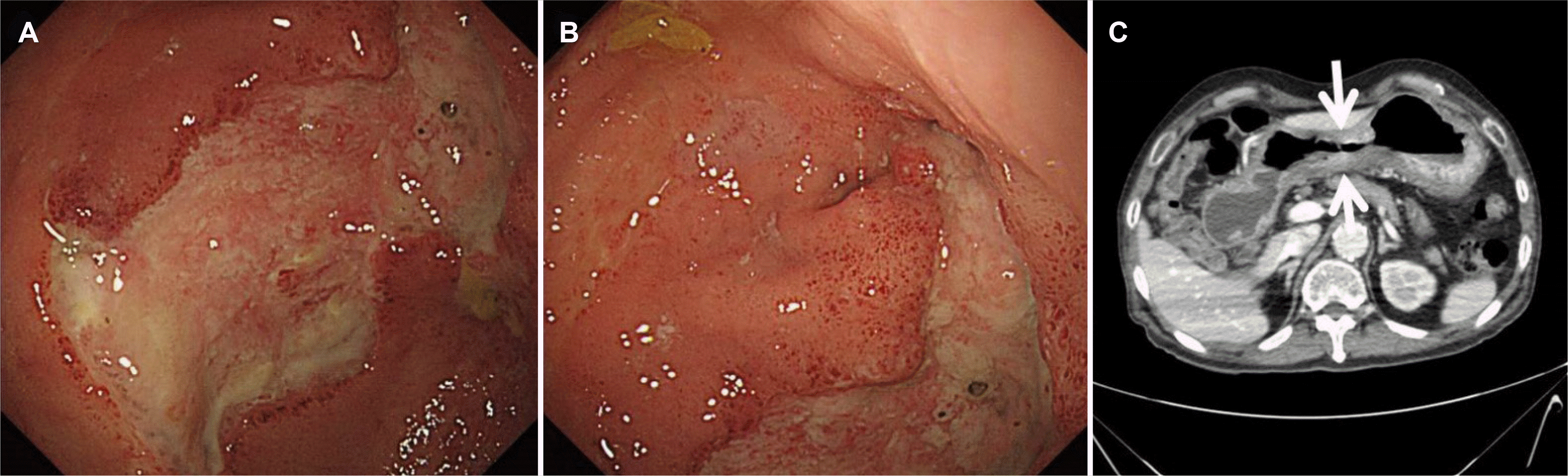
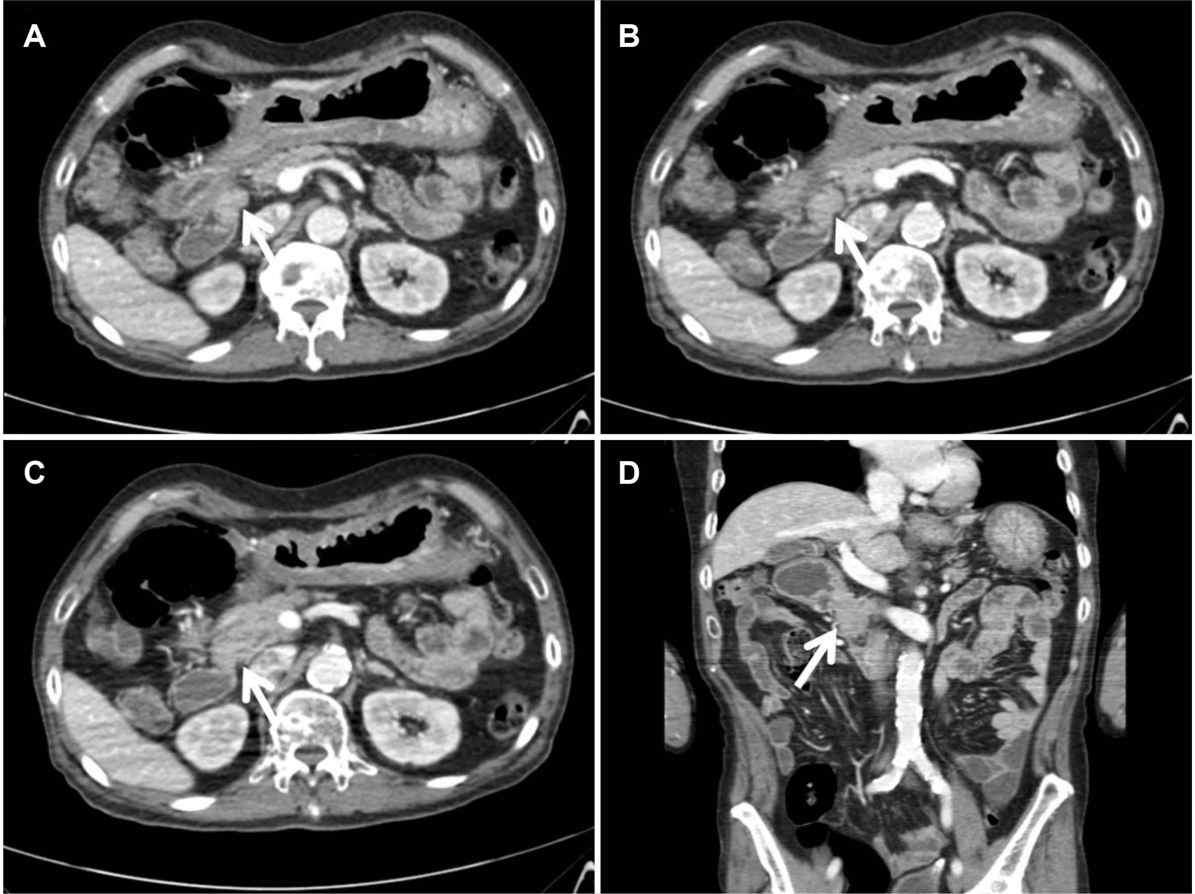
A 61-year-old man presented to the emergency department with melena that started 3 days previously. He was diagnosed with a gastric ulcer one year earlier but had no other medical history. He had been experiencing epigastric pain, postprandial fullness, and dyspepsia for several years. He appeared dehydrated with conjunctival pallor suggestive of anemia. A physical examination revealed minimal tenderness in the right lower abdomen. He had a blood pressure of 124/74 mmHg, a heart rate of 91 beats/min, respiration rate of 21 breaths/min, and a body temperature of 37℃. His abdominal radiography, chest radiography, and electrocardiography findings were normal. A laboratory workup revealed that his white blood cell count was 19,400/mm3, hemoglobin 9.8 g/dL, AST 49 U/L, ALT 25 U/L, total bilirubin 0.67 mg/dL, amylase 57 U/L, lipase 84 U/L, BUN 146 mg/dL, creatinine 6.07 mg/dL, and sodium 124 mmol/L. Upper endoscopy revealed pyloric stenosis and an active-stage ulcer in the antrum, which was suspected to be Bormann type III advanced gastric cancer (Fig. 1A, B). Microscopically, specimens collected from the antrum revealed a chronic ulcer. Abdominal contrast-enhanced CT showed diffuse stomach wall thickening, suggestive of gastric cancer (Fig. 1C). In addition, there was an abrupt narrowing of the duodenal bulb with a 1.9-cm lobular enhancing lesion, indicative of annular pancreas (Fig. 2).
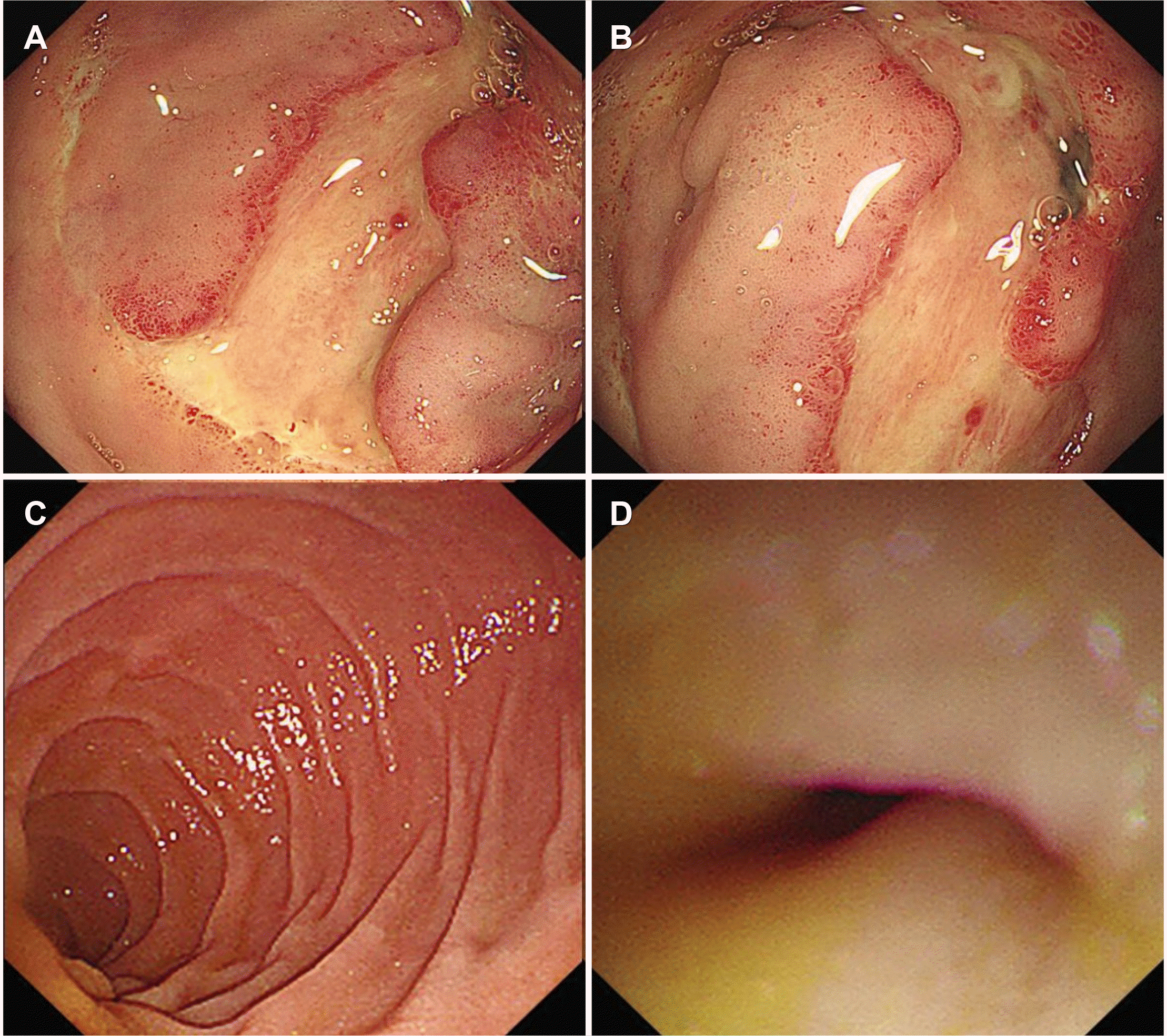
He was conservatively treated with high-dose intravenous pantoprazole, fluid therapy, and empirical antibiotics. Seven days later, a repeat upper endoscopy showed an improvement in the gastric ulcer, although the pyloric stenosis persisted (Fig. 3A, B). A pediatric endoscope was then passed through the pyloric ring, and it revealed duodenal bulb stenosis (Fig. 3C, D). Biopsy samples were taken from the ulcer in the antrum, and the pathologic diagnosis remained the same as in the previous biopsy. He was prescribed a soft diet and an oral proton pump inhibitor. He commenced a regular diet. Although he did not complain of any specific discomfort and did not experience upper gastrointestinal bleeding following conservative care, we recommended surgery because of the duodenal bulb obstruction. He, however, declined surgery and wanted to continue conservative care. He was therefore discharged and we decided to observe the patient’s progress in the out-patient clinic.
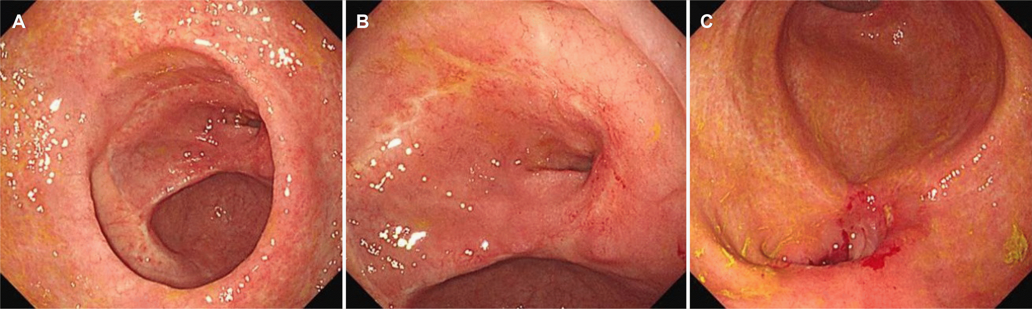
Five days after discharge, he returned to the emergency department with a 1-day history of melena and abdominal pain. Physical examination revealed right upper abdominal tenderness but the vital signs were stable. Blood tests showed a hemoglobin level of 8.2 g/dL. The other laboratory findings were within the normal range. Endoscopy demonstrated an ulcer in the antrum and a large amount of food residue in the stomach. We decided to proceed with a surgical procedure to resolve the complication. Gastrojejunostomy with jejunojejunostomy was performed without any postoperative complications. During surgery, an annular pancreas encircling the duodenal bulb was found. Two weeks after surgery, the patient was able to tolerate food and was discharged. Six months after surgery, the patient underwent upper endoscopy in an outpatient clinic which revealed ulcer scars (Fig. 4A, B) and an anastomosis site (Fig. 4C) in the antrum. Additionally, biopsy samples were taken from the ulcer scars in the antrum to exclude the possibility of gastric cancer and a diagnosis of chronic gastritis was made.
DISCUSSION
Annular pancreas is a rare embryologic anomaly, which affects approximately one in 20,000 newborns.5 This congenital anomaly was first reported by Tiedemann in 1818 and was later named ‘annular pancreas’ by Ecker in 1862.1 The reported incidence in adults is 0.050-0.015%,5 but the exact prevalence is unknown as the majority of patients with an annular pancreas have no symptoms.6 Several theories have been proposed to explain the development of annular pancreas. Lecco’s theory states that the left ventral bud degenerates and the end of the right ventral bud adheres to the duodenal wall before its rotation, thereby resulting in the pancreatic obstructive ring,7 whereas Baldwin postulated that the persistence and development of the left ventral bud is the cause of the annular formation of the pancreatic tissue surrounding the duodenum.8 However, the pathogenesis of annular pancreas in adults remains unclear.
The clinical presentation of annular pancreas is variable, and it may be asymptomatic or symptomatic.2 About two-thirds of the patients develop no symptoms in their lifetime.6 Infants and children commonly present with symptoms related to duodenal obstruction, such as vomiting, abdominal distension, and food intolerance.4 Most of the symptomatic adults aged 20-60 years show symptoms of upper gastrointestinal tract obstruction, such as epigastric pain, nausea and vomiting, postprandial fullness, and early satiety.9 The associated complications in adult patients include gastric outlet obstruction, peptic ulcer disease with upper gastrointestinal hemorrhage, acute or chronic pancreatitis, pancreatic head carcinoma, and biliary obstruction.9 In the present case, the patient could tolerate the discomfort for a long time because the partial duodenal obstruction allowed the passage of food through his stomach and duodenum. However, when he developed severe upper gastrointestinal symptoms and melena, he visited the hospital and was diagnosed with a gastric ulcer due to annular pancreas.
Cases of annular pancreas manifesting with upper gastrointestinal bleeding are rarely reported. In a case similar to that in our report, a 12-year-old boy diagnosed with annular pancreas had severe duodenal obstruction, developed a duodenal ulcer, and presented with a sudden onset of hematemesis.10 Our patient presented with melena due to a gastric ulcer caused by chronic partial duodenal obstruction. The pattern of upper gastrointestinal bleeding can differ based on the degree of duodenal obstruction. Further, as most patients visit the hospital with symptoms such as abdominal pain and vomiting before presenting with peptic ulcer bleeding, the reported incidence of upper gastrointestinal bleeding has been rare in patients with annular pancreas.
Peptic ulcer disease is reported in 25% of adults with annular pancreas.1 It is thought to be caused by a gastric outlet obstruction, which results in gastric stasis, leading to gastric acid hypersecretion and reduced access to alkaline secretions.11 In our case, the patient was diagnosed with annular pancreas surrounding the first part of the duodenum, and endoscopy showed an active-stage gastric ulcer and stricture of the pylorus. As a gastric ulcer is difficult to distinguish from advanced gastric cancer, biopsy samples were taken from the ulcer in the antrum before and after surgery. Consequently, the lesion in the antrum was confirmed to be a chronic ulcer and not gastric cancer.
Although surgery is the gold standard for diagnosing annular pancreas,12 some diagnostic modalities can enable the preoperative diagnosis of annular pancreas.13 Ultrasound, simple abdominal radiography, and upper gastrointestinal series are used to detect duodenal obstruction.14,15 Abdominal contrast- enhanced CT and MRI can reveal the pancreatic head encircling the descending part of the duodenum.16 ERCP, MRCP, and EUS can also provide an accurate preoperative evaluation of the pancreatic tissue surrounding the duodenum, along with the bile and pancreatic ducts.17,18 Endoscopy confirms gastric outlet obstruction and excludes malignancy, which is a common cause of pyloric obstruction.19
Surgery is required in a symptomatic patient with an annular pancreas. Bypass surgeries such as duodeno-duodenostomy, duodenojejunostomy, and gastrojejunostomy are preferred.20 Removing the annular ring is not recommended because complications such as pancreatic fistula, duodenal fistula, pancreatitis, and pancreatic failure may occur.20 In our case, after bypass surgery, the patient’s discomfort was relieved, and the gastric emptying time was decreased.
In conclusion, annular pancreas is rarely diagnosed in adults and is a rare cause of upper gastrointestinal bleeding, but it should be considered when evaluating a patient with a combination of peptic ulcer and duodenal stricture. Furthermore, as annular pancreas may be associated with periampullary malignancies, it is crucial to obtain a thorough medical history in addition to performing a complete physical examination so as not to miss the diagnosis of annular pancreas.
 XML Download