Journal List > Cancer Res Treat > v.54(3) > 1516078526

Yoo, Yu, Cho, Han, Oh, Lim, Nam, Lee, Sung, and Shin: Chronological Analysis of Acute Hematological Outcomes after Proton and Photon Beam Craniospinal Irradiation in Pediatric Brain Tumors

Abstract

Purpose

This study aimed to compare the early hematological dynamics and acute toxicities between proton beam craniospinal irradiation (PrCSI) and photon beam craniospinal irradiation (PhCSI) for pediatric brain tumors.

Materials and Methods

We retrospectively reviewed patients with pediatric brain tumors who received craniospinal irradiation (CSI). The average change in hemoglobin levels (ΔHbavg), absolute lymphocyte counts (ΔALCavg), and platelet counts (ΔPLTavg) from baseline values was evaluated and compared between the PrCSI and PhCSI groups at 1 and 2 weeks after the initiation of CSI, 1 week before and at the end of radiotherapy, and 3–4 weeks after the completion of radiotherapy using t-test and mixed-model analysis.

Results

The PrCSI and PhCSI groups consisted of 36 and 30 patients, respectively. There were no significant differences in ΔHbavg between the two groups at any timepoint. However, ΔALCavg and ΔPLTavg were significantly lower in the PhCSI group than in PrCSI group at every timepoint, demonstrating that PrCSI resulted in a significantly lower rate of decline and better recovery of absolute lymphocyte and platelet counts. The rate of grade 3 acute anemia was significantly lower in the PrCSI group than in in the PhCSI group.

Conclusion

PrCSI showed a lower rate of decline and better recovery of absolute lymphocyte and platelet counts than PhCSI in the CSI for pediatric brain tumors. Grade 3 acute anemia was significantly less frequent in the PrCSI group than in the PhCSI group. Further large-scale studies are warranted to confirm these results.

Introduction

Craniospinal irradiation (CSI) is the standard treatment for pediatric brain tumors prone to leptomeningeal spread [1]. Owing to the large field size, a significant volume of normal organs is included in the irradiated area [2]. Therefore, various side effects, including hematological and gastrointestinal toxicities, are observed during and after CSI [37]. In an effort to reduce the potential toxicities related to CSI, advanced techniques have been adopted. Among these, proton beam therapy (PBT) has received attention due to its dosimetric advantage [8,9]. In contrast to X-ray therapy, in which the dose deposition gradually decreases along the beam path as the beam passes over the target volume, PBT involves a sharp rise and fall in energy deposition, known as the Bragg peak, that stops at the end of the finite beam range [10]. Therefore, there is no exit dose, and fewer acute and late toxicities are expected compared to X-ray therapy.
Despite the physical advantages of PBT, studies comparing the clinical benefits of CSI with PBT (PrCSI) to those of CSI with photon beams (PhCSI), particularly with respect to the reduction of toxicities, are very limited [2,9]. While some studies have reported data showing a potential reduction in acute and late toxicities, the superiority of PrCSI over PhCSI for the treatment of pediatric brain tumors has not been well elucidated at the clinical level [11]. A limited number of studies that have retrospectively compared acute hematological and gastrointestinal toxicities between PrCSI and PhCSI [2,9]. However, none of these studies have performed a detailed analysis of the dynamics of various hematological profiles over time, which reflect patterns in the reduction and recovery of blood cell levels. Furthermore, toxicity profiles other than those for gastrointestinal toxicities have not been compared. Therefore, we performed a study to compare the early hematological dynamics and various toxicity profiles between PrCSI and PhCSI for pediatric brain tumors.

Materials and Methods

1. Study population

We retrospectively reviewed patients with pediatric brain tumors who received CSI between January 2010 and June 2019. The inclusion criteria were as follows: age at CSI of 20 years or less, no history of radiation therapy (RT), no concurrent chemotherapy, and no subsequent chemotherapy within 3 weeks after the completion of RT. Of the 115 patients who were screened, 49 were excluded due to a previous history of RT (n=22), CSI with a combination of proton and photon beams (n=5), chemotherapy within 3 weeks of completion of RT (n=20), lack of hematological data (n=1), and the interruption of CSI for more than 1 week (n=1). Finally, 66 patients were included in the analysis.

2. Treatment

Initially only PhCSI was performed. The facility and protocol for PrCSI were established in March 2016. After the initiation of PrCSI, CSI was mainly performed with PBT. The fraction size, prescribed dose of CSI, and boost RT to the high-risk area were determined according to the histological diagnosis or clinical situation. The boost RT was followed after the completion of CSI in all patients.
The simulations for PhCSI (n=30) were performed using nonenhanced computed tomography (CT) scans with a slice thickness of 5 mm for 3-dimensional conformal RT (3DCRT; n=29) and 2.5 mm for helical tomotherapy (HT; n=1). Prone (n=18) or supine positions (n=12) were adapted according to the patients’ performance, ability to cooperate, or the need for sedation, in which the prone position was inappropriate. Chloral hydrate was administered orally for sedation, if needed (n=4). The clinical target volume (CTV) for CSI included the entire cranial and spinal meninges with an inferior limit of S2 or S3 level according to the T2 sequence of whole spine magnetic resonance images. The planning target volume (PTV) was expanded from the CTV by 0.5–0.7 cm. For 3DCRT, bilateral fields corresponding to the cranial and cervical spinal meninges were applied to the PTV, and the PTV of the spinal level below the cervical spine was covered by posteroanterior (PA) field(s). If the range of the PA field was insufficient to cover the rest of the PTV, two or more PA fields were applied. Margins of the fields were adjusted between bilateral fields and the PA field or two adjacent PA fields considering the beam divergence. The field junctions were moved daily alternatively with a gap of approximately 1 cm to attenuate the uncertainty. For treatment planning for 3DCRT, we used Pinnacle (Phillips Healthcare, Andover, MA). A linear accelerator generating 6 MV X-ray (Varian Clinac 6Ex, Varian Medical Systems, Palo Alto, CA) was used for beam delivery. For verification of the patient setup, a weekly portal image was obtained using an electronic portal imaging device. The junction moving technique was not required for PhCSI with HT. Megavoltage CT was performed for setup verification before every session of CSI. The treatment plan for HT was developed by TomoTherapy (Accuray, Sunnyvale, CA).
PrCSI (n=36) has been adopted since March 2016. Simulations for PrCSI, consisting of nonenhanced CT scans with a slice thickness of 2.5 mm, were also used. The delineations of the CTV were performed in the same manner as those for PhCSI and PTV was generated with expansion of CTV by 0.5–0.7 cm, too. However, for patients in the growth age, the PTV was additionally extended to encompass the entire vertebral body when PrCSI was applied (n=24) to prevent radiation-induced kypholordosis according to the guideline from the European Society for Paediatric Oncology [12]. We used the PBT with the continuous line scanning method, which has been described in detail in a previous report [13]. Line scanning proceeded in the lateral direction, moved to the next line at the lateral edge, and proceeded again in the opposite direction. Each layer was painted once without any repainting. To achieve optimal dosimetry, line spacing, spot size, and spot speed were modulated. The depth of the layers could be adjusted by 0.1 g/cm2 and the time interval for beam energy change was 2 seconds or less. Similar to PhCSI, two bilateral fields and PA field(s) were applied for PrCSI with two or three isocenters. We developed the dose gradients near the junctions and overlapped them with adjacent fields to overcome the junctional problem and maintain robustness against uncertainties. The treatment plan was developed by RayStation (RaySearch Laboratories, Stockholm, Sweden). In the treatment room, daily image guidance was performed before the sessions with orthogonal kilovoltage X-ray images. The first three consecutive patients were treated in the prone position due to dosimetric uncertainty in the region of the couch. However, the supine position was chosen after this uncertainty was resolved. For patients who were unable to cooperate with the treatment due to performance status or age, total intravenous anesthesia with 1% propofol was administered during the simulations and treatments (n=13). S1 and S2 Figs. present examples of PhCSI and PrCSI plans.

3. Evaluation of acute toxicities

The change in serum hemoglobin levels (ΔHb), absolute lymphocyte counts (ΔALC), and platelet counts (ΔPLT) from baseline values was evaluated 1 and 2 weeks after the initiation of CSI (T1 and T2, respectively), 1 week before and at the end of local boost RT following CSI (T3 and T4, respectively), and 3–4 weeks after the completion of total RT (T5). Each timeline was allowed the time variation up to 3 days. To adjust for the effect of transfusion, we assumed that transfusion of 1 U increased the hemoglobin level by 1 g/mL and the platelet count by 12,000 cells/μL, according to previous literature [2]. The laboratory methodology for measurement of blood count was described in a previous study [14]. We used a hematology analyzer (XN-9000, Sysmex, Kobe, Japan) for electronic cell counting. The normal ranges for hemoglobin level, absolute lymphocyte count, and platelet count in our institute are summarized in S3 Table. Hematological and other toxicities were graded according to the Common Terminology Criteria for Adverse Events ver. 5.0.

4. Statistical analysis

Categorical variables were compared between the PrCSI and PhCSI groups using Pearson’s chi-square test or Fisher exact test. For the comparison of continuous variables at baseline, an unpaired t-test was used. Paired t-tests and mixed-model analyses were used to compare the changes in hematological variables over time. The estimation and comparison of survival curves was performed using the Kaplan-Meier method and log-rank test, respectively. Hematological variables were also analyzed for subgroups stratified according to the CSI dose. A two-sided p-value < 0.05 was considered statistically significant. For statistical analyses, SPSS ver. 22.0 (IBM Corp., Armonk, NY) was used.

Results

The comparison of patient characteristics between the PrCSI and PhCSI groups is summarized in Table 1. The proportions of male patients (p=0.044), median body mass index (p=0.012), patients who received a CSI dose < 23.4 Gy or GyRBE (p=0.008), and patients who received a fraction size of 1.5 Gy (p < 0.001) were significantly higher in the PhCSI group than in the PrCSI group (Table 1). The median number of total RT fractionations (p=0.005) and total dose prescribed to the brain (p=0.002) were significantly higher in the PrCSI group than in the PhCSI group (Table 1). However, there were no significant differences in the median age; and median hemoglobin levels, absolute lymphocyte counts, or platelet counts at baseline between the two groups. The median follow-up duration was 38 months (range, 1 to 114 months). While more patients with medulloblastoma were included in PrCSI group, proportion of germinoma is higher in PhCSI group despite of the statistical insignificance (p=0.094). The survival curves and causes of death are shown in S4 Fig. The accumulated radiation doses at each timeline were summarized in S5 Table and compared in S6 Table showing that there were no significant differences in the median accumulated radiation dose from T1 to T4 but that was significantly higher in PrCSI group at T5 (p=0.002) (S6 Table).
There were no significant differences in the average ΔHb levels (ΔHbavg) at any timepoint from T1 to T5 between the two groups. However, the average ΔALC (ΔALCavg) and ΔPLT (ΔPLTavg) were significantly lower in the PhCSI group than in PrCSI group the at every timepoint from T1 to T5 (Fig. 1). The changes in ΔHbavg over time showed irregular patterns or minimal changes in both groups. However, ΔALCavg declined through T1 and T2 and then increased from T3 to T5 in both groups (Fig. 1). ΔPLTavg also declined from T1 to T2 in both groups. However, in the PrCSI group, it increased from T3 to T5, and in the PhCSI group, it increased from T4 to T5 (Fig. 1). Mixed-model analyses revealed that the beam modalities did not significantly differentiate the chronological trends of ΔHb (p=0.538) and ΔALC (p=0.930), but it significantly differentiated the chronological trends of ΔPLT between groups (p=0.042). In addition, only ΔALC (p < 0.001) and ΔPLT (p=0.002) were significantly higher in the PrCSI group when the variable of time was fixed.
The subgroups were stratified according to the cutoff CSI dose of 23.4 Gy or GyRBE, which was the median CSI dose. Fig. 2 shows the ΔHb, ΔALC, and ΔPLT of each subgroup according to the CSI modality. In the lower dose subgroup (≤ 23.4 Gy or GyRBE), ΔALCavg and ΔPLTavg were significantly higher in the PrCSI group through the timelines, except for ΔALCavg at T4. In this subgroup, ΔALCavg decreased through T1 and T2 and increased from T3 to T5 in both groups. ΔPLTavg also decreased from T1 to T2 in both groups, with rebounding occurring at T3 and T4 in the PrCSI and PhCSI groups, respectively, which was similar to the trends observed in the total cohort (Fig. 2). Mixed-model analyses revealed that the trends over time for ΔHb (p=0.580), ΔALC (p=0.768), and ΔPLT (p=0.635) were not significantly different between the groups. Only ΔALC (p=0.018) and ΔPLT (p=0.007) were significantly different between the groups if the time variable was fixed. In the higher CSI dose subgroup (> 23.4 Gy or GyRBE), the ΔHbavg, ΔALCavg, and ΔPLTavg patterns were conserved and resembled those of the total cohort and the lower CSI dose subgroup (Fig. 2). However, a significant difference between the beam modalities was observed only in ΔPLTavg at T3 and T4 (Fig. 2). Mixed-model analyses showed that the trends over time were significantly different between the groups only for ΔPLTavg (p=0.015).
In the PrCSI group, the hematological outcomes were compared according to whether the PTV encompassed the entire vertebral body (large PTV), or not (small PTV). The hematological outcomes were inconsistent showing the tendency of higher ΔHbavg and average ΔPLTavg in small PTV group but higher average ΔALCavg in large PTV group especially in T4 and T5 (S7 Fig.).
The acute toxicity profiles are summarized in Table 2. The incidence of acute toxicities was higher in the PhCSI group than in the PrCSI group, although only grade 3 anemia showed a statistically significant difference.

Discussion

CSI is an essential treatment for pediatric brain tumors that tend to spread to the leptomeningeal space [1]. However, the volume irradiated by CSI is very large and a range of toxicities, including hematological and gastrointestinal complications, are observed during and after CSI [37]. Due to the absence of an exit dose for CSI compared with photon beams, it has been expected that PBT would reduce these toxicities [3,9]. However, limited relevant studies have been performed [2,9]. In particular, a comparison of the hematological dynamics has not been well investigated.
Our study showed that PrCSI resulted in a significantly lower rate of decline and better recovery of absolute lymphocyte counts and platelet counts than PhCSI. The platelet count recovered earlier in the PrCSI group than in the PhCSI group. The significance of the superiority of PrCSI with respect to ΔALCavg and ΔPLTavg was maintained when the time variable was fixed. The trend was also conserved in both the CSI dose subgroups. The PrCSI group showed significantly lower median body mass index and higher the median fractionation number which could associated with mitigation in the recovery of blood cell level. Furthermore, the proportion of medulloblastoma was higher in the PrCSI group while that of germinoma was higher in PhCSI group, leading to higher potential for performance of more intensive chemotherapy in PrCSI [15,16]. Considering these patients’ characteristics, our results could favor the PrCSI in terms of the early hematological outcome. While ΔALCavg was decreased though T1 and T2, it seemed to rebound from T3. Considering the median number of CSI fractionations was 13 (range, 10 to 17) which was apparently included in the window of T2, T2 could be regarded as the timeline for the end of CSI. In this point of view, recovery of ΔALCavg in both groups occurred at T3, a few weeks later after the end of CSI even during the local boost RT. The recovery of ΔALCavg seem to be earlier comparing with other studies reporting that the recovery of lymphocyte level occurred several weeks later after the complete of RT [17,18]. However, populations of those studies received concurrent chemotherapy during the RT which was the exclusion criteria of the study. In addition, the boost RT in our study was focused on the limited volume in the brain or, in some patients, spinal cord where the boost irradiation could not affect the significantly in the lymphocyte generation. The causes of the earlier ΔALCavg recovery are unclear and need further investigation.
The hematological and gastrointestinal toxicities between PrCSI and PhCSI have previously been compared in patients with pediatric brain tumors [2]. This study reported that the recovery rates of absolute leukocyte and platelet counts were significantly higher in patients who underwent PrCSI than in those who underwent PhCSI. However, this study evaluated only the recovery rates by measurements of variables only at baseline and 1 month after RT, and did not compare the dynamics and their trends of blood cell count both during and after RT which were the main finding of ours. Furthermore, that study did not include the analysis for the dynamics of absolute leukocyte counts which were also an important finding in the present study. Another study compared acute toxicities between PrCSI and PhCSI in adult patients with medulloblastoma and reported that reduction of peripheral white blood cells, hemoglobin levels, and platelet counts was significantly lower with PrCSI than with PhCSI [9]. However, this study only evaluated the nadir of the hematological parameters and a comparison of dynamics over time was lacking. To our knowledge, our study is the first to evaluate the dynamics of various blood counts over time with the tendency analyses.
As various dosimetric studies have demonstrated, PrCSI is beneficial in protecting organs at risk from even low-dose irradiation due to the absence of an exit dose [8,10]. Above all, PrCSI can spare the bone marrow of the vertebral body in the spinal column [9]. The activity of bone marrow in the whole spine is estimated to be approximately 40% of the total bone marrow activity [19]. Therefore, by sparing the bone marrow within the vertebral bodies, PrCSI can potentially prevent bone marrow suppression by irradiation. This is supported by the study by Brown et al. [9], which reported that in adult patients, reductions in blood count after CSI were associated with the mean vertebral dose, which tended to be lower with PrCSI. However, in our study, the effect of bone marrow sparing on the improved hematological outcomes observed with PrCSI could not be determined because whole vertebral bodies were included in the PTV in 66.7% patients in the PrCSI group due to concerns of radiation-induced kypholodorsis resulting from asymmetric growth in the vertebral body. In addition, the comparison of hematological dynamics between the large and small PTV groups in PrCSI subgroup was performed showing that the results were inconsistent according to the blood cell types (S7 Fig.). This inconsistency might be resulted from the demographic discrepancy in the large and small PTV group because large PTVs were applied only in the patients with growth age whose ability of hematopoiesis or diagnoses would be different from the small PTV group. Therefore, there is limitation in the present result to confirm the bone marrow sparing effect in the improvement of hematological outcomes. Various studies have reported that partial spinal RT showed more frequent and more severe abnormalities after higher dose irradiation and longer follow-up duration in pediatric cancer patients [20]. In particular, the steep gradients in vertebral body dose can potentially increase the incidence of spinal deformities [21]. Therefore, when the strategy of irradiating the whole vertebral body to mitigate radiation-induced kypholodorsis is employed, the effect of bone marrow suppression could be diluted [22]. However, researchers from Loma Linda University investigated the long-term effects of vertebral body-sparing PrCSI on the spine of young patients with medulloblastoma and reported that although a decrease in the growth of the posterior portions of vertebral bodies was observed, it was compensated by hypertrophy of posterior intervertebral discs, leading to the absence of increased severe spinal abnormalities [22]. If a strategy for vertebral body-sparing PrCSI is available, the effect of PrCSI on the preservation of bone marrow function would be enhanced. Despite the indeterminate effect on bone marrow sparing, PrCSI showed better hematological outcomes. Therefore, factors other than bone marrow sparing should be considered in the interpretation of the results. During PhCSI, irradiation to the heart, liver, gastrointestinal organs, great vessels, and whole vertebral body is not avoidable. Most of the abovementioned organs are organs of hematopoiesis or circulation. Therefore, the protection of these organs even from low-dose irradiation is essential for the preservation of blood cells that are highly sensitive to radiation [23].
Approximately 85% lymphocytes exist outside the bone marrow and migrate via the circulatory system between the spleen, lymph nodes, and non-lymphoid tissues such as the liver, gastrointestinal tract, and other tissues. Therefore, the various organs in which lymphocytes stay or circulate need to be considered for the preservation of lymphocytes. The significance of the radiation dose to circulating lymphocytes has been reported recently. The integral dose to large blood vessels, the heart, and lymphoid or non-lymphoid organs, including the lungs or liver, is associated with the reduction in the number of lymphocytes and the subsequent development of lymphopenia [2426]. Furthermore, because lymphocytes are the effector of immune response against the tumor cells, lymphopenia is expected to be associated with poor outcomes, such as those reported in various types of malignancies [18,27,28]. Therefore, PrCSI is superior to PhCSI in sparing blood cells outside of the bone marrow as well as those within the bone marrow, and it can be hypothesized that PrCSI will show better hematological and oncologic outcomes than PhCSI in patients with pediatric brain tumors. Further long-term follow-up studies with a large population are required to confirm this hypothesis.
The reduction of platelet counts can also be related to organs other than the bone marrow. The action of the humoral regulator thrombopoietin is essential for thrombopoiesis. Thrombopoietin is primarily produced in the liver and kidneys [18]. In previous studies, patients with poor liver function or thrombocytopenia showed reduced levels of thrombopoietin [29]. PhCSI results in irradiation to certain portions of the liver parenchyma, mainly the lateral segments. Therefore, in contrast to PrCSI, PhCSI may induce radiation-induced damage to the liver, which could lead to reduced thrombopoietin production, thereby resulting in a decrease in the level of thrombopoietin. However, no study has investigated the relationship between dosimetric parameters in the liver and thrombopoietin production or platelet counts. Additional quantitative studies are necessary to investigate the severity of thrombocytopenia according to the dosimetric differences between PrCSI and PhCSI.
While the ΔHbavg values were not significantly different at any timepoint between the modalities, the rates of grade 3 anemia, transfusions, and granulocyte colony-stimulating factor administration were lower in the PrCSI group than in the PhCSI group, although the difference was statistically significant only for grade 3 anemia. In addition, gastrointestinal toxicities were less frequent in the PrCSI group than in the PhCSI group, although the difference was not statistically significant. Due to the small sample size, the statistical power was insufficient. However, the tendency of PrCSI to be associated with fewer incidences of acute toxicity is consistent with the dosimetric advantages of PrCSI compared with PhCSI as well as with previous findings. A large-scale study is required with a sufficient statistical power to clearly confirm the study results.
There are several limitations in this study. This is a retrospective study with a small sample size from a single institution; therefore, selection bias was inevitable. Due to the small sample size, the statistical power was insufficient. Especially, in the subgroup analysis, the sample size of higher dose subgroup quite smaller under 10 in both PrCSI and PhCSI groups. Therefore, to generalize the result showing the trend of the better acute hematological outcome in PrCSI group regardless of the CSI doses, the samples with larger size would be required to generalize the result. Diversity in the types of primary tumor is also a limitation of the present study because differences in the chemotherapeutic regimens administered before CSI may have affected the activity of subsequent hematopoiesis.
In conclusion, PrCSI was associated with a significantly lower rate of decline and better recovery of absolute lymphocyte counts and platelet counts compared with PhCSI in patients with pediatric brain tumors, regardless of the CSI dose. Acute grade 3 anemia was also significantly less frequent in the PrCSI group than in PhCSI patients. Further large-scale clinical studies are warranted to confirm the study results.

Notes

Ethical Statement

This study was approved by the institutional review board (SMC-2020-10-145). The waiver of informed consent was approved by the institutional review board.

Author Contributions

Conceived and designed the analysis: Yu JI, Lim DH.

Collected the data: Yoo GS, Cho S, Han Y, Oh Y, Lim DH, Lee JW, Sung KW, Shin HJ.

Contributed data or analysis tools: Yoo GS.

Performed the analysis: Yoo GS.

Wrote the paper: Yoo GS, Lim DH.

Critical review: Yu JI, Cho S, Han Y, Lim DH, Nam HR, Lee JW, Sung KW, Shin HJ.

Supervision: Lim DH.

Conflicts of Interest

Conflict of interest relevant to this article was not reported.

ACKNOWLEDGMENTS

This work was supported by a grant from the Creative Research Program (SMO1210181) funded by Samsung Medical Center, Sungkyunkwan University School of Medicine.

References

1. DeNunzio NJ, Yock TI. Modern radiotherapy for pediatric brain tumors. Cancers (Basel). 2020; 12:1533.
crossref
2. Song S, Park HJ, Yoon JH, Kim DW, Park J, Shin D, et al. Proton beam therapy reduces the incidence of acute haematological and gastrointestinal toxicities associated with craniospinal irradiation in pediatric brain tumors. Acta Oncol. 2014; 53:1158–64.
crossref
3. Seravalli E, Bosman M, Lassen-Ramshad Y, Vestergaard A, Oldenburger F, Visser J, et al. Dosimetric comparison of five different techniques for craniospinal irradiation across 15 European centers: analysis on behalf of the SIOP-E-BTG (radiotherapy working group). Acta Oncol. 2018; 57:1240–9.
crossref
4. Cox MC, Kusters JM, Gidding CE, Schieving JH, van Lindert EJ, Kaanders JH, et al. Acute toxicity profile of craniospinal irradiation with intensity-modulated radiation therapy in children with medulloblastoma: a prospective analysis. Radiat Oncol. 2015; 10:241.
crossref
5. Chang EL, Allen P, Wu C, Ater J, Kuttesch J, Maor MH. Acute toxicity and treatment interruption related to electron and photon craniospinal irradiation in pediatric patients treated at the University of Texas M. D. Anderson Cancer Center. Int J Radiat Oncol Biol Phys. 2002; 52:1008–16.
crossref
6. Song KJ, Park JH, Im HJ, Ahn SD. Survival and long-term toxicities of pediatric Hodgkin lymphoma after combined modality treatment: a single institute experience. Radiat Oncol J. 2020; 38:198–206.
crossref
7. Lee J, Chung SY, Han JW, Kim DS, Kim J, Moon JY, et al. Treatment outcome of anaplastic ependymoma under the age of 3 treated by intensity-modulated radiotherapy. Radiat Oncol J. 2020; 38:26–34.
crossref
8. Barney CL, Brown AP, Grosshans DR, McAleer MF, de Groot JF, Puduvalli V, et al. Technique, outcomes, and acute toxicities in adults treated with proton beam craniospinal irradiation. Neuro Oncol. 2014; 16:303–9.
crossref
9. Brown AP, Barney CL, Grosshans DR, McAleer MF, de Groot JF, Puduvalli VK, et al. Proton beam craniospinal irradiation reduces acute toxicity for adults with medulloblastoma. Int J Radiat Oncol Biol Phys. 2013; 86:277–84.
crossref
10. Yoon M, Shin DH, Kim J, Kim JW, Kim DW, Park SY, et al. Craniospinal irradiation techniques: a dosimetric comparison of proton beams with standard and advanced photon radiotherapy. Int J Radiat Oncol Biol Phys. 2011; 81:637–46.
crossref
11. Miralbell R, Lomax A, Cella L, Schneider U. Potential reduction of the incidence of radiation-induced second cancers by using proton beams in the treatment of pediatric tumors. Int J Radiat Oncol Biol Phys. 2002; 54:824–9.
crossref
12. Ajithkumar T, Horan G, Padovani L, Thorp N, Timmermann B, Alapetite C, et al. SIOPE - Brain tumor group consensus guideline on craniospinal target volume delineation for high-precision radiotherapy. Radiother Oncol. 2018; 128:192–7.
crossref
13. Yoo GS, Yu JI, Cho S, Jung SH, Han Y, Park S, et al. Comparison of clinical outcomes between passive scattering versus pencil-beam scanning proton beam therapy for hepatocellular carcinoma. Radiother Oncol. 2020; 146:187–93.
crossref
14. Yu JI, Park HC, Yoo GS, Paik SW, Choi MS, Kim HS, et al. Clinical significance of systemic inflammation markers in newly diagnosed, previously untreated hepatocellular carcinoma. Cancers (Basel). 2020; 12:1300.
crossref
15. Martin AM, Raabe E, Eberhart C, Cohen KJ. Management of pediatric and adult patients with medulloblastoma. Curr Treat Options Oncol. 2014; 15:581–94.
crossref
16. Lee JW, Lim DH, Sung KW, Cho HW, Ju HY, Yoo KH, et al. Induction chemotherapy reduces radiation therapy dose and volume in the treatment of intracranial germinoma: results of the SMC-G13 trial. Int J Radiat Oncol Biol Phys. 2020; 108:649–56.
crossref
17. Deng W, Xu C, Liu A, van Rossum PS, Deng W, Liao Z, et al. The relationship of lymphocyte recovery and prognosis of esophageal cancer patients with severe radiation-induced lymphopenia after chemoradiation therapy. Radiother Oncol. 2019; 133:9–15.
crossref
18. Liu H, Wang H, Wu J, Wang Y, Zhao L, Li G, et al. Lymphocyte nadir predicts tumor response and survival in locally advanced rectal cancer after neoadjuvant chemoradiotherapy: Immunologic relevance. Radiother Oncol. 2019; 131:52–9.
crossref
19. Hayman JA, Callahan JW, Herschtal A, Everitt S, Binns DS, Hicks RJ, et al. Distribution of proliferating bone marrow in adult cancer patients determined using FLT-PET imaging. Int J Radiat Oncol Biol Phys. 2011; 79:847–52.
crossref
20. Hoeben BA, Carrie C, Timmermann B, Mandeville HC, Gandola L, Dieckmann K, et al. Management of vertebral radiotherapy dose in paediatric patients with cancer: consensus recommendations from the SIOPE radiotherapy working group. Lancet Oncol. 2019; 20:e155–66.
crossref
21. Paulino AC, Fowler BZ. Risk factors for scoliosis in children with neuroblastoma. Int J Radiat Oncol Biol Phys. 2005; 61:865–9.
crossref
22. MacEwan I, Chou B, Moretz J, Loredo L, Bush D, Slater JD. Effects of vertebral-body-sparing proton craniospinal irradiation on the spine of young pediatric patients with medulloblastoma. Adv Radiat Oncol. 2017; 2:220–7.
crossref
23. Jin JY, Mereniuk T, Yalamanchali A, Wang W, Machtay M, Spring Kong FM, et al. A framework for modeling radiation induced lymphopenia in radiotherapy. Radiother Oncol. 2020; 144:105–13.
crossref
24. So TH, Chan SK, Chan WL, Choi H, Chiang CL, Lee V, et al. Lymphopenia and radiation dose to circulating lymphocytes with neoadjuvant chemoradiation in esophageal squamous cell carcinoma. Adv Radiat Oncol. 2020; 5:880–8.
crossref
25. Xu C, Jin JY, Zhang M, Liu A, Wang J, Mohan R, et al. The impact of the effective dose to immune cells on lymphopenia and survival of esophageal cancer after chemoradiotherapy. Radiother Oncol. 2020; 146:180–6.
crossref
26. Xie X, Lin SH, Welsh JW, Wei X, Jin H, Mohan R, et al. Radiation-induced lymphopenia during chemoradiation therapy for non-small cell lung cancer is linked with age, lung V5, and XRCC1 rs25487 genotypes in lymphocytes. Radiother Oncol. 2021; 154:187–93.
crossref
27. Lambin P, Lieverse RIY, Eckert F, Marcus D, Oberije C, van der Wiel AM, et al. Lymphocyte-sparing radiotherapy: the rationale for protecting lymphocyte-rich organs when combining radiotherapy with immunotherapy. Semin Radiat Oncol. 2020; 30:187–93.
crossref
28. Ng SP, Bahig H, Jethanandani A, Pollard C, Berends J, Sturgis EM, et al. Lymphopenia during radiotherapy in patients with oropharyngeal cancer. Radiother Oncol. 2020; 145:95–100.
crossref
29. Miller JB, Figueroa EJ, Haug RM, Shah NL. Thrombocytopenia in chronic liver disease and the role of thrombopoietin agonists. Gastroenterol Hepatol (N Y). 2019; 15:326–32.
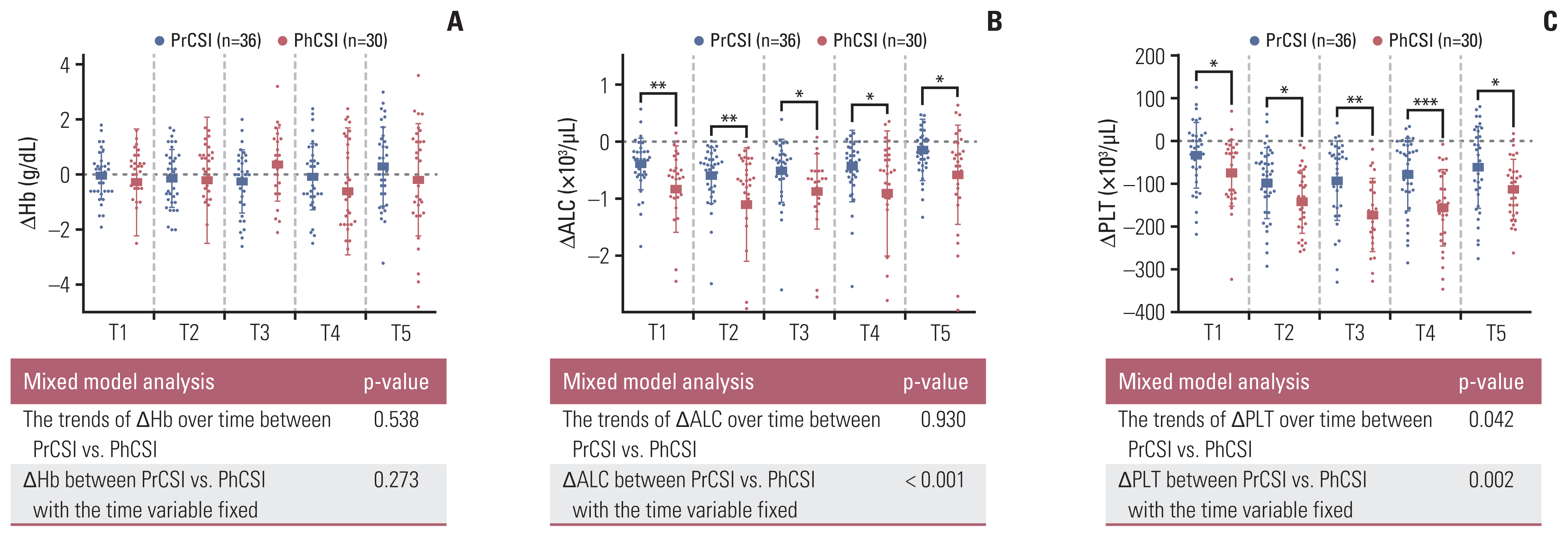
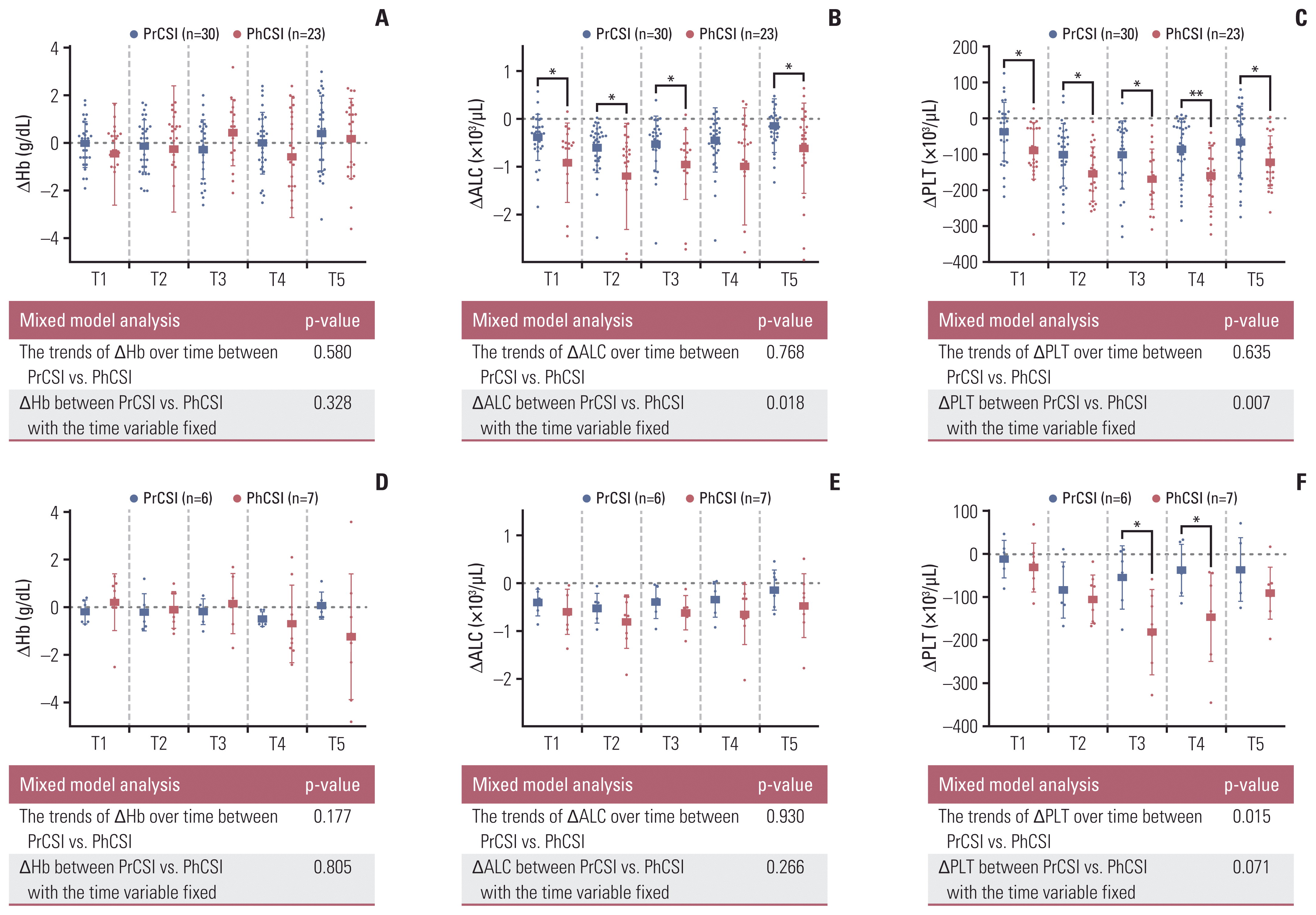
Fig. 1
Box plots and mixed analyses of the comparison of changes in hemoglobin levels (ΔHb) (A), absolute lymphocyte counts (ΔALC) (B), and platelet counts (ΔPLT) (C) from baseline values at each timepoint (*0.01 ≤ p < 0.05, **0.001 ≤ p < 0.01, ***p < 0.001). The changes in hematological variables were calculated 1 and 2 weeks after the initiation of craniospinal irradiation (T1 and T2, respectively), 1 week before the completion of radiotherapy (T3), at the end of radiotherapy (T4), and 3–4 weeks after the completion of radiotherapy (T5). PhCSI, photon beam craniospinal irradiation; PrCSI, proton beam craniospinal irradiation.
crt-2021-332f1.gif
Fig. 2
Box plots and mixed analyses of changes in hemoglobin levels (ΔHb) (A, D), absolute lymphocyte counts (ΔALC) (B, E), and platelet counts (ΔPLT) (C, F) from baseline values at each timepoint in subgroups with lower (≤ 23.5 Gy or GyRBE) (A–C) and higher doses (> 23.5 Gy or GyRBE) (D–F). The changes in hematological variables were calculated 1 and 2 weeks after the initiation of craniospinal irradiation (T1 and T2, respectively), 1 week before the completion of radiotherapy (T3), at the end of radiotherapy (T4), and 3–4 weeks after the completion of radiotherapy (T5). PhCSI, photon beam craniospinal irradiation; PrCSI, proton beam craniospinal irradiation. * 0.01 ≤ p < 0.05, **0.001 ≤ p < 0.01, ***p < 0.001.
crt-2021-332f2.gif
Table 1
Patients’ characteristics
Characteristic PrCSI (n=36) PhCSI (n=30) p-value
Age (yr) 10 (3–20) 13 (2–20) 0.536
Sex
 Male 19 (52.8) 24 (80.0) 0.044
 Female 17 (47.2) 6 (20.0)
Histology
 Medulloblastoma 20 (55.6) 10 (33.3) 0.094
 Germinoma 6 (16.7) 15 (50.0)
 NGGCT 3 (8.3) 2 (5.7)
 Mixed germ cell tumor 2 (5.6) 0
 AT/RT 3 (8.3) 1 (3.3)
 Ependymoma 0 2 (6.7)
 Others 2 (5.6) 0
ECOG performance status
 0, 1 32 (88.9) 26 (86.7) > 0.99
 2 4 (11.1) 4 (13.3)
Leptomeningeal metastasis
 No 33 (91.7) 28 (93.3) > 0.99
 Yesa) 3 (8.3) 2 (6.7)
Body mass index (kg/m2) 16.80 (12.81–24.32) 19.66 (12.29–29.18) 0.012
Baseline hemoglobin level (g/dL) 11.3 (8.8–13.8) 11.3 (9.5–14.7) 0.733
Baseline ALC level (×103/μL) 2.19 (0.61–6.13) 2.48 (0.88–7.21) 0.661
Baseline platelet level (×103/μL) 232 (50–459) 242 (107–484) 0.666
Previous chemotherapy
 No 1 (2.8) 0 > 0.99
 Yes 35 (97.2) 30 (100)
No. of chemotherapy cycles 4 (0–8) 4 (2–8) 0.195
Interval between chemotherapy to CSI start (day) 30 (21–136) 33 (20–402) 0.197
High-dose chemotherapy and auto-PBSCT before CSI
 No 29 (80.6) 24 (80.0) 0.955
 Yes 7 (19.4) 6 (20.0)
Cytopenia during the previous chemotherapyb)
 No 6 (16.7) 10 (33.3) 0.153
 Yes 30 (83.3) 20 (66.7)
CSI dose (GyRBE or Gy)
 < 23.4 7 (19.4) 12 (40.0) 0.008
 23.4–30.0 23 (63.9) 12 (40.0)
 30.6 6 (16.7) 6 (20.0)
Dose per fx of CSI (GyRBE or Gy)
 1.5 0 9 (30.0) < 0.001
 1.8 36 (100) 21 (70.0)
No. of CSI fractionations 13 (10–17) 13 (10–17) 0.336
No. of local boost RT fractionations 17 (0–20) 11 (0–17) < 0.001
No. of total RT (day) 30 (13–30) 24 (13–30) 0.005
Dose to brain (GyRBE or Gy) 54.0 (23.4–54.0) 36.0 (19.5–58.4) 0.002

Values are presented as median (range) or number (%). ALC, absolute lymphocyte count; AT/RT, atypical teratoid rhabdoid tumor; auto-PBSCT, autologous peripheral blood stem cell transplantation; CSI, craniospinal irradiation; ECOG, Eastern Cooperative Oncology Group; NGGCT, non-germinomatous germ cell tumor; PhCSI, photon beam craniospinal irradiation; PrCSI, proton beam craniospinal irradiation; RBE, relative biological effectiveness; RT, radiation therapy.

a) All the patients with leptomeningeal metastasis received boost irradiation on the level of metastases,

b) Cytopenia includes anemia, thrombocytopenia, and/or neutropenia with grade 3 or more which are defined in the Common Terminology Criteria for Adverse Events 5.0.

Table 2
Comparisons of acute toxicities between the modalities of cerebrospinal irradiation
Category PrCSI (n=36) PhCSI (n=30) p-value
Nausea grade 2 10 (27.8) 14 (46.7) 0.131
Vomiting grade 2 15 (41.7) 13 (43.3) > 0.99
Anorexia 3 (8.3) 7 (23.3) 0.166
Diarrhea 0 10 (3.3) 0.455
Dyspepsia 2 (5.6) 4 (13.3) 0.399
Fatigue 1 (2.8) 3 (10.0) 0.323
Esophagitis 1 (2.8) 2 (6.7) 0.587
Cough 1 (2.8) 0 > 0.99
Anemia grade 3 0 4 (13.3) 0.038
Neutropenia grade 3 13 (36.1) 12 (40.0) 0.802
Lymphopenia grade 4 11 (30.6) 13 (43.3) 0.314
Thrombocytopenia grade 3 4 (11.1) 6 (20.0) 0.154
RBC transfusion 9 (25.0) 9 (30.0) 0.355
Platelet transfusion 2 (5.6) 4 (13.3) 0.600
G-CSF administration 4 (11.1) 6 (20.0) 0.235

Values are presented as number (%). G-CSF, granulocyte colony-stimulating factor; PhCSI, photon beam craniospinal irradiation; PrCSI, proton beam craniospinal irradiation; RBC, red blood cell transfusion.

TOOLS
Similar articles