

 PDF
PDF Citation
 Print
Print


INTRODUCTION
Fungal ball is the most common form of fungal sinusitis, usually occurring in adults with a normal immune system, and is the most commonly developed in maxillary sinuses. It occurs more frequently with increasing age and occurs more in women than men [1-3]. In one study, the percentage of fungal ball of all sinusitis patients showed upward trend, displaying the proportion of fungal ball patients increased from 1996 to 2000 to 2.8%, from 2001 to 2005 to 5.1%, and from 2006 to 2010 to 8.3% [4].
Corresponding to the increasing trend, radiological and clinical studies have been conducted recently on fungal ball in the maxillary sinus, and studies comparing the diagnosis of fungal ball based on sinus tomography and the postoperative results according to the lesion site of the maxillary sinus have been reported [5,6].
The maxillary sinus fungal ball is usually removed using various forceps and suction tubes through a middle meatal antrostomy (MMA). In some cases, saline washing is very helpful. However, since the entire maxillary sinus cannot be observed through the MMA, the fungal ball may remain. Therefore, the necessity of the inferior meatal antrostomy (IMA) as an additional procedure has been reported [7,8].
The purpose of this study is to classify the radiological findings of patients diagnosed with and undergone surgery due to fungal ball in the maxillary sinus, and to analyze whether there are differences in surgical approaches and postoperative results.
METHODS
Subjects
From January 2007 to December 2018, a total of 221 patients (216 unilaterally, 5 bilaterally) who underwent surgery by one otolaryngologist for the diagnosis of fungal ball in the maxillary sinus and were confirmed to have fungal ball in the maxillary sinus by pathological examination were included to be studied retrospectively. At the time of diagnosis, the sinus computed tomography findings and surgical approach were analyzed, and if follow-up was possible for more than six months, the maintenance status of the MMA was examined. This study was conducted under the approval of the Wonkwang University Hospital Institutional Review Board (IRB No. 2021-6-014).
Sinus computed tomography analysis
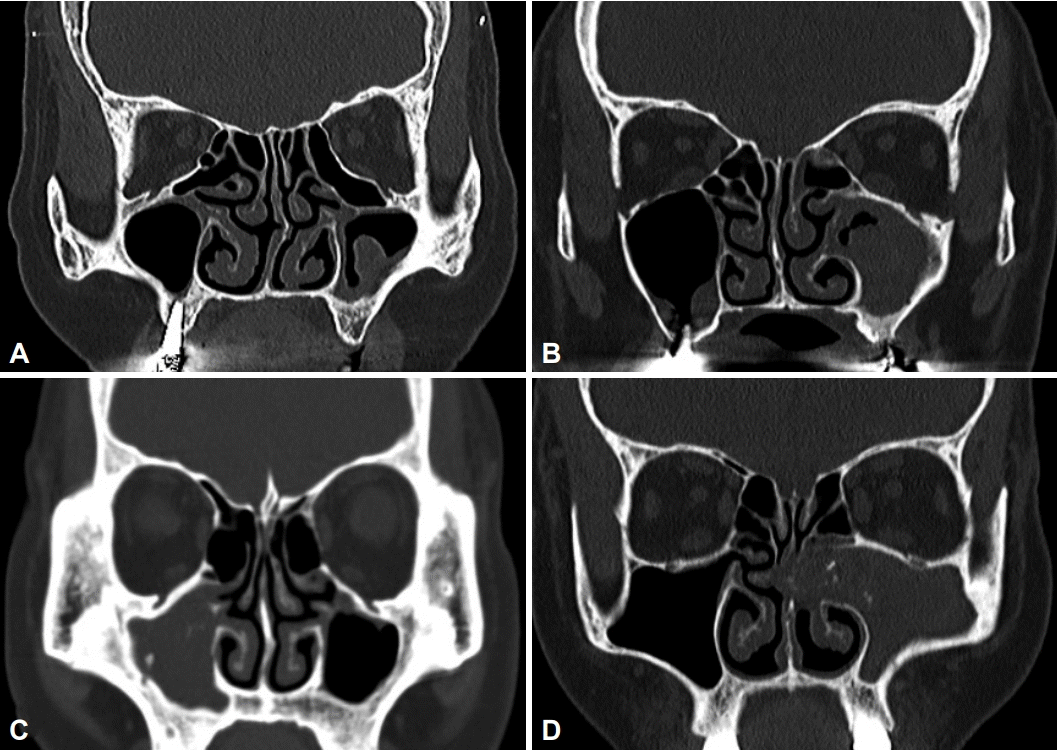
First, the degree of opacity within the maxillary sinus through sinus tomography, the presence of protrusions on an irregular surface or part of the surface in case of air-shaded areas, and protrusion of the maxillary sinus lesion to the mid-nasal level were examined. The classification of this study was devised by modifying the classification of Cha et al. [5], and it was classified from grade I to grade IV depending on the severity of the lesion caused by fungal ball in the maxillary sinus. Grade I is defined as the opacity of the maxillary sinus is less than 50% of the total sinus and there is an irregular surface or a protrusion on a part of the surface in the air-shaded area where there is no opacity. If the opacity of the maxillary sinus is more than 50%, but is not overall manifestation and there is an irregular surface or a protrusion on a part of the surface in the air shade area, it is defined as grade II. Grade III has overall opacity of the maxillary sinus and no protrusion into the middle nasal passage, grade IV has overall opacity of the maxillary sinus and protrusion into the middle nasal passage (Fig. 1).
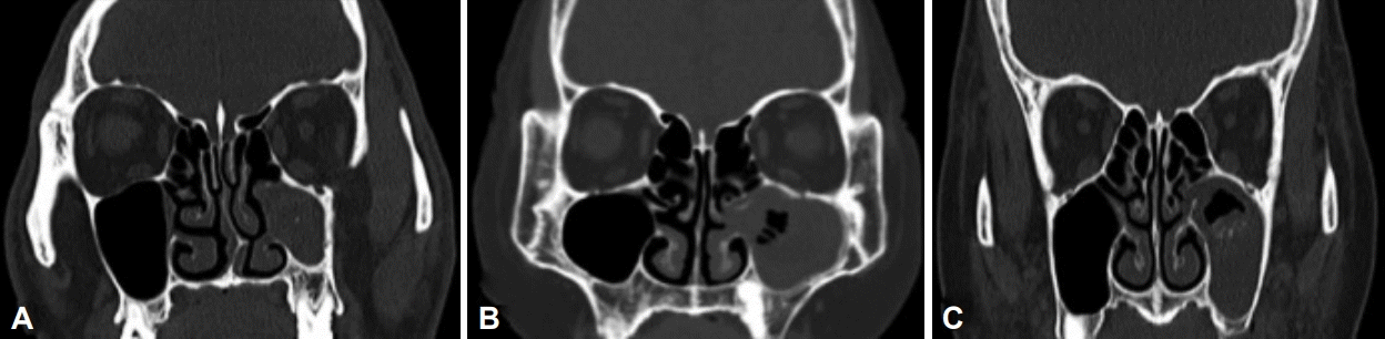
Second, the presence of calcification was examined. Third, the condition of maxillary sinus pneumatization on the side where lesion with the fungal ball located was examined. Having nasal floor as a baseline, the case where the maxillary sinus pneumatization did not reach the nasal floor was classified as grade I, the case where it reached to the nasal floor was classified as grade II, and the case where the pneumatization was reached below the nasal floor was classified as grade III (Fig. 2).
Surgical approach
In the case of using the middle and IMA, it was analyzed whether they were approached individually or in combination. As a basic surgical approach, a single approach to the middle nasal passage was performed. Cases where fungal ball was able to be removed through MMA, and additional IMA was performed to remove fungal ball remaining on the anterior or lower wall of the maxillary sinus even after removal through the MMA and saline wash.
The surgical approach was to first check where the uncus was attached to the sidewall of the nasal cavity before resection of the uncus, and then detach the uncus from the sidewall of the nasal cavity using a sickle knife and freer elevator, then remove the entire uncus. Then with cutting forceps or microdebrider, the MMA was performed as wide as possible by widening it posteriorly and downward. For grades I–III in sinus tomography, only the above surgical methods were used, and for grade IV, if the middle nasal passage and the anterior ethmoidal sinus were invaded through the maxillary sinus, an anterior ethmoidal sinus resection was also performed. In the IMA, the inferior nasal concha is first moved inward, and then the external wall of the nasal meatus is punctured with a curved curette, or if the bone of the external wall of the nasal meatus is hard, a hole is made using a curved penetrating needle and a hammer, and then various forceps are used to widen the cavity to remove the remaining fungal balls in the maxillary sinus.
Maintenance of MMA
For patients who could be followed up for more than six months after fungal ball removal, the case where a 4 mm curved suction tip was inserted into the maxillary sinus was considered as maintenance of the maxillary sinus antrostomy, and the case where it was not inserted was considered stenosis.
Statistics
Statistical analysis was performed using SPSS version 24.0 program (IBM Corp., Armonk, NY, USA). A chi-square test and a linear versus linear combination were used for categorical variable data. Statistical significance was determined to be statistically significant when the p-value was less than 0.05.
RESULTS
Characteristics of the subject group
The characteristics of the study subjects were a total of 221 patients, with 70 males and 151 females by sex, and the overall average age was 63.79±10.94 years. A total of 226 cases were included in the study, 216 patients of them unilaterally and 5 bilaterally.
Total four classifications according to sinus tomography
There were 52 cases of grade I, 59 cases of grade II, 35 cases of grade III, and 80 cases of grade IV, and the frequencies were IV, II, I, and III in order (Table 1).
Presence of calcification observed in sinus tomography
Calcification within the maxillary sinus was seen in 155 cases (68%) out of a total of 226 cases, and in 71 cases (32%) without visible calcification.
The relationship between the classification of sinus tomography and the condition of the antrostomy site
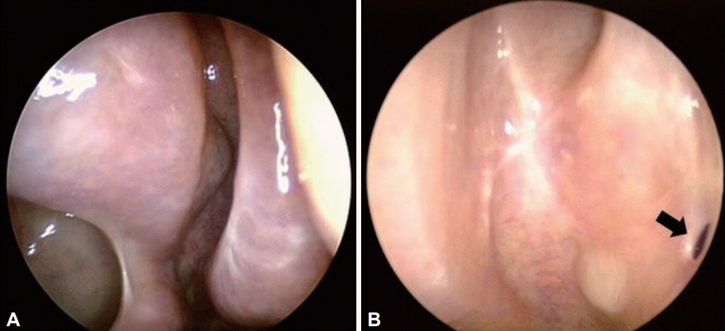
Follow-up for more than six months after surgery was conducted for 202 cases in the study group, 194 cases maintained MMA and 8 cases showed stenosis (Fig. 3). Complete occlusion was seen in one of eight cases classified as stenosis. Among 49 the cases where follow-up was conducted, 47 cases maintained the antrostomy site, and two showed stenosis for the grade I based on the classification of sinus tomography. For grade II 50 cases out of 51 maintained and one showed stenosis. For grade III, 28 cases out of 31 maintained and one showed stenosis. For grade IV, 69 out of 71 cases maintained and two showed stenosis (Table 1). There was no statistically significant difference between the maintenance of the maxillary sinus antrostomy or stenosis according to the four classifications (p=0.328).
In the case of IMA, the size of the antrostomy decreased as time elapsed during the 6-month follow-up period after the surgery, but the maxillary sinus could be observed through the IMA site with a 2.7 mm endoscope.
Pneumatization of the maxillary sinuses and analysis of the procedure
There were 6 cases of grade I, 47 cases of grade II, and 173 cases of grade III which showed pneumatization. As a result of analyzing the cases of MMA alone or a combination of MMA and IMA as a surgical approach, 6 cases showed single approach in grade I, 47 cases for grade II. There was no combination use for grade I and II. In grade III, 159 cases used single approach and 14 cases used combination approach (Table 2). There was a significant difference in the surgical approach according to the maxillary sinus pneumatization (p=0.042).
DISCUSSION
In the four classifications of sinus tomography in this study, 115 cases (51% of the total) had overall opacity in the maxillary sinus. 111 cases (49%) showed presence of irregular surfaces or protrusions in the air-shaded area. In the diagnosis of fungal ball in the maxillary sinus through sinus tomography, the mentioned characteristic was an important finding. In general, observation of calcification has been known to be a decisive finding in diagnosis of fungal ball, but calcification is not observed in all fungal ball patients. In previous studies, calcification within the maxillary sinus was observed in 50%–83% of patients with fungal ball [9,10] and, in this study, 155 out of 226 cases (68%) showed calcification in the maxillary sinus.
The authors attempted to confirm findings that would be helpful in diagnosis in sinus fungal ball patients in addition to calcification through a retrospective study involving patients with fungal ball in maxillary sinus. Depending on the presence of fungal cells in the maxillary sinus, 49% of the subjects of this study showed irregular surfaces or protrusions in the air-shaded area, which was a very important clue of the presence of the fungal ball when calcification was not observed.
Endoscopic MMA is often restricted in the access to the anterior and inferior medial regions of the maxillary sinus, including the anterior wall and prelacrimal recess. In this study, among 226 cases, 212 cases were able to remove fungal ball by MMA, and 14 cases needed IMA to remove fungal balls in the anterior and inferior walls. Therefore, there is a limit to securing the entire vision within the maxillary sinus only with endoscopic MMA, and IMA was required in some cases.
What is noteworthy in this study is that in grade III–when maxillary sinus pneumatization was reached below the nasal cavity–the combined approach of middle and IMA was used more often in a statistically significant way than MMA alone approach used in grades I and II. The above analysis was not previously reported, and the severe the pneumatization is, the more careful attention should be paid to the residual fungal cells.
Ju et al. [6] analyzed the patients with postoperative maxillary sinus antrostomy narrowing and found that there was a correlation with the range of maxillary sinus lesions caused by fungal ball. When the inflammatory lesion became larger and reached the natural foramen of the maxillary sinus, the probability of narrowing of the natural foramen of the maxillary sinus was increased compared to less severe lesion where is locates in the lower side of the maxillary sinus. Kim et al. [11] reported that the stenosis of the MMA for maxillary sinusitis after sinusitis surgery can be related to various factors such as the degree of inflammation and purulent secretion of the maxillary sinus during surgery, the degree of mucosal thickening, the pathological mucous membrane in the maxillary sinus and the mucosal treatment process around the natural foramen during surgery by the operator, presence of purulent discharge, and the formation of granulation tissue around the natural foramen of the maxillary sinus.
In this study, there was no significant difference between the four classifications according to sinus tomography and the maintenance and stenosis of the MMA. The study of Ju et al. [6] was analyzed at a relatively short time of three months after surgery, whereas this study was conducted more than six months. Kim et al. [11] set the standard for stenosis of MMA to be 5 mm or less, and reported that stenosis was observed within six months after surgery. The reason for setting the standard for stenosis in this study to be the capability of 4 mm curved suction tip inserted into the maxillary sinus was based on a judgment of whether maxillary sinus saline washing was possible on an outpatient basis. Unlike fungal ball, the targeted disease of this study, the study group of Kim et al. [11] studied chronic sinusitis. In this study, the fungal ball was removed, and saline wash was conducted to remove the accumulated purulent secretions, whereas in the study of Kim et al. [11] removed the thickened maxillary sinus mucosa with a micro-cutter through the canine fossa in most cases.
According to the study results of Ju et al. [6], the more severe the inflammatory lesion of the maxillary sinus, the higher the probability of a narrowing of the maxillary sinus antrostomy site. It was expected that grade IV, which is the most severe cases based on the classification of sinus topography, has a possibility of narrowing down in maxillary sinus antrostomy site, but there was no statistically significant difference. However grade III showed more narrowing. Grade IV is the most severe case of maxillary sinus lesions, and because fungal cells are often pushed into the natural foramen of the maxillary sinus, it extends not only to the bones constituting the uncus inside the natural foramen of the maxillary sinus, but also to the vertical plate of the palatine at the rear of the natural foramen of the maxillary sinus. Therefore, many of these cases required auto-MMA. Therefore, the authors hypothesized that there was less stenosis in grade IV due to the wide auto- MMA site. In a study on postoperative isthmus cysts, the possibility of occlusion of the IMA site over time was higher if the postoperative isthmus cyst did not protrude enough than if it had protruded sufficiently into the inferior sinus [12]. Based on these evidences, the authors carefully inferred that the case where the maxillary sinus lesion protruded to the middle nasal passage was more likely to maintain the maxillary sinus meatal antrostomy site than the case where the maxillary sinus lesion did not protrude.
Following is a summary of the results of this study and its limitations. First, the observation of calcification in the maxillary sinus and the presence of protrusions on the irregular surface or part of the surface of the maxillary sinus lesion were important findings in the diagnosis of fungal ball in the maxillary sinus through sinus tomography. However, a comparative study with radiographic findings with unilateral sinusitis such as acute sinusitis and odontogenic sinusitis may be necessary in the future for the cases where no calcification is observed among the radiographic findings of fungal ball in the maxillary sinus. Second, as the maxillary sinus pneumatization develops in patients with fungal ball in the maxillary sinus, an additional IMA may be required. However, depending on the location of the fungal cells, IMA can be conducted even when pneumatization does not manifest. Third, there was no association between the extent of the maxillary sinus lesion caused by fungal cells and the stenosis of the MMA site after surgery. However, as various factors such as the patient’s underlying disease, the surgeon’s procedure, and postoperative management may be involved, additional research may be required.
 XML Download