

 PDF
PDF Citation
 Print
Print


INTRODUCTION
Central sleep apnea (CSA) is defined as an absence of breathing without respiratory drive during sleep [1], unlike obstructive sleep apnea (OSA), which is accompanied by efforts to breathe. Various conditions such as high altitude, drug use, and cerebrovascular disease, can lead to central apnea. As with OSA, complications like frequent nocturnal awakenings, daytime sleepiness, and an increased risk of cardiovascular diseases also occur with CSA [1]. Literature has shown considerable overlap between obstructive and central apnea in several lines of evidence [2], and an increased propensity to develop central apnea was demonstrated in patients with OSA [3]. In a particular group of patients CSA appears after treatment of OSA, usually with positive airway pressure (PAP), a phenomenon called treatment-emergent central sleep apnea (TECSA). In this report we describe a patient who developed transient CSA after multi-level surgery for OSA.
CASE REPORT
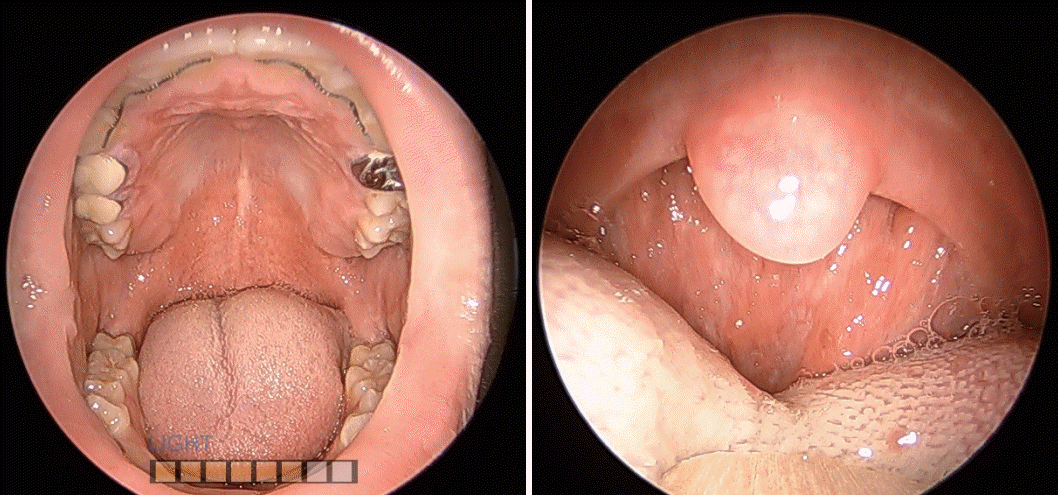
A 23-year-old male presented to our outpatient clinic with snoring and occasional sleep apnea. He also complained of nasal obstruction, morning headache with lightheadedness, and daytime sleepiness. The patient previously underwent adenoidectomy sixteen years ago, and septoplasty with bilateral tonsillectomy six years ago. His medical history was unremarkable and he was not taking any drug. Physical examination showed a body mass index of 27.16 kg/m2 , hypertrophy of left inferior turbinate, and modified Mallampati class III oropharynx (Fig. 1). Müller maneuver showed a complete collapse at velum and partial obstruction at the tongue base level of the oropharynx.
The patient underwent overnight laboratory-based polysomnography (PSG). The results revealed an apnea-hypopnea index (AHI) of 50.5 without central components and a respiratory distress index of 62.5, indicating a severe degree of sleep apnea. Multiple obstructive respiratory events occured throughout the sleeping time and the nadir oxygen saturation was 85%. Positional apnea was seen with a supine AHI of 62.8 and a non-supine index of 16.2. With a left lateral decubitus position, respiratory events along with snoring, desaturation, and arousal stopped, and N3 sleep stage was achieved.
The patient preferred a surgical treatment as he was going to join the army soon. He underwent modified uvulopalatopharyngoplasty (UPPP), robot-assisted tongue base resection, and bilateral coblator-assisted turbinoplasty, and was discharged eight days later without any complication. The patient reported an improvement of symptoms during his follow-up visits, specifically a decrease in the number of snoring and apneic events observed by his family. However, PSG performed ten months after the surgery showed an aggravated AHI of 82.6 and a central apnea index (CAI) of 13.5, indicating the emergence of CSA. Automatic PAP therapy was prescribed and three-week data showed an average AHI of 4.14 with high compliance. Follow-up PSG eight months after application of PAP showed an improved AHI of 58.2 and oxygen nadir of 91.0%, with disappearance of central apnea. Pertinent details of the pre- and postoperative PSG data are summarized in Table 1. Subsequent PAP data consistently showed excellent compliance with decreased AHI, accompanied by a further subjective improvement of symptoms.
DISCUSSION
TECSA is typically diagnosed when a CAI is 5 or greater after initiating treatment for OSA. This phenomenon has most commonly been seen with continuous positive airway pressure (CPAP) therapy. Javaheri et al. [4] suggested in their large retrospective study that 6.5% of patients with OSA had TECSA after initiation of CPAP therapy, which disappeared within eight weeks with continued treatment.
Altough the exact mechanism of TECSA is unclear, high loop gain and relief of inspiratory flow limitation are highly possible explanations. In OSA patients, due to limitations of pharyngeal structures, including anatomical impairment and incompetent control of upper airway muscles, increased ventilatory drive with elevated PCO2 levels may not lead to effective responses to maintain adequate upper airway patency [2,5]. This in turn results in insufficiently increased airflow and an augmented response to PCO2, and thus high loop gain. As inspiratory flow limitation is relieved with treatment, ventilatory overshoot follows and the PCO2 level decreases. Nocturnal hypercapnia is resolved even to the level below apnea threshold, and then ventilatory drive decreases and central apnea arises. In addition, improvement of TECSA and ventilatory control abnormalities after PAP therapy was reported, also suggesting that OSA may lead to ventilatory instability, or elevated loop gain [3].
TECSA also appears in patients whose upper airway obstruction is relieved with non-PAP treatments, including tracheostomy, maxillomandibular advancement surgery, and mandibular advancement devices [6-9]. Central apnea following UPPP as in our patient, however, has rarely been reported [10]. As in many of PAP-emergent sleep apnea patients, CSA disappeared with time also in these cases [6-9].
In our patient, obstructive indices and arousal remained high after pharyngeal and nasal surgery, despite slight improvements of subjective responses. As ventilation increases after arousal, it is likely that frequent arousal and high loop gain of our patient led to a ventilatory overshoot, followed by repetitive drops in the PCO2 level below apnea threshold and resultant CSA [2]. PAP therapy then ameliorated residual airway obstruction and the ventilatory instability resolved over time, which led to the elimination of CSA and possibly a rise in oxygen nadir in turn.
The increased oxygen nadir has significance, since the AHI also increased compared with the one before surgery. Possible reasons for this increased AHI even after disappearance of CSA may include the followings: longstanding incompetency of pharyngeal muscles that may still have existed after surgery; incomplete adaptation of pharyngeal muscles to the altered anatomical structure; and a possibility of postoperative nasopharyngeal stenosis, which was not evident on examinations on clinic visits. We surmise that, although these possibilities could and did cause an elevated AHI, they were not severe enough to cause significant desaturation and led to the elevated oxygen nadir. The ventilatory overshoot induced from high loop gain might also have played a role by increasing oxygen intake, but not to the degree that could induce CSA.
The importance of postoperative PSG monitoring for detection of CSA is highlighted in this case. In addition, performing PSG early after surgery is also important and necessary. There was a discrepancy between our patient’s report on improved symptoms and the actual PSG data after surgery; earlier postoperative PSG would have helped determine the accuracy of the patient’s report and check his state correctly, allowing earlier detection and treatment of CSA. Furthermore, along with the AHI and specific obstructive indices, the arousal index may play an important role in prediction of CSA. Quantification of loop gain and monitoring changes in arterial PCO2 levels after surgery may also aid in this process. In conclusion, this case suggests that postoperative monitoring should include multiple follow-up PSG tests from early on and that determining effectiveness of treatment of OSA should be based on long-term careful observation, especially in patients at high risk of TECSA.
 XML Download