

 PDF
PDF Citation
 Print
Print


Endoscopic resection of a polyp involving a colonic diverticulum is challenging. As there is no muscularis propria within a diverticulum, the potential risk of perforation associated with endoscopic resection must be considered.1,2
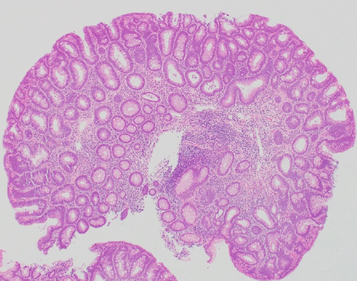
A 72-year-old male patient underwent follow-up colonoscopy after surgery for rectal cancer. A sessile polyp, 3 mm in size, was found in a cecal diverticulum. The underwater method eliminated halation and expanded the diverticulum cavity, making it possible to observe the lesion stably (Fig. 1A, Supplementary Video 1). Subsequently, underwater cold forceps polypectomy (CFP) was performed using Radial Jaw4-Jumbo cold polypectomy forceps (Boston Scientific, Tokyo, Japan) (Fig. 1B, C). Observation using narrow-band imaging and continuous irrigation confirmed that there were no residual lesions or perforations (Fig. 1D), and the wound was closed using clips. The pathological result was a tubular adenoma (Fig. 2).
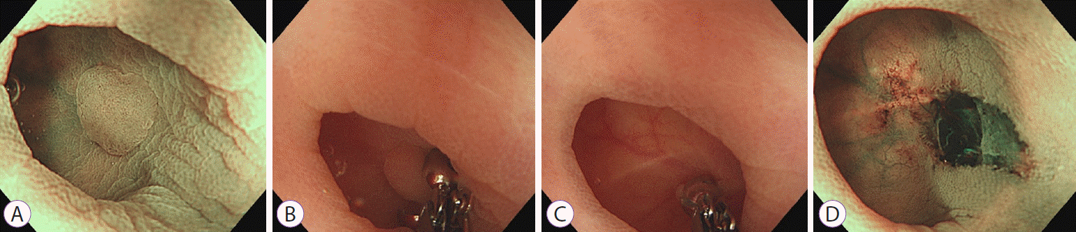 | Fig. 1.Underwater cold forceps polypectomy. (A) A sessile polyp 3 mm in size was found in a cecal diverticulum. The underwater method improved polyp visualization. Narrow-band imaging (NBI). (B, C) Underwater cold forceps polypectomy was performed. (D) Observation using NBI and continuous irrigation confirmed that there were no residual lesions or perforation.
|
The underwater method made it possible to expand the diverticulum cavity and float the lesion, allowing CFP to be performed safely.3 Cold biopsy forceps polypectomy was performed to avoid the risk of delayed perforation associated with hot polypectomy.4 This technique may be useful for cold snare polypectomy of larger polyps extending into a diverticulum.5 However, it would not be safe for large polyps involving more than one-fourth of a diverticulum. To the best of our knowledge, this is the first reported case of underwater CFP for an adenoma within a colonic diverticulum.

 XML Download