Abstract
Background and Objectives
It is important to identify variations of paranasal sinuses during sinus surgery. The Onodi cell (OC), a variant of the paranasal sinuses, is the most posterior ethmoid cell with a close relationship with the optic nerve (ON). The purpose of this study is to evaluate the prevalence of OCs and analyze the relationship of OCs with ON in Koreans.
Subjects and Method
This retrospective study utilized CT images of 526 slides from 263 Korean adults. The prevalence of the OCs and the degree of indentation of the ON within the OC was determined using binary logistic regression analysis.
Results
The OCs were observed in 37.3% of 263 subjects and in 27.6% of 526 slides. The OCs are found more frequently in males than in females (p=0.01), and also more frequently in the right side than in the left side (p=0.001). Binary logistic regression analysis revealed that the ON protrusion in female was 0.339 times lower than in male and 1.052 times higher with the increased age. The ON protrusion within the OC in the postsellar type was 10.214 times higher than that in the presellar.
Go to : 
오노디 세포는 1904년 아돌프 오노디에 의해 처음 기술되었으며, 가장 후방에 존재하는 후사골동 세포로 접형동의 상외측으로 함기화되어 시신경에 해부학적으로 인접한다[1]. 임상적으로 오노디 세포의 존재가 매우 중요한 이유로, 첫째, 오노디 세포는 시신경의 인접한 해부학적 관계로 부비동 내시경 수술 혹은 뇌하수체 종양 제거를 위한 내시경하 경접형동 접근 과정에서 시신경 손상의 위험을 증가시킨다. 둘째, 오노디 세포에 부비동염 발생 시 시각적 문제를 일으킬 수 있다[2,3]. 셋째, 오노디 세포는 오노디 세포가 없는 경우에 비해 접형동을 하내측으로 위치하게 하여 수술 중에 접형동 자연공을 찾는데 어려움을 초래할 수 있고, 오노디 세포를 접형동으로 오인할 수 있어 접형동 수술이 불완전하게 진행될 수 있다[4].
한국인을 대상으로 부비동 전산화단층촬영(CT)을 이용한 오노디 세포에 대한 발생률에 대한 연구는 몇 차례 보고되었으나 어떤 연구들은 인원수를 기준으로, 다른 연구들은 부비동의 한 측을 기준으로 보고하여 전체적으로 쉽게 파악하기에는 어려운 실정이다. 보고된 결과들은 연구자에 따라 162명에서 53명(32.7%) [5)], 129명에서 61명(47.3%) [6], 258측에서 88측 (34.1%) [6], 220측에서 28측(12.7%) [7], 200측에서 64측(32%) [8]이었다.
부비동 내시경 수술 과정에서 오노디 세포 존재 시 시신경에 대한 손상은 주요 합병증으로, 오노디 세포 내로 특히 시신경 직경이 돌출될수록 고위험군이 된다[9,10]. 본 연구는 부비동 CT를 이용하여 첫째, 과거의 연구들보다 더 많은 한국인을 대상으로 오노디 세포 발생률을 알아보았다. 둘째, 시신경의 오노디 세포 내로의 돌출 정도를 4단계로 나누어 분류하고 돌출 정도에 따라 인접 혹은 돌출로 구분하였다. 셋째, 성별, 연령, 접형동 함기화 같은 요인들이 오노디 세포 내로 시신경 돌출에 영향을 주는지 확인하고자 하였다.
Go to : 
2017년 1월부터 2018년 12월까지 본 병원에 내원하여 부비동 CT를 했던 18세에서 85세까지 총 263명, 각각 양측을 대상으로 총 526측에 대해 연구를 수행하였다. 대상군은 남자는 156명, 여자는 107명, 전체 평균나이는 48.3±16.3세였다. 연구대상군 선정에서 부비동 단층촬영을 통한 분석에 혼돈을 줄 수 있는 사골동 혹은 접형동의 혼탁, 코 수술 기왕력 혹은 과거 안면 골절로 변형이 있는 경우는 제외하였다. 본 연구는 원광대학교병원 기관심의위원회의 승인(2020-06-011- 001)을 받고 수행되었다.
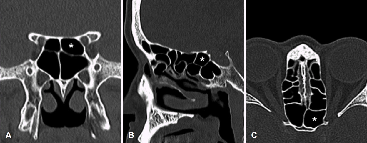
첫째, 후사골동 세포가 접형동의 상, 외측으로 함기화가 진행된 경우를 오노디 세포로 정의하였으며, 각각 부비동 CT의 축상면, 관상면, 시상면를 통해 오노디 세포가 일측 혹은 양측에 있는지 분석하였다(Fig. 1).
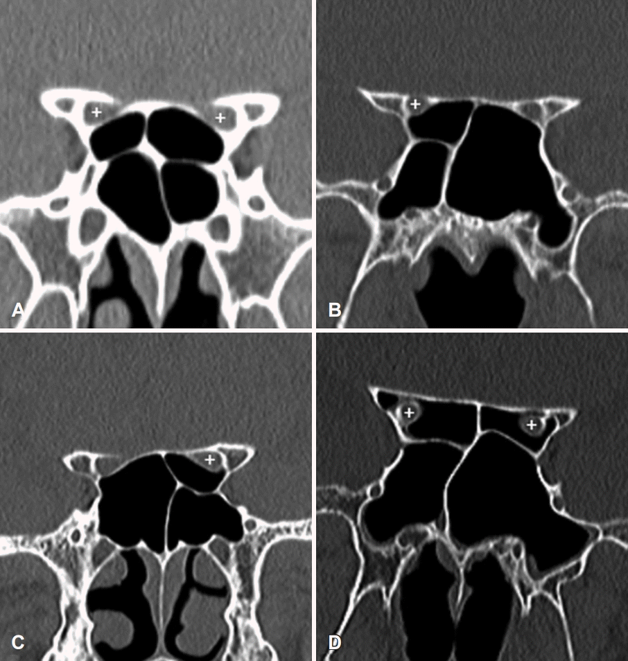
둘째, 오노디 세포와 관련된 시신경의 분류는 접형동과의 시신경 관계에 대한 Itagi 등 [11]의 분류를 수정 고안하였는데, 그들의 연구에서 시신경이 접형동 내의 돌출 정도에 따라 1단계에서 4단계로 구분하였다. 1단계는 접형동 내로 시신경이 인접하였으나 돌출이 없는 경우, 2단계는 접형동 내로 시신경 돌출이 시신경 직경(circumference)의 50%를 넘지 않는 경우, 3단계는 접형동 내로 시신경 돌출이 시신경 직경의 50%를 넘는 경우, 4단계는 오노디 세포가 있는 경우로 분류하였다. 본 연구의 1단계는 오노디 세포 내로 시신경이 인접하였으나 돌출이 없는 경우, 2단계는 오노디 세포 내로 시신경 돌출이 시신경 직경의 50%를 넘지 않는 경우, 3단계는 오노디 세포 내로 시신경 돌출이 시신경 직경의 50%를 넘는 경우, 4단계는 오노디 세포 내로 시신경이 관통하는 경우로 하였다(Fig. 2). 그리고 본 연구에서는 1과 2단계를 오노디 세포와 관련하여 시신경의 인접, 3, 4단계를 시신경의 돌출로 분류하였다.
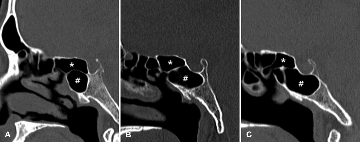
셋째, 오노디 세포가 관찰된 경우에 오노디 세포의 하측에 있는 접형동 함기화 정도를 시상면 영상에서 확인하였다. 1단계는 presellar 타입으로 터키안 전방까지 함기화된 경우이며, 2단계는 sellar 타입으로 터키안 후방까지 함기화된 상태, 3단계는 postsellar 타입으로 터키안 후방을 지나서 함기화된 상태로 하였다(Fig. 3). 그에 대한 결과를 통해 접형동 함기화 정도에 따라서 오노디 세포과 시신경의 관련성을 분석하였다.
통계학적 분석은 SPSS version 24.0 program (IBM Corp., Armonk, NY, USA)을 이용하였다. 오노디 세포의 유무에 대한 좌, 우측(편측) 간, 남녀 간의 차이는 카이 제곱 검정, 남녀 간의 오노디 세포와 시신경의 4단계 차이의 분석은 선형 대 선형 결합, 그리고 남녀 간의 오노디 세포와 시신경의 2타 입(인접, 돌출)의 분석은 카이 제곱 검정을 사용하였다. 오노디 세포에 대한 시신경의 2타입(인접, 돌출)에 영향을 줄 수 있는 요인들을 분석하기 위해 이항 로지스틱 회귀분석을 사용하였다. 통계학적 유의성은 p값이 0.05 미만인 경우에 통계적으로 유의하다고 판정하였다.
Go to : 
연구대상군 총 263명 중 98명(37.3%)에서 오노디 세포가 관찰되었으며, 우측에서만 오노디 세포가 관찰된 경우는 30명(11.4%), 좌측은 21명(8.0%), 양측은 47명(17.9%)이었다. 남자 156명 중 65명(41.6%)에서 오노디 세포가 관찰되었으며, 우측에서만 관찰된 경우는 21명(13.4%), 좌측은 10명(6.4%), 양측은 34명(21.8%)이었다. 여자 107명 중 33명(30.8%)에서 오노디 세포가 관찰되었으며, 우측에서만 관찰된 경우는 9명(8.4%), 좌측은 11명(10.3%), 양측은 13명(12.1%)이었으며 남녀 간에 유의한 차이는 없었다(p=0.673) (Table 1).
Comparison of the prevalence of Onodi cells in the total and in the male and female
연구대상군 263명의 526측 중 145측(27.6%)에서 오노디 세포가 있었으며, 남자의 312측에서 오노디 세포가 있는 경우는 99측, 여자의 214측에서 오노디 세포가 있는 경우는 46측에서 있었다. 남자에서 여자에 비해 양측에서 오노디 세포가 관찰된 경우가 많아서 전체적으로 남자가 여자에 비해 발생이 많았다(p=0.010) (Table 1).
좌, 우측 각각 263측으로 우측에서 오노디 세포가 있는 경우는 77측, 없는 경우는 186측, 좌측에서 오노디 세포가 있는 경우는 68측, 없는 경우는 195측이었으며, 우측이 좌측에 비해 발생이 많았다(p=0.001) (Table 2).
남자에서 시신경의 오노디 세포로의 돌출 정도에 따라 1단 계는 50측, 2단계는 14측, 3단계는 16측, 4단계는 19측, 여자 에서 1단계는 24측, 2단계는 9측, 3단계는 7측, 4단계는 6측 으로 유의한 차이는 보이지 않았다(p=0.475) (Table 3).
시신경의 오노디 세포로의 인접 혹은 돌출에 대해 남자에서 인접은 64측, 돌출은 35측, 여자에서 인접은 33측, 돌출은 13측이 관찰되었다(p=0.398) (Table 3).
연구대상군 526측 중 오노디 세포가 있었던 145측의 접형동의 함기화는 presellar 타입은 25예(17.2%), sellar 타입은 67예(46.2%), postsellar 타입은 53예(36.6%)였다.
이항 로지스틱 회귀분석을 하였으며 여자가 남자에 비해 시신경 돌출의 위험성이 0.339배 감소되었으며, 연령이 증가될수록 1.052배 증가되었다. 접형동의 함기화 정도에 따라 postsellar 타입이 presellar 타입에 비해 10.214배로 증가하였다(Table 4).
Logistic regression analysis of the factors that may affect protrusion of the optic nerve into Onodi cells
Go to : 
배아 발달 과정에서 후상방 사골동 세포가 상부 접형골까지 성장하여 시신경을 둘러싸면서 터키안까지 확장되는 오노디 세포가 생길 수 있다[12]. 이러한 오노디 세포에 환기와 배출이 잘 되지 않아 부비동염이 발생되면 시신경이 근접해 있기 때문에 시각과 관련된 증상을 일으킬 수 있다[13]. 오노디 세포에 발생한 점액낭종은 시신경을 지속적으로 압박하여 심한 경우 시력 소실을 일으킬 수 있다[14].
CT를 이용한 연구에서 2010년 이전에 보고된 오노디 세포의 발생률은 7%-24%로 비교적 낮은 발생률이 보고되었으나[15-19], 2010년 이후에서의 한 외국 문헌[9]의 보고는 65.3%로 현재까지 가장 높은 발생률의 연구였다. 이러한 발생률의 큰 차이는 축상면과 관상면 영상보다는 시상면을 포함한 3차원으로 재구성된 영상을 이용해 보다 많은 오노디 세포를 발견하는 결과를 보였는데 영상 기술의 발달이 기여한 것으로 생각된다.
한국인을 대상으로 한 오노디 세포의 발생률은 2010년 이전 연구보다는 거의 대부분 2010년 이후의 연구들로, 한 연구[7)를 제외하면 2010년 이전의 외국문헌의 7%-24%보다 높았으며, 가장 최근 발생률은 47.3% [6]로 지금까지 한국인의 연구에서 가장 높았다. 본 연구에서도 CT의 축상면, 관상면, 시상면 영상을 이용하여 오노디 세포의 존재를 확인하였으며, 총 연구대상군 263명 중 98명(37.3%)으로, Lee 등[6]의 47.3% 보다는 적었으나, Shin 등[5]의 162명 중 53명(32.7%)과 가장 유사하였다. 본 연구의 526측 중 145측(27.6%)으로, Shin 등[7] 의 220측에서 28측(12.7%)보다는 많았으나, Lee 등[6]의 258측에서 88측(34.1%), Hwang 등[8]의 200측에서 64측(32%)보다는 적었다. 위의 연구들을 평균적으로 보면, 한국인 대상의 연구들에서 인구의 약 30%대에서 오노디 세포의 발생률을 보였다.
본 연구에서 오노디 세포는 여자보다 남자에서 더 높은 발생률을 보였다. 여자에 비해 남자에서 양측의 오노디 세포가 관찰된 경우가 많아서 전체적으로 남자에서 발생이 많았던 것으로 생각된다. 남자에서의 부비동 함기화가 여자에 비해 전체적으로 더 크다는 연구와 상악동과 접형동외의 부비동에서 남자에서의 함기화가 크다는 연구 결과가 있다[20,21]. 그래서 저자들은 남자와 여자의 후사골동 함기화의 차이로 인해 남자에서의 오노디 세포의 발생이 보다 많았던 것으로 추정하였다.
본 연구에서 우측에서 오노디 세포가 있는 경우는 29.3%, 좌측에서는 25.8%로 우측 발생률이 높았다. 한국인을 대상으로 한 과거 연구에서 우측은 29.5%, 좌측은 26.2%에서 관찰되어 본 연구와 같이 우측에서 발생률이 높았다[6]. 외국 문헌에 의하면, 우측은 51.7%, 좌측은 48.3%로 우측에서 발생률이 높았다[4]. 편측 간의 발생 차이는 첫째, 두개안면 해부학 연구들에 따르면 두개안면골의 비대칭은 성인에서 흔히 관찰되는 소견으로 아마 태아의 발달 과정에서 발생하는 것으로 추정된다[22,23]. 둘째, 우측과 좌측 접형동 함기화를 비교하는 연구에서 우측에 비해 좌측이 유의하게 증가되었다[24]. 오노디 세포가 있는 경우 접형동 크기가 감소될 뿐만 아니라 접형동이 하내측으로 위치하게 되는데 좌측에 비해 우측 접형동 함기화의 감소는 오노디 세포의 존재 가능성을 보다 시사할 수 있다[25].
시신경에 대한 손상은 부비동 수술 과정에서 꼭 피해야 할 주요 합병증으로 오노디 세포 내로 시신경 직경의 50% 이상이 돌출된 경우에 골결손의 동반 가능성이 높아 손상 위험성이 증가된다[6]. 본 연구에서 오노디 세포 내로 시신경의 돌출이 여자보다 남자에서, 연령이 증가할수록 많았다. 오노디 세포의 발생이 여자보다 남자에서 더 많아 남자에서의 시신경 돌출 위험성이 증가된 것으로 생각되며, 연령이 증가할수록 오노디 세포 내로 시신경 돌출이 1.052배로 증가하나 다른 독립변수들에 비해 위험성이 높지 않아 더 많은 연구대상군이 포함된 추가적인 연구가 필요할 수 있다. 본 연구에서는 접형동의 함기화 정도에 따라 postsellar 타입이 presellar 타입에 비해 10.214배로 돌출 위험성이 매우 증가하였다. 종합하면, 남자와 접형동의 postsellar 타입이 각각 오노디 세포 내로 시신경 돌출에 영향을 준다.
본 연구의 강점으로는 첫째, 지금까지 한국인을 대상으로 한 연구는 162명이 가장 많은 연구대상군였으나 이번 연구에서는 그보다 많은 연구대상군 263명, 526측을 분석하였다. 둘째, 한국인 대상의 연구에서 오노디 세포에 대한 시신경의 돌출 정도에 대한 세부적인 분류가 지금까지 있지 않았다. 본 연구에서 처음 4단계로 구분하여 오노디 세포와 시신경과의 관계를 파악하는 데 의미 있는 자료로 활용될 수 있다. 셋째, 로지스틱 회귀분석을 한 결과, 오노디 세포 내 시신경 돌출과 접형동 함기화 정도가 서로 연관성이 있음을 알 수 있었다. 접형동의 postsellar 타입에서 오노디 세포로의 시신경 돌출의 위험성이 높았으며, 내시경 부비동 수술뿐만 아니라 뇌하수체 종양 제거를 위한 내시경 경접형동 접근 시 오노디 세포를 제거하는 과정에 시신경 손상 가능성의 근거를 제시하였다.
본 연구의 제한점으로는 접형동의 postsellar 타입과 오노디 세포 내로의 시신경 돌출과의 연관성을 밝힌 연구로, 접형동의 postsellar 타입과 오노디 세포 내로의 시신경 돌출과의 원인 관계를 규명한 것은 아니다. 차후 접형동의 함기화와 오노디 세포의 함기화와 연관성이 있을지, 혹은 오노디 세포의 함기화에 따른 시신경 돌출의 연관성을 포함한 다양한 요인들에 대해 추가적인 연구가 필요할 것으로 생각된다.
수술 전 영상학적 검사를 이용하여 오노디 세포의 존재 유무를 확인하고 존재하는 경우 시신경과의 관계를 파악하여 수술하는 과정에서 시신경 손상과 같은 합병증을 방지해야 겠다.
Go to : 
Notes
Author Contribution
Conceptualization: Jung-Hun Kown, Jae-Hoon Lee. Data curation: Jung-Hun Kown, Hyung-Bon Koo. Formal analysis: SeungYoon Han, Myeongsin Kang, Jin Lee. Methodology: Jung-Hun Kown, Jae-Hoon Lee. Supervision: Jae-Hoon Lee. Writing—original draft: Jung-Hun Kown, Jae-Hoon Lee. Writing—review & editing: Jae-Hoon Lee
Go to : 
REFERENCES
1. Onodi A. Die Sehstörungen und Erblindung nasalen Ursprunges, bedingt durch Erkrankungen der hinteren Nebenhöhlen. Ophthalmologica. 1904; 12(1):23–46.

2. Yanagisawa E, Weaver EM, Ashikawa R. The Onodi (sphenoethmoid) cell. Ear Nose Throat J. 1998; 77(8):578–80.

3. Tan HK, Ong YK. Sphenoid sinus: An anatomic and endoscopic study in Asian cadavers. Clin Anat. 2007; 20(7):745–50.

4. Liu J, Yuan J, Dai J, Wang N. The whole lateral type of the sphenoethmoidal cell and its relevance to endoscopic sinus surgery. Ear Nose Throat J. 2021; 100(9):NP416-23.

5. Shin JH, Kim SW, Hong YK, Jeun SS, Kang SG, Kim SW, et al. The Onodi cell: An obstacle to sellar lesions with a transsphenoidal approach. Otolaryngol Head Neck Surg. 2011; 145(6):1040–2.
6. Lee SJ, Kang YK, Lee ES, Kim JS. Prevalence of Onodi cells in Korean based on computed tomography. Korean J Otorhinolaryngol-Head Neck Surg. 2015; 58(12):855–8.

7. Shin JM, Jang WI, Baek BJ. Analysis of sphenoid sinus and surrounding structures using multidetector computed tomography. Korean J Otorhinolaryngol-Head Neck Surg. 2012; 55(2):95–100.

8. Hwang SH, Joo YH, Seo JH, Cho JH, Kang JM. Analysis of sphenoid sinus in the operative plane of endoscopic transsphenoidal surgery using computed tomography. Eur Arch Otorhinolaryngol. 2014; 271(8):2219–25.

9. Tomovic S, Esmaeili A, Chan NJ, Choudhry OJ, Shukla PA, Liu JK, et al. High-resolution computed tomography analysis of the prevalence of Onodi cells. Laryngoscope. 2012; 122(7):1470–3.

10. Cherla DV, Tomovic S, Liu JK, Eloy JA. The central Onodi cell: A previously unreported anatomic variation. Allergy Rhinol (Providence). 2013; 4(1):e49–51.

11. Itagi RM, Adiga CP, Kalenahalli K, Goolahally L, Gyanchandani M. Optic nerve canal relation to posterior paranasal sinuses in indian ethnics: Review and objective classification. J Clin Diagn Res. 2017; 11(4):TC01-3.

12. Lim CC, Dillon WP, McDermott MW. Mucocele involving the anterior clinoid process: MR and CT findings. AJNR Am J Neuroradiol. 1999; 20(2):287–90.
13. Chee E, Looi A. Onodi sinusitis presenting with orbital apex syndrome. Orbit. 2009; 28(6):422–4.

14. Toh ST, Lee JC. Onodi cell mucocele: Rare cause of optic compressive neuropathy. Arch Otolaryngol Head Neck Surg. 2007; 133(11):1153–6.
15. Driben JS, Bolger WE, Robles HA, Cable B, Zinreich SJ. The reliability of computerized tomographic detection of the Onodi (Sphenoethmoid) cell. Am J Rhinol. 1998; 12(2):105–11.

16. Jones NS, Strobl A, Holland I. A study of the CT findings in 100 patients with rhinosinusitis and 100 controls. Clin Otolaryngol Allied Sci. 1997; 22(1):47–51.

17. Keast A, Yelavich S, Dawes P, Lyons B. Anatomical variations of the paranasal sinuses in Polynesian and New Zealand European computerized tomography scans. Otolaryngol Head Neck Surg. 2008; 139(2):216–21.

18. Tan KL, Harvinder S. Prevalence of Onodi cells in hospital Raja Permaisuri Bainun, Ipoh, Malaysia. Med J Malaysia. 2010; 65(2):101–2.
19. Pérez-Piñas I, Sabaté J, Carmona A, Catalina-Herrera CJ, Jiménez-Castellanos J. Anatomical variations in the human paranasal sinus region studied by CT. J Anat. 2000; 197(Pt 2):221–7.

20. Kang M, Mo J, Chung Y. The differences in paranasal sinus pneumat izat ion af ter adolescence in Korean. Korean J Otorhinolaryngol-Head Neck Surg. 2019; 62(7):395–403.

21. Karakas S, Kavakli A. Morphometric examination of the paranasal sinuses and mastoid air cells using computed tomography. Ann Saudi Med. 2005; 25(1):41–5.

23. Vig PS, Hewitt AB. Asymmetry of the human facial skeleton. Angle Orthod. 1975; 45(2):125–9.
Go to : 


 PDF
PDF Citation
 Print
Print


 XML Download