

 PDF
PDF Citation
 Print
Print


Introduction
Tinnitus is commonly observed in individuals who had hearing loss. Somatic tinnitus (ST) is a subtype of subjective tinnitus, it is defined when tinnitus appears to be preceded or strictly linked to a somatic disorder, and therefore related to problems of the musculoskeletal system rather than of the ear [1]. Glossopharyngeal neuralgia (GPN) is a rare type of neuralgia presented with recurrent lancinating pain of the tongue, throat, ear, and tonsils, the areas where glossopharyngeal nerve innervates. The pain especially happens during swallowing, chewing, talking, coughing, yawning or laughing [2]. GPN is known to be caused by stimulation of glossopharyngeal nerve, and the most common cause is compression of the glossopharyngeal nerve by irregularly positioned blood vessels, skull base tumor, or infection of the throat and mouth [3]. According to the neurological model of somatic (craniocervical) tinnitus, sensory inputs from common spinal tract (CST) of facial nerve, glossopharyngeal nerve, and vagus nerve converge to medullary somatosensory nuclei (MSN), and the nerve fibers projecting from MSN to dorsal cochlear nucleus (DCN) can finally cause disinhibition of DCN, resulting in ST [4].
Herein, we report a case of tiny tonsillolith-induced GPN and ST, which was resolved after radiofrequency tonsil ablation, and speculated the mechanism of ST.
Go to : 
Case
This study was approved by in accordance with the Helsinki Declaration.
A previously healthy 66-year-old male presented with bilateral high-pitched tinnitus and recurrent sharp pain of his right throat, jaw and ear which persisted over than 1 year. His pain started whenever he initiated swallowing food, and it lasted for several minutes. As the pain repeatedly occurred during the daytime, he consequently developed phobia of food intake. Medication of analgesics was not effective for controlling his pain. His past medical histories were unremarkable. There was no history of the use of ototoxic drugs or exposure to noise.
On physical examination, the right ear canal and the tympanic membrane were intact. His oral cavity, oropharynx and larynx examination showed no remarkable findings. Under the impression of GPN, he was prescribed with 300 mg/day of gabapentin and 180 mg/day of roxoprofen.
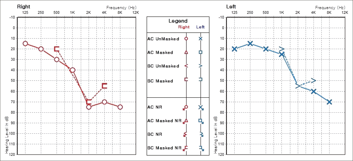
Pure tone audiometry showed bilateral high frequency hearing loss (Fig. 1). After two weeks of medication, the intensity of pain was slightly decreased, but still intolerable. Along the process of physical examination, manual compression of his right submandibular gland or right tonsil subsequently induced severe pain. Therefore, to find the secondary lesion causing GPN, neck ultrasonography and neck CT were preceded.
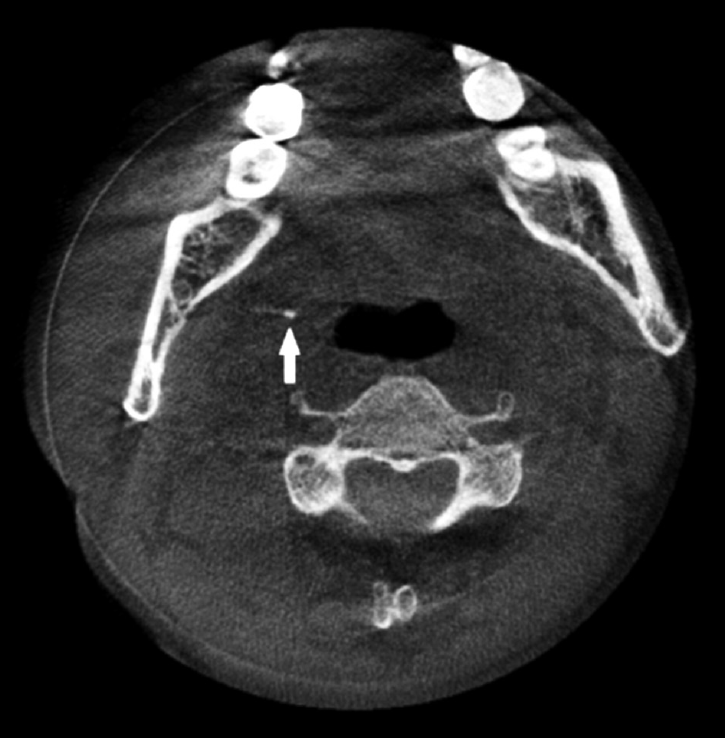
Neck ultrasonography showed normal findings. Neck CT revealed a tiny radio-opaque lesion in his right tonsil (Fig. 2). The lesion was diagnosed as tonsillolith. However, his tonsillolith was too small to be regarded as a cause of his pain, considering the previous case of large tonsillolith-caused GPN. However, as his pain did not subside, we at first tried extracting tonsillolith, supposedly the potential cause of persisting pain. But the first trial of extraction was failed. Then, we decided to perform right sided tonsillectomy to remove tonsillolith. Considering the side effects of conventional tonsillectomy and uncertainty of the effect of tonsillectomy on relieving his current symptoms, we planed radiofrequency tonsil ablation. The patient was explained thoroughly about the procedure of tonsillectomy using radiofrequency system (Celon AG medical instruments, Teltow, Germany) and also under local anesthesia, injection of dental lidocaine (1.8 mL of lidocaine HCl 2% and epinephrine 1:50000) was applied to right tonsil bed. Immediately after lidocaine injection, he presented prompt alleviation and his pain dramatically subsided, indicating that the tonsillolith was the main cause of GPN. Tonsil ablation was achieved with the insertion of a radiofrequency probe into the tonsil till submucosal level and ablation was done on three points. There was no immediate postoperative complication, and vital sign was stable.
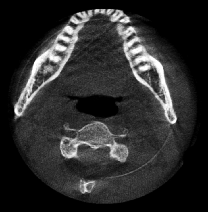
After tonsil ablation, his pain was completely disappeared, and did not recur for over 1 year. Follow-up CT scan taken 1 month after operation showed reduction of tonsil size and complete disappearance of tonsillolith (Fig. 3). In addition, his tinnitus of both ears that has been persisting for more than 1 year has surprisingly disappeared after tonsil ablation.
Go to :
Discussion
ST indicates that the tinnitus originated from the somatic disorder of the head and neck area. Tinnitus can be evoked or modulated by inputs from the somatosensory, somatomotor and visuomotor systems in some individuals. This has led to the term somatosensory modulation of tinnitus [4]. Such modulations may occur immediately following stimuli such as forceful muscle contractions of the temporomandibular joint (TMJ), head and neck, and limbs [5]. Animal studies have shown that projections from auditory nerve and trigeminal and dorsal column ganglia and brainstem nuclei integrates in the DCN [6]. And according to the neurological model of somatic (craniocervical) tinnitus, sensory inputs from CST of cranial nerve VII, IX, and X converge to MSN, and the nerve fibers projecting from MSN to DCN can finally cause disinhibition of DCN, resulting in ST [4,6].
Based on above ST model, we can explain that his tonsillolith has been the cause of his tinnitus. This case is about ST which showed somatic modulation by tonsil manipulation.
Clinically ST was defined by a positive history for TMJ and/or head and neck dysfunction and/or a positive modulation of tinnitus following somatic maneuvers [1]. There was a case in which tinnitus and ear pain occurred after abrasion of nasopharyx and modulated with forceful contraction of sternocleidomastoid muscle [7]. In our case, the high-pitched hearing loss is different from the previous case, and we did not tested somatic modulation before surgery. It is the limitation of our study. But the tinnitus disappeared after treatment of GPN is believed to be related to somatosensory modulation. Comparison of previous studies on somatic modulation of tinnitus, the average prevalence of modulation was 69%. The main somatic regions resulting in tinnitus modulation were the TMJ and head and neck region, followed by the eyes and limbs [8].
Some studies have shown that bilateral ST was more common in modulated by head and neck maneuver more than TMJ maneuver and combined hyperacusis [1,9].
Hence, we thought that neck disease can cause bilateral ST and it resolved by treatment of the disease. However, more research will be needed on bilateral tinnitus, which was associated with single side GPN.
GPN is rare symptomatic entity characterized by severe pain in the region where glossopharyngeal nerve innervates. Glossopharyngeal nerve sends out multiple branches. Pharyngeal branch contains taste sensory fibers from posterior one-third of tongue, and general sensory fibers of oropharynx. Tympanic branch of glossopharyngeal nerve, as known as Jacobson’s nerve, derives from the inferior ganglion and puts out parasympathetic fibers of parotid gland and general sensory fibers of middle ear. Thus, based on neuroanatomy, referred otalgia can develop from the stimulation of anywhere along the glossopharyngeal nerve pathway, including pharynx, tonsil, soft palate, and posterior one-third of tongue.
We can use gabapentin, carbamazepine and pregabalin as primary medical options, and non-steroidal anti-inflammatory drugs can also be effective. Glosspharyngeal nerve block can also be considered when the pain is extremely intractable.
Once the patient becomes refractory or intolerant to medications, surgery is the next treatment option. In idiopathic GPN, microvascular decompression of the nerve roots can be effective. However, in secondary GPN, underlying pathology should be removed. Various surgical procedures including intracranial tumor resection of cerebellopontine angle tumor, posterior fossa decompression of Chiari malformation, carotid stent in carotid megabulb, coagulation of choroid plexus overgrowth, tonsillectomy in chronic tonsillitis or tonsillolithiasis, and stylectomy for Eagle’s syndrome can be performed [3,10,11].
In conclusion, small tonsillolith may cause ST and GPN. They can be treated by eliminating tonsillolith and radiofrequency tonsil ablation was good choice of treatment.
Go to :
 XML Download