Journal List > J Pathol Transl Med > v.56(4) > 1516078308

Koh, Shin, Lee, and Jeon: Lymphoproliferative disorder involving body fluid: diagnostic approaches and roles of ancillary studies

Abstract

Lymphocyte-rich effusions represent benign reactive process or neoplastic condition. Involvement of lymphoproliferative disease in body cavity is not uncommon, and it often causes diagnostic challenge. In this review, we suggest a practical diagnostic approach toward lymphocyte-rich effusions, share representative cases, and discuss the utility of ancillary tests. Cytomorphologic features favoring neoplastic condition include high cellularity, cellular atypia/pleomorphism, monomorphic cell population, and frequent apoptosis, whereas lack of atypia, polymorphic cell population, and predominance of small T cells usually represent benign reactive process. Involvement of non-hematolymphoid malignant cells in body fluid should be ruled out first, followed by categorization of the samples into either small/medium-sized cell dominant or large-sized cell dominant fluid. Small/medium-sized cell dominant effusions require ancillary tests when either cellular atypia or history/clinical suspicion of lymphoproliferative disease is present. Large-sized cell dominant effusions usually suggest neoplastic condition, however, in the settings of initial presentation or low overall cellularity, ancillary studies are helpful for more clarification. Ancillary tests including immunocytochemistry, in situ hybridization, clonality test, and next-generation sequencing can be performed using cytologic preparations. Throughout the diagnostic process, proper review of clinical history, cytomorphologic examination, and application of adequate ancillary tests are key elements for successful diagnosis.

Effusions in the body cavities occur as the common complication in patients with various types of diseases including benign condition and malignancy. Among the cytologically proven malignant body fluid effusions, involvement of malignant lymphoma is not uncommon. It is reported that about 10%–15% of all malignant pleural effusions and ascites were caused by malignant lymphoma [1-4], after more common causes including carcinomas of lung, breast, and ovary.
Lymphocyte-rich effusions pose a diagnostic challenge in cytopathology, because effusions can be caused by non-neoplastic etiology [2], consisting of predominantly small lymphocytes, thereby mimicking low-grade indolent lymphomas [3-5]. It is often hard to discriminate lymphoma cells from reactive activated lymphocytes based on cytomorphology and cellular atypia. In addition, lymphoma is a very heterogeneous disease composed of tens of entities, and thus their cytomorphogical features are highly variable across disease entities and even in a single disease entity as well. Therefore, careful review of clinical history and precise cytologic examination along with proper use of ancillary testings are essential for accurate diagnosis of lymphoproliferative disease (LPD) involving body fluid. In this review, we aim to discuss the clinical pathological characteristics, diagnostic strategies, ancillary studies, and special considerations regarding LPD involving body fluid. It should be noted that specific diagnosis of hematolymphoid malignancy diagnosis should be based on tissue material and is not necessarily the scope of cytologic diagnosis in practice, except for a few rare conditions (i.e., lymphoma initially or solely presented with effusion, such as primary vitreoretinal lymphoma). However, understanding of unique cytomorphology of lymphoma entities may be very helpful and thus will be partly addressed in this review.

CLINICOPATHOLOGICAL CHARACTERISTIC OF LYMPHOPROLIFERATIVE DISEASE INVOLVING BODY FLUID

Body fluid effusions are common complication of LPD and are generally associated with poor prognosis [6-8]. Pleural effusion complicates about 6%–20% of patients with non-Hodgkin lymphoma (NHL), where the proportion is higher with mediastinal involvement [9,10]. Significant age variation is reported in lymphomatous effusions; LPD accounted for the most common cause of malignant body fluid in the children under age 18 years, while only a minority of malignant pleural effusion was caused by LPD in the patients over 40 years of age [3].
Lymphomatous effusions can be either primary or secondary. Primary effusion lymphoma (PEL) is a rare disease entity presenting as serous effusions without detectable nodal disease or tumor masses, which will be discussed later in this review. Secondary involvement of body fluid by LPDs are much more common than PEL. A study in a tertiary medical center in Taiwan [11] reported that the lymphomatous effusions were more commonly caused by B-cell NHL (27/36, 75.0%) than T-cell NHL (8/36, 22.2%) or classic Hodgkin lymphoma (1/36, 2.8%). Most common causes included diffuse large B-cell lymphomas (DLBCL) and high-grade B-cell lymphomas (16/36, 44.4%), followed by peripheral T-cell lymphomas (6/36, 16.7%) and mantle cell lymphomas (MCL) (5/36, 13.9%) [11].
To have a glance at the real world data of lymphomatous effusions, we collected the consecutive cases of cytologically diagnosed LPDs involving body fluid and cerebrospinal fluid (CSF) in Seoul National University Hospital in the year 2020 (Table 1). Samples definitely diagnosed with involvement of hematolymphoid malignancy were included, and those with indeterminate diagnosis such as ‘atypical cells’ were excluded from the collection to provide clearer view. Our results on body fluid also showed the predominance of B-cell NHLs (12/24, 50.0%) including DLBCLs and high-grade B-cell lymphomas. On the other hand, CSF involvement by leukemic conditions (9/17, 52.9%) outweighed that by lymphoid neoplasm (7/17, 41.2%) or myeloma (1/17, 5.9%).

DIAGNOSTIC APPROACH TO LYMPHOPROLIFERATIVE DISEASES INVOLVING BODY FLUID

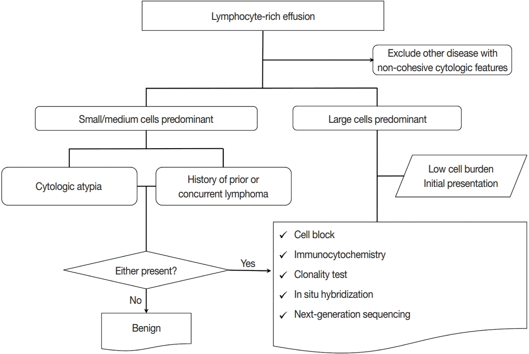
Previous studies have proposed various diagnostic algorithms for lymphocyte-rich effusions. Chen et al. [12] presented an algorithmic approach based on the cell size (large, intermediate, and small) and morphologic features (Reed-Sternberg like and plasmacytoid), along with immunohistochemical panel and genomic assays for proper subtyping of LPDs in body fluid. Gochhait et al. [13] also suggested cell sized based triage using a 3-tier (large, intermediate, and small) system for initial classification, whereas more practical 2-tier (small and large) scheme was recently introduced [5]. We consider that 2-tier scheme would be easier to apply in daily practice, particularly for pathologist other than hematopathologist, therefore, we suggest a modified diagnostic algorithm as depicted in Fig. 1.
First, it is important to acknowledge that non-hematolymphoid malignant cells in body fluid specimens can masquerade as lymphocyte-rich effusions. Specific subtypes of non-hematologic cancers often showing discohesive cells include invasive lobular carcinoma of breast [14], small cell lung carcinoma [15], gastric signet ring cell carcinoma [16,17], malignant melanoma [18], and small round cell sarcomas [19] including Ewing sarcoma and rhabdomyosarcoma. Careful review of clinical history and cytomorphology is crucial for differential diagnosis; the cytomorphology of discohesive non-hematolymphoid malignant effusion will not be elaborated in this review. Adjunctive testings including immunocytochemistry (ICC) using cell blocks (CB) could be helpful for differential diagnosis, which will be discussed in detail in ancillary tests section.
After ruling out the possibility of non-hematolymphoid malignant cells in body fluid specimens, we suggest starting with the assessment of the size of the lymphocytes in the effusion fluid and categorize the samples into either small/medium-sized cell dominant or large-sized cell dominant fluid (Table 2, Fig. 1). The criteria for defining cell size are proposed as follows: small/medium- sized cells have diameters less than those of 2 mature lymphocytes or red blood cells (RBCs), while large-sized cells usually have diameters exceeding 3 to 4 times of those of lymphocytes or RBCs.
Representative neoplastic conditions presenting as small/medium- sized cell dominant or large-sized cell dominant fluid are summarized in Table 2 (right column). However, it should be emphasized that classification of hematolymphoid malignancy other than PEL using fluid is beyond the scope of cytologic diagnosis in practice.

Body fluid rich in small/medium-sized lymphoid cells

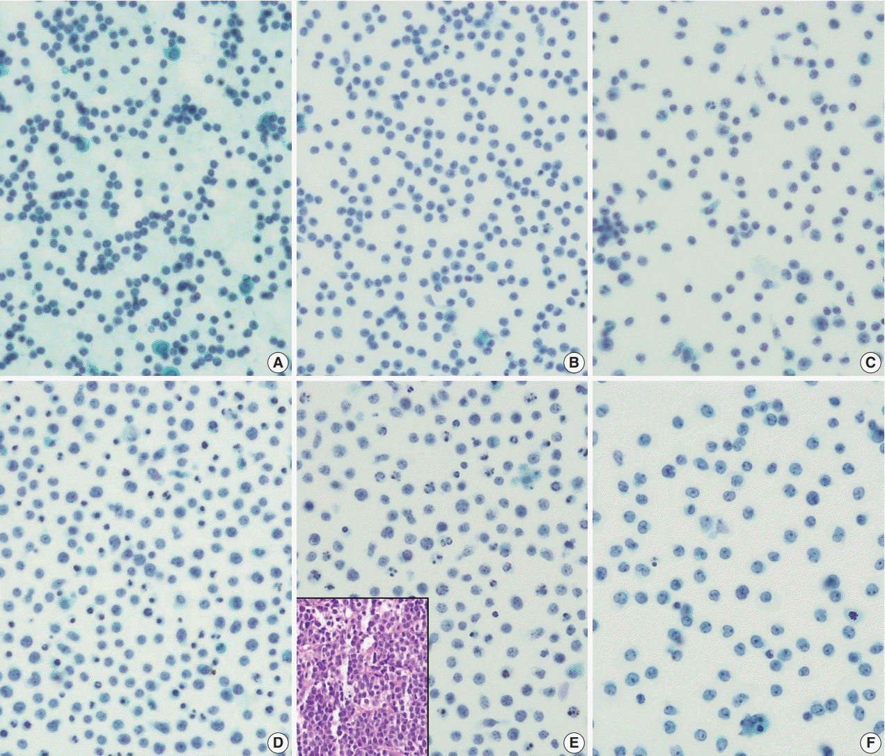
When dealing with the effusions having predominantly small/medium-sized cells, the distinction between neoplastic conditions from reactive lymphocytosis is often very difficult. Numerous physiologic and pathologic processes can produce reactive lymphocytosis, and commonly encountered causes are described in Table 2 and Fig. 2AC. Careful review of clinical history should be taken to rule out the possibilities of reactive lymphocytosis; conversely, if the patients have history of previous or suspected concurrent LPD, this warrants further investigation with ancillary studies.
Cytomorphological clues indicating a malignant process in small/medium-sized effusions are suggested in Table 3. Cellular atypia usually refers to the following features: irregular nuclear membrane, nuclear folding or cleavage, coarse chromatin or unusual chromatin patterns (e.g., clotted or soccer-ball-like) [5].
However, one should note that these atypical features are hardly recognizable in small/medium-sized lymphocytes in effusion cytology, especially on liquid-based cytology (LBC) specimens [20] where the cells and their nuclei appear smaller than in conventional smears. Nevertheless, when there is significant and obvious cellular atypia in the small/medium-sized cell predominant effusions, further workup including ancillary tests is strongly recommended.
On the other hand, monomorphic appearance of small/medium- sized lymphoid cells with high cellularity usually represents a neoplastic process (Fig. 2DF), therefore justifying the use of ancillary tests even in the absence of clinical history of LPD. In addition, frequent apoptotic figures also indicate a likelihood of malignant fluid (Fig. 2E).
When small/medium-sized lymphoid cells are dispersed in the polymorphous cellular background, it usually suggests benign reactive process, especially when combined with large centroblasts, immunoblasts, and plasma cells. However, when medium to large plasmacytoid cells are predominant cellular components, outnumbering all other cell types, careful consideration should be taken to rule out the possibility of plasma cell neoplasm. Thorough review of clinical features and assessment of nuclear atypia would aid in the differential diagnosis.

Body fluid rich in large-sized lymphoid cells

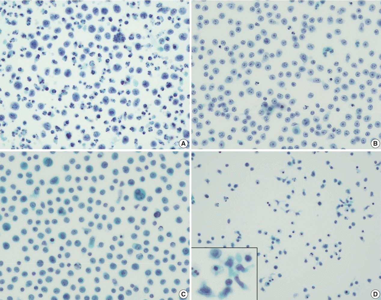
Large cell dominant (particularly monomorphic) lymphoid effusions almost always suggest pathologic conditions, therefore diagnostic steps on these fluids are easier to approach. In many cases, large cells show significant cytologic atypia and the background usually exhibit degenerated cell debris, necrosis and apoptosis. DLBCL is the most common cause of secondary large cell predominant lymphoid effusions, and often suggest poor prognosis (Fig. 3A, B) [7]. Less common causes of body fluids rich in large cells include leukemic conditions (Fig. 3C), and T- or natural killer (NK)-cell lineage lymphomas of certain entities which often show twisted, convoluted or multi-lobated nuclei and nuclear folding or grooving (Fig. 3D). In many cases, the diagnosis of large cell predominant lymphoid effusions is followed by ancillary studies for accurate subtyping, if required, and prognostic stratification, which will be further discussed in ancillary tests section.

SPECIFIC DISEASE ENTITIES

Primary effusion lymphoma

PEL is a unique type of large B-cell lymphoma presenting solely as serous effusion in body cavity without tumor masses, lymphadenopathy or organomegaly, usually associated with immunodeficient condition (as represented by human immunodeficiency virus [HIV] infection). PEL is almost always associated with human herpes virus 8 (HHV8) and may be co-infected with Epstein-Barr virus (EBV). It usually confers extremely unfavorable prognosis, with expected median survival of less than 6 months [21]. According to a recent nationwide study, PEL accounts for 0.1% of all malignant lymphoma in Republic of Korea [21,22]. Typically, PEL is composed of large size B-cell with immunoblastic, plasmablastic or anaplastic morphology and shows post-germinal center plasmablastic differentiation. A typical example is shown in the Fig. 4A. A 79-year-old male patient with a history of prostate cancer presented with dyspnea on exertion, where large amount of pleural effusion was noted. On systemic work-up, there was no evidence of solid tumor mass or lymphadenopathy. Drained pleural fluid was composed of numerous large lymphoid cells with many apoptotic bodies, and subsequent ICC on CB revealed CD45-positive, CD20-negative, CD38-positive and HHV8-positive immunophenotype, consistent with PEL.
Recently, further subtyping of PEL was suggested, namely PEL type I and type II [23]. In contrast to the classic type I PEL, type II PEL (also called effusion-based lymphoma) is characterized by frequent expression of pan B-cell markers (e.g., CD20 and CD79a) and less plasmablastic differentiation; most importantly, type II is often HHV8-negative and rarely associated with HIV. Clinically, type II PEL is rather associated with medial conditions which lead to fluid overload states, including hepatitis C viral infection. We also experienced a case of HHV8-negative effusionbased large B-cell lymphoma (Fig. 4B). Type II PEL also confers poor prognosis [23-25], despite a little better than the type I.

Dasatinib-induced effusion

Another unique entity presenting as body cavity-based lymphocyte- rich effusion is dasatinib-induced effusion. Dasatinib is the second-generation tyrosine kinase inhibitor used for the treatment of patients with BCR-ABL translocation positive chronic myelogenous leukemia (CML) [26]. The abnormal accumulation of body fluid occurs in about 20% to 40% of patients with CML receiving dasatinib, where severe forms develop in 3% to 7% of patients [27,28]. The effusion is composed of clonal proliferation of cytotoxic T- or NK-cells. Although the pathogenesis is unclear, several hypotheses are suggested including increased vascular permeability due to kinase inhibition or immune-mediated reaction [27]. Temporary reduction of dasatinib dosage or short-term steroid therapy usually alleviate the effusion, therefore, it is very important to recognize the lympho-dominant nature of the fluid, thus, not to be confused with the secondary leukemic involvement in patients with CML treated with dasatinib.

Breast implant associated anaplastic large cell lymphoma

Breast implant associated anaplastic large cell lymphoma (BIALCL) is a rare type of non-Hodgkin T-cell lymphoma occurring in patients who underwent breast augmentation procedure [29-31]. About two thirds of patients present with effusion around the implant, after 9 years from implantation on average [29]. Aspiration fluid cytology evaluation is an effective and minimally invasive method for the diagnosis of BI-ALCL [32].
Aspirated fluid from the patients with BI-ALCL usually shows hypercellular population of large atypical lymphoid cells with lobulated nuclei [33]. Cytoplasmic vacuolizations can be extensive, and necrotic or fibrinoid background can be a distinguishing feature from ‘benign seroma’ composed of transudate fluid. When BI-ALCL is suspected, CB preparation is recommended for ancillary studies [33].

ANCILLARY TESTS

Concerns regarding the cytology preparation

Body fluid specimen drawn from the patients can be processed for adequate evaluation using variable methods including conventional smears, cytospins, CBs and LBC [34]. Most of the reports comparing the diagnostic performance of variable preparations showed that the newer technology—LBC—is comparable to that of conventional methods [34-37]. Specifically, LBC usually result in better cell yield, provide easier examination since cells are evenly dispersed in a small area, with minimal drying artifact and erythrocytes obscuring examination. However, some technical disadvantages of LBC preparation should be kept in mind. During the process, morphologic alterations including cell/nuclear size shrinkage and architecture disruption can occur [38]. Despite these disadvantages, stored LBC material can be processed for CBs, immunostainings and molecular genetic analyses up to 3 or 4 months [34], therefore, LBC preparation is a preferred method with many additional advantages with comparable diagnostic efficacy.

CB and hematoxylin and eosin stain

When dealing with effusions from patients with known history or clinical suspicion of LPD, or those rich in large-sized lymphoid cells, it is important to preserve a portion of specimen to establish CB with appropriate preparation method. Since the most of procedures in pathology laboratories are based on the use of formalin fixed paraffin embedded (FFPE), CBs can readily be handled with ease. In addition, CB specimens retain the cytomorphologic features and antigenicity for further microscopic examination and ICC studies [39,40].

Immunocytochemistry

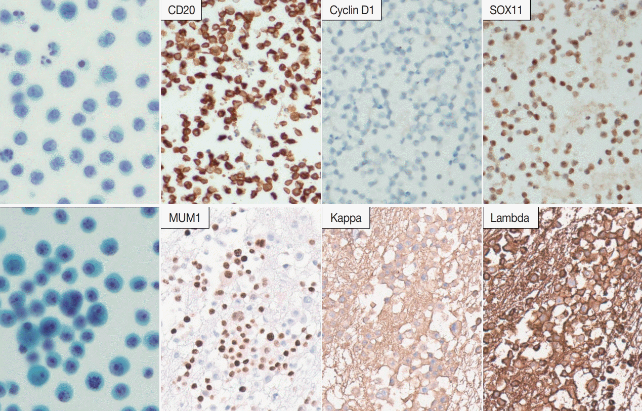
ICC is the most commonly used ancillary study for the diagnosis of lymphocyte-rich effusion. The ICC stains retain the cytomorphologic features and expression of certain markers help the confirmatory diagnosis in some entities, i.e., HHV8 expression in PELs. In many cases, panels of multiple items including lineage markers are required for the diagnosis of LPD in effusion cytology.
Fig. 5A shows pleural effusion fluid from a 77-year-old man, where medium-sized lymphoid cells were predominant. CB was prepared and subsequent ICC revealed CD20-positivity, suggesting the possibility of B-cell lymphoma. The cells were negative for cyclin D1 but SOX11 positive, rendering the diagnosis of lymphoma, consistent with MCL. Fig. 5B is another pleural fluid specimen drained from a 46-year-old male, and numerous MUM1 positive plasmacytoid cells having abundant cytoplasm and eccentric nuclei are observed. The cells were diffusely positive for lambda while negative for kappa; this light chain restriction revealed by ICC aids in confirming the neoplastic nature of the fluid and involvement of patient’s underlying multiple myeloma.

Flow cytometry

Body fluid effusions are often suitable samples for flow cytometry (FC), given the samples are not fixed by alcohol or formalin. FC-based analysis is an extremely useful method when dealing with differential diagnosis of effusion-based hematolymphoid malignancy, because this technology provides a reproducible, quantitative and sensitive immunophenotyping of cells, using fluorescent- tagged antibodies targeting surface markers [5,41-44]. Similar to ICC, FC analysis using a panel of antibodies can provide diagnostic clues in lymphoid malignancies. One important consideration should be noted; since the reactive lymphocyte-rich effusions are often T-cell predominant, T-cell abundancy shown by FC does not guarantee the diagnosis of T-cell lymphoma [5,41].
Though rarer than lymphoid diseases, FC also serves important roles in leukemic conditions [41,43]. For instance, involvement of chronic lymphocytic leukemia (CLL) on body fluid can be very difficult to distinguish from reactive lymphocytosis. When there is clinical history or suspicion for CLL, FC is strongly recommended, and documentation of CD19+, CD20+ B-cell with CD5 or CD23 expression can qualify the diagnosis of CLL involvement [43].

Clonality studies

Many studies have shown the efficacy of polymerase chain reaction (PCR) for the assessment of immunoglobulin heavy chain (IgH) gene rearrangements or TCRγ gene rearrangements. Recently, clonality tests in many pathology laboratories are readily performed using BIOMED-2 assay [45,46]. Many studies have reported that detecting clonality is useful for the differential diagnosis between reactive lymphocytosis and lymphomatous fluid [47-51].
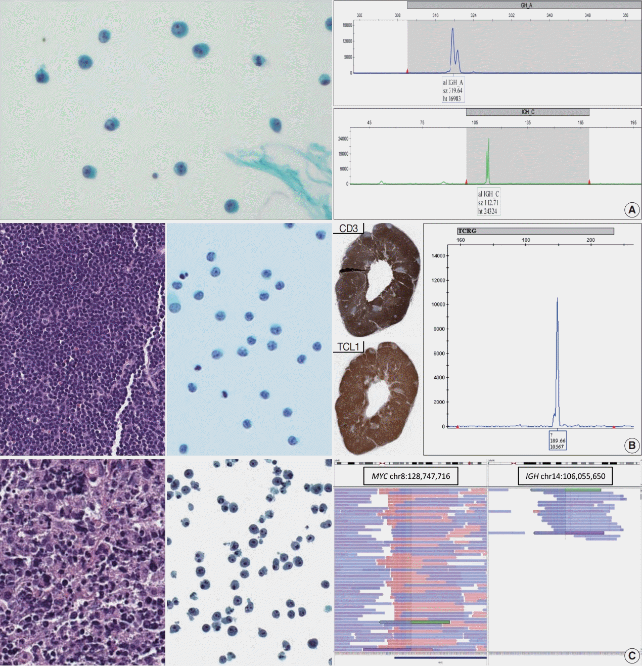
Fig. 6A shows a case where clonality testing was very helpful in the diagnosis. A 61-year-old female was diagnosed with primary central nervous system (CNS) DLBCL by stereotactic biopsy, and her CSF contained a few atypical lymphoid cells (Fig. 6A). B-cell monoclonality was found in IgH gene rearrangement study, strongly suggesting the involvement of DLBCL.
Reactive effusions tend to be T-cell predominant, therefore, T-cell clonality test is often required to confirm involvement of T-cell LPD in body fluids [5,52]. T-cell prolymphocytic leukemia (T-PLL) can rarely involve extramedullary sites [53], and nodal involvement was diagnosed in a 63-year-old female patient (Fig. 6B). The patient developed ascites, and leukemic involvement was suspected. Cytomorphologically, some small/medium-sized lymphocytes were observed and the overall cellularity was not remarkable; however, TCRγ gene rearrangement test proved monoclonal T-cell proliferation, supporting the involvement of T-PLL of patient.
One of the practically useful tips for correct interpretation is to consider comparing the clonality testing result from cytology specimen with that from the tissue specimen. If the both results show clonal peaks/bands with approximately same size, the diagnostic power of lymphomatous effusion could be strengthened.
Clonality test could be useful for the diagnosis of CSF cytology in patients with malignant lymphoma. Review of 101 CSF samples from patients with B-cell lymphoma diagnosed in Seoul National University Hospital from 2015 to 2017 showed that monoclonality by IgH gene rearrangement study using BIOMED-2 assay was detected in 42.9% of cytomorphologically atypical/ positive CSF and as much as 18.2% of cytomorphologically negative CSF specimens (Table 4). While clonality tests are very helpful, a specific consideration should be taken when interpreting the test performed on CSF specimen. Since the overall cellularity in CSF specimen is usually very low, high sensitivity of PCR test may result in false positive pseudoclonality due to selective amplification of certain non-neoplastic clones. According to our internal review of CSF cytology from73 patients with B-cell lymphoma and active brain (leptomeningeal and/or parenchymal) disease on neuroimaging (i.e., magnetic resonance imaging), rate of positive/atypical cytology was 35.6% and B-cell monoclonality was detected in 32.9%. The combined interpretation of both cytomorphology and clonality test resulted in a higher concordance up to 50.7% with neuroimaging results. Thus, retrieval of proper clinical and radiological information should accompany the interpretation of molecular study when dealing with CSF cytology.

In situ hybridization

As in histopathologic diagnosis using FFPE specimens, EBV in situ hybridization can readily be performed on cytological preparations including CB [54]. Identification of EBV is extremely valuable in characterization of certain EBV-associated LPDs, for example, EBV-positive DLBCLs, extranodal NK/T-cell lymphomas or PEL.
Fluorescent in situ hybridization (FISH) is another valuable tool for documentation of genetic alterations. Feasibility of FISH on cytologic preparation is well studied in several studies on solid tumors [51,55-65]. Detection of hallmark genomic variations direct leads to final diagnosis, for example, documentation of BCL2 translocation in follicular lymphoma or MYC translocation in Burkitt lymphoma. Prognostic stratification of high-grade B-cell lymphomas can also be achieved by finding MYC or BCL2 translocation. Development of many commercially available diagnostic FISH probes would enable characterization of less common structural variations, for instance, IRF4/DUSP22 or TP63 translocations.

Mutational analysis and next-generation sequencing

Recent advances in genomic profiling technologies dramatically accelerated precision diagnosis in daily practice. Multiple techniques including direct sequencing, real-time PCR, pyrosequencing have been performed on cytologic preparations, most notably on effusion fluids from the patients with non-small cell lung cancer [65-70]. Clinically meaningful disease-specific genetic alterations have been uncovered in the majority of lymphoma entities [71]. For instance, detection of MYD88 L265P mutation using allele-specific PCR assay has been proven its efficacy on differentiating lymphoplasmacytic lymphomas [72,73] from other types of low-grade B-cell lymphomas. Recently, genetic classification of DLBCL has been recently introduced, and unique genomic landscape of certain DLBCL entities has been identified. For example, primary CNS lymphoma (PCNSL) and primary vitreoretinal lymphoma are characterized by frequent mutations in MYD88 and CD79B. Thus, clonality test and/or MYD88 mutation analysis would be helpful to determine the CSF involvement of PCNSL in equivocal cases based on cytomorphology only. In a similar vein, clonality test and/or MYD88 mutation analysis would be informative in the diagnosis of vitreoretinal lymphoma using cytology specimen (e.g., vitreoretinal fluid) [74-76].
Single gene assays are being gradually replaced by next-generation sequencing (NGS), which enables testing for multiple genetic alterations on a single platform. Accumulating evidences suggest that various types of cytological preparations—smears, cytospins, liquid-based preparations and CBs—can be used for NGS-based assays with equivalent performance [77,78]. Some recommend that a portion of cytology specimens be reserved before CB preparation, for formalin fixation during the process my cause DNA cross-linkage and fragmentation [79-81].
Clinical utility of NGS-based assay on LPDs has been suggested by recent studies, both in diagnostic and prognostic perspectives [82-85]. One example is a report of angioimmunoblastic T-cell lymphoma [86], of which the primary diagnosis was made on pleural effusion fluid by detecting pathognomic RHOA G17V mutation by targeted NGS as well as immunophenotyping on CB. Fig. 6C shows another example that NGS-based assay helped in the genomic characterization and prognostic stratification of a patient with lymphoma. A 74-year-old male was diagnosed with gastric DLBCL of non-germinal center B-cell immunophenotype, and the patient developed ascites during the course. Targeted NGS using customized panel encompassing 121 lymphoma- related genes was performed, which revealed IGH-MYC translocation. In addition, TET2 truncating mutations and heterozygous deletions of CDKN2A and CDKN2B were also found.
In some instances, cytologic specimens may be the only available resource for diagnosis and genomic profiling, therefore, the importance of cytologic preparation in NGS-based practice would be further expanded [81,87].

CONCLUSION

LPDs can frequently involve body cavities, which can pose significant diagnostic challenge to pathologists. Along with proper review of clinical history, cytomorphologic examination is the key process. Triaging the lymphocyte-rich effusions into small/medium- sized cell predominant and large-cell predominant patterns is a useful diagnostic approach. Since the distinction between reactive lymphocytosis and pathologic LPD is seldom possible based on cytologic atypia alone, ancillary tests guided by cytomorphology and clinical information are crucial. In addition to conventional ancillary testings including ICC and clonality assays, recent advances in NGS technology will further aid in retrieval of precious information from cytology specimens.

Notes

Ethics Statement

Not applicable.

Availability of Data and Material

The datasets generated or analyzed during the study are available from the corresponding author on reasonable request.

Code Availability

Not applicable.

Author contributions

Conceptualization: YKJ. Figures and data: SAS, JAL. Writing—original draft: JK. Writing—review & editing: JK, YKJ. Approval of final manuscript: all authors.

Conflicts of Interest

The authors declare that they have no potential conflicts of interest.

Funding Statement

No funding to declare.

References

1. Johnston WW. The malignant pleural effusion. A review of cytopathologic diagnoses of 584 specimens from 472 consecutive patients. Cancer. 1985; 56:905–9.
crossref
2. Valdes L, Alvarez D, Valle JM, Pose A, San Jose E. The etiology of pleural effusions in an area with high incidence of tuberculosis. Chest. 1996; 109:158–62.
crossref
3. Das DK. Serous effusions in malignant lymphomas: a review. Diagn Cytopathol. 2006; 34:335–47.
crossref
4. Tong LC, Ko HM, Saieg MA, Boerner S, Geddie WR, da Cunha Santos G. Subclassification of lymphoproliferative disorders in serous effusions: a 10-year experience. Cancer Cytopathol. 2013; 121:261–70.
5. Liu CY, Chuang SS. A simple and practical guide for triaging lymphocyte- rich effusions for ancillary studies. Adv Anat Pathol. 2021; 28:94–104.
crossref
6. Hunter BD, Dhakal S, Voci S, Goldstein NP, Constine LS. Pleural effusions in patients with Hodgkin lymphoma: clinical predictors and associations with outcome. Leuk Lymphoma. 2014; 55:1822–6.
crossref
7. Chen YP, Huang HY, Lin KP, Medeiros LJ, Chen TY, Chang KC. Malignant effusions correlate with poorer prognosis in patients with diffuse large B-cell lymphoma. Am J Clin Pathol. 2015; 143:707–15.
crossref
8. Yanamandra U, Deo P, Sahu KK, et al. Clinicopathological profile of myelomatous pleural effusion: single-center real-world experience and review of literature. Clin Lymphoma Myeloma Leuk. 2019; 19:183–9.
crossref
9. Weick JK, Kiely JM, Harrison EG Jr, Carr DT, Scanlon PW. Pleural effusion in lymphoma. Cancer. 1973; 31:848–53.
crossref
10. Alexandrakis MG, Passam FH, Kyriakou DS, Bouros D. Pleural effusions in hematologic malignancies. Chest. 2004; 125:1546–55.
crossref
11. Wang RC, Chen YH, Chen BJ, Chuang SS. The cytopathological spectrum of lymphomas in effusions in a tertiary center in Taiwan. Diagn Cytopathol. 2021; 49:232–40.
crossref
12. Chen L, Zhang JS, Liu DG, Cui D, Meng ZL. An algorithmic approach to diagnose haematolymphoid neoplasms in effusion by combining morphology, immunohistochemistry and molecular cytogenetics. Cytopathology. 2018; 29:10–21.
crossref
13. Gochhait D, Balakrishnan K, Venkatesan D, et al. The cytological evaluation of ascitic fluid led to the diagnosis of mediastinal T-lymphoblastic lymphoma: a simplified algorithmic approach to diagnosis of lymphoid cell-rich effusion. Cytopathology. 2020; 31:359–61.
crossref
14. Menet E, Becette V, Briffod M. Cytologic diagnosis of lobular carcinoma of the breast: experience with 555 patients in the Rene Huguenin Cancer Center. Cancer. 2008; 114:111–7.
crossref
15. Domagala-Kulawik J, Gornicka B, Krenke R, Mich S, Chazan R. The value of cytological diagnosis of small cell lung carcinoma. Pneumonol Alergol Pol. 2010; 78:203–10.
16. Wang J, Katz RL, Stewart J, Landon G, Guo M, Gong Y. Fine-needle aspiration diagnosis of lymphomas with signet ring cell features: potential pitfalls and solutions. Cancer Cytopathol. 2013; 121:525–32.
17. Ainechi S, Pambuccian SE, Wojcik EM, Barkan GA. Cytomorphologic features and differential diagnosis of neoplasms with small cell features in liquid-based urinary tract cytologic specimens. J Am Soc Cytopathol. 2015; 4:295–306.
crossref
18. Murali R, Loughman NT, McKenzie PR, Watson GF, Thompson JF, Scolyer RA. Cytological features of melanoma in exfoliative fluid specimens. J Clin Pathol. 2009; 62:638–43.
crossref
19. Wong JW, Pitlik D, Abdul-Karim FW. Cytology of pleural, peritoneal and pericardial fluids in children: a 40-year summary. Acta Cytol. 1997; 41:467–73.
20. Murro D, Slade JM, Gattuso P. Cytologic findings of hematologic malignancies in bronchoalveolar lavage fluid. J Am Soc Cytopathol. 2015; 4:267–75.
crossref
21. Kim Y, Park CJ, Roh J, Huh J. Current concepts in primary effusion lymphoma and other effusion-based lymphomas. Korean J Pathol. 2014; 48:81–90.
crossref
22. Jung HR, Huh J, Ko YH, et al. Classification of malignant lymphoma subtypes in Korean patients: a report of the 4th nationwide study. J Hematopathol. 2019; 12:173–81.
crossref
23. Chen BJ, Wang RC, Ho CH, et al. Primary effusion lymphoma in Taiwan shows two distinctive clinicopathological subtypes with rare human immunodeficiency virus association. Histopathology. 2018; 72:930–44.
crossref
24. Kim M, An J, Yoon SO, et al. Human herpesvirus 8-negative effusion- based lymphoma with indolent clinical behavior in an elderly patient: a case report and literature review. Oncol Lett. 2020; 20:343.
25. Alexanian S, Said J, Lones M, Pullarkat ST. KSHV/HHV8-negative effusion-based lymphoma, a distinct entity associated with fluid overload states. Am J Surg Pathol. 2013; 37:241–9.
crossref
26. Paydas S. Dasatinib, large granular lymphocytosis, and pleural effusion: useful or adverse effect? Crit Rev Oncol Hematol. 2014; 89:242–7.
crossref
27. Cortes JE, Jimenez CA, Mauro MJ, Geyer A, Pinilla-Ibarz J, Smith BD. Pleural effusion in dasatinib-treated patients with chronic myeloid leukemia in chronic phase: identification and management. Clin Lymphoma Myeloma Leuk. 2017; 17:78–82.
crossref
28. Brixey AG, Light RW. Pleural effusions due to dasatinib. Curr Opin Pulm Med. 2010; 16:351–6.
crossref
29. Quesada AE, Medeiros LJ, Clemens MW, Ferrufino-Schmidt MC, Pina-Oviedo S, Miranda RN. Breast implant-associated anaplastic large cell lymphoma: a review. Mod Pathol. 2019; 32:166–88.
crossref
30. Leberfinger AN, Behar BJ, Williams NC, et al. Breast implant-associated anaplastic large cell lymphoma: a systematic review. JAMA Surg. 2017; 152:1161–8.
crossref
31. Chai SM, Kavangh S, Ooi SS, et al. Anaplastic large-cell lymphoma associated with breast implants: a unique entity within the spectrum of peri-implant effusions. Diagn Cytopathol. 2014; 42:929–38.
crossref
32. Granados R, Lumbreras EM, Delgado M, Aramburu JA, Tardio JC. Cytological diagnosis of bilateral breast implant-associated lymphoma of the ALK-negative anaplastic large-cell type: clinical implications of peri-implant breast seroma cytological reporting. Diagn Cytopathol. 2016; 44:623–7.
crossref
33. Barbe E, de Boer M, de Jong D. A practical cytological approach to the diagnosis of breast-implant associated anaplastic large cell lymphoma. Cytopathology. 2019; 30:363–9.
crossref
34. Gabriel C, Achten R, Drijkoningen M. Use of liquid-based cytology in serous fluids: a comparison with conventional cytopreparatory techniques. Acta Cytol. 2004; 48:825–35.
35. Fetsch PA, Simsir A, Brosky K, Abati A. Comparison of three commonly used cytologic preparations in effusion immunocytochemistry. Diagn Cytopathol. 2002; 26:61–6.
crossref
36. Ylagan LR, Zhai J. The value of ThinPrep and cytospin preparation in pleural effusion cytological diagnosis of mesothelioma and adenocarcinoma. Diagn Cytopathol. 2005; 32:137–44.
crossref
37. Lee YM, Hwang JY, Son SM, et al. Comparison of diagnostic accuracy between CellprepPlus(R) and ThinPrep(R) liquid-based preparations in effusion cytology. Diagn Cytopathol. 2014; 42:384–90.
38. Hoda RS. Non-gynecologic cytology on liquid-based preparations: a morphologic review of facts and artifacts. Diagn Cytopathol. 2007; 35:621–34.
crossref
39. Jain D, Mathur SR, Iyer VK. Cell blocks in cytopathology: a review of preparative methods, utility in diagnosis and role in ancillary studies. Cytopathology. 2014; 25:356–71.
crossref
40. Nambirajan A, Jain D. Cell blocks in cytopathology: an update. Cytopathology. 2018; 29:505–24.
crossref
41. Gupta P, Pandey T, Gautam U, et al. Lymphoreticular malignancies in serous effusions: cytomorphologic, flow cytometric and immunocytochemical analysis. Diagn Cytopathol. 2021; 49:647–56.
crossref
42. Zheng Y, Wan X, Gui X, et al. Value of multi-parameter flow cytometry immunophenotyping in T/NK-cell neoplasms in cytology specimens: a retrospective study in Chinese patients. Pathol Res Pract. 2020; 216:152921.
crossref
43. Kaur K, Patel T, Patra S, Trivedi P. Cytomorphology, Immunophenotype, and cytogenetic profile of leukemic serous effusions. Diagn Cytopathol. 2021; 49:948–58.
crossref
44. Bhardwaj N, Gupta N, Gupta P, Malhotra P. Flow cytometric immunophenotyping in a liquid-based cytology sample of pleural fluid: connecting the dots. Cytopathology. 2022; 33:269–72.
crossref
45. Jeon YK, Yoon SO, Paik JH, et al. Molecular testing of lymphoproliferative disorders: current status and perspectives. J Pathol Transl Med. 2017; 51:224–41.
crossref
46. van Dongen JJ, Langerak AW, Bruggemann M, et al. Design and standardization of PCR primers and protocols for detection of clonal immunoglobulin and T-cell receptor gene recombinations in suspect lymphoproliferations: report of the BIOMED-2 Concerted Action BMH4-CT98-3936. Leukemia. 2003; 17:2257–317.
crossref
47. Alkan S, Lehman C, Sarago C, Sidawy MK, Karcher DS, Garrett CT. Polymerase chain reaction detection of immunoglobulin gene rearrangement and bcl-2 translocation in archival glass slides of cytologic material. Diagn Mol Pathol. 1995; 4:25–31.
crossref
48. Mihaescu A, Gebhard S, Chaubert P, et al. Application of molecular genetics to the diagnosis of lymphoid-rich effusions: study of 95 cases with concomitant immunophenotyping. Diagn Cytopathol. 2002; 27:90–5.
crossref
49. Li J, Zhang W, Wang W, et al. Forty-nine cases of acute lymphoblastic leukaemia/lymphoma in pleural and pericardial effusions: a cytological- histological correlation. Cytopathology. 2018; 29:172–8.
crossref
50. Murphy M, Signoretti S, Nasser I, Sherburne B, Loda M. Detection of concurrent/recurrent non-Hodgkin’s lymphoma in effusions by PCR. Hum Pathol. 1999; 30:1361–6.
crossref
51. Pinto D, Schmitt F. Current applications of molecular testing on body cavity fluids. Diagn Cytopathol. 2020; 48:840–51.
crossref
52. Bode-Lesniewska B. Flow cytometry and effusions in lymphoproliferative processes and other hematologic neoplasias. Acta Cytol. 2016; 60:354–64.
crossref
53. Chen X, Cherian S. Immunophenotypic characterization of T-cell prolymphocytic leukemia. Am J Clin Pathol. 2013; 140:727–35.
crossref
54. Garady C, Saieg MA, Ko HM, Geddie WR, Boerner SL, da Cunha Santos G. Epstein-Barr virus encoded RNA detected by in situ hybridization using cytological preparations. Cytopathology. 2014; 25:101–7.
crossref
55. Minca EC, Lanigan CP, Reynolds JP, et al. ALK status testing in nonsmall- cell lung carcinoma by FISH on ThinPrep slides with cytology material. J Thorac Oncol. 2014; 9:464–8.
56. Sundling KE, Cibas ES. Ancillary studies in pleural, pericardial, and peritoneal effusion cytology. Cancer Cytopathol. 2018; 126 Suppl 8:590–8.
crossref
57. Walts AE, Hiroshima K, McGregor SM, Wu D, Husain AN, Marchevsky AM. BAP1 immunostain and CDKN2A (p16) FISH analysis: clinical applicability for the diagnosis of malignant mesothelioma in effusions. Diagn Cytopathol. 2016; 44:599–606.
58. Wang W, Tang Y, Li J, Jiang L, Jiang Y, Su X. Detection of ALK rearrangements in malignant pleural effusion cell blocks from patients with advanced non-small cell lung cancer: a comparison of Ventana immunohistochemistry and fluorescence in situ hybridization. Cancer Cytopathol. 2015; 123:117–22.
59. Chen Z, Wang DD, Peier A, Stone JF, Sandberg AA. FISH in the evaluation of pleural and ascitic fluids. Cancer Genet Cytogenet. 1995; 84:116–9.
crossref
60. Savic S, Franco N, Grilli B, et al. Fluorescence in situ hybridization in the definitive diagnosis of malignant mesothelioma in effusion cytology. Chest. 2010; 138:137–44.
crossref
61. Matsumoto S, Nabeshima K, Kamei T, et al. Morphology of 9p21 homozygous deletion-positive pleural mesothelioma cells analyzed using fluorescence in situ hybridization and virtual microscope system in effusion cytology. Cancer Cytopathol. 2013; 121:415–22.
crossref
62. Betz BL, Dixon CA, Weigelin HC, Knoepp SM, Roh MH. The use of stained cytologic direct smears for ALK gene rearrangement analysis of lung adenocarcinoma. Cancer Cytopathol. 2013; 121:489–99.
63. Tsai TH, Wu SG, Hsieh MS, Yu CJ, Yang JC, Shih JY. Clinical and prognostic implications of RET rearrangements in metastatic lung adenocarcinoma patients with malignant pleural effusion. Lung Cancer. 2015; 88:208–14.
crossref
64. Jardin F, Jais JP, Molina TJ, et al. Diffuse large B-cell lymphomas with CDKN2A deletion have a distinct gene expression signature and a poor prognosis under R-CHOP treatment: a GELA study. Blood. 2010; 116:1092–104.
crossref
65. Cai G, Wong R, Chhieng D, et al. Identification of EGFR mutation, KRAS mutation, and ALK gene rearrangement in cytological specimens of primary and metastatic lung adenocarcinoma. Cancer Cytopathol. 2013; 121:500–7.
66. Lozano MD, Zulueta JJ, Echeveste JI, et al. Assessment of epidermal growth factor receptor and K-ras mutation status in cytological stained smears of non-small cell lung cancer patients: correlation with clinical outcomes. Oncologist. 2011; 16:877–85.
crossref
67. Bellevicine C, Malapelle U, Vigliar E, de Luca C, Troncone G. Epidermal growth factor receptor test performed on liquid-based cytology lung samples: experience of an academic referral center. Acta Cytol. 2014; 58:589–94.
crossref
68. Scarpa A, Sikora K, Fassan M, et al. Molecular typing of lung adenocarcinoma on cytological samples using a multigene next generation sequencing panel. PLoS One. 2013; 8:e80478.
crossref
69. Gailey MP, Stence AA, Jensen CS, Ma D. Multiplatform comparison of molecular oncology tests performed on cytology specimens and formalin-fixed, paraffin-embedded tissue. Cancer Cytopathol. 2015; 123:30–9.
crossref
70. Lindeman NI, Cagle PT, Aisner DL, et al. Updated molecular testing guideline for the selection of lung cancer patients for treatment with targeted tyrosine kinase inhibitors: guideline from the College of American Pathologists, the International Association for the Study of Lung Cancer, and the Association for Molecular Pathology. Arch Pathol Lab Med. 2018; 142:321–46.
crossref
71. Moffitt AB, Dave SS. Clinical Applications of the genomic landscape of aggressive non-Hodgkin lymphoma. J Clin Oncol. 2017; 35:955–62.
crossref
72. Pan ST, Wang RC, Kuo CC, Hsieh YC, Su YZ, Chuang SS. MYD88 L265P mutation analysis is a useful diagnostic adjunct for lymphoplasmacytic lymphoma with pleural effusion. Pathol Int. 2019; 69:601–7.
crossref
73. Varettoni M, Arcaini L, Zibellini S, et al. Prevalence and clinical significance of the MYD88 (L265P) somatic mutation in Waldenstrom’s macroglobulinemia and related lymphoid neoplasms. Blood. 2013; 121:2522–8.
74. Hiemcke-Jiwa LS, Ten Dam-van Loon NH, Leguit RJ, et al. Potential diagnosis of vitreoretinal lymphoma by detection of MYD88 mutation in aqueous humor with ultrasensitive droplet digital polymerase chain reaction. JAMA Ophthalmol. 2018; 136:1098–104.
crossref
75. Bonzheim I, Giese S, Deuter C, et al. High frequency of MYD88 mutations in vitreoretinal B-cell lymphoma: a valuable tool to improve diagnostic yield of vitreous aspirates. Blood. 2015; 126:76–9.
crossref
76. Tanaka R, Kaburaki T, Taoka K, et al. More accurate diagnosis of vitreoretinal lymphoma using a combination of diagnostic test results: a prospective observational study. Ocul Immunol Inflamm. 2021; Apr. 1. [Epub]. https://doi.org/10.1080/09273948.2021.1873394.
crossref
77. Roy-Chowdhuri S, Goswami RS, Chen H, et al. Factors affecting the success of next-generation sequencing in cytology specimens. Cancer Cytopathol. 2015; 123:659–68.
crossref
78. Roy-Chowdhuri S, Chen H, Singh RR, et al. Concurrent fine needle aspirations and core needle biopsies: a comparative study of substrates for next-generation sequencing in solid organ malignancies. Mod Pathol. 2017; 30:499–508.
crossref
79. Williams C, Ponten F, Moberg C, et al. A high frequency of sequence alterations is due to formalin fixation of archival specimens. Am J Pathol. 1999; 155:1467–71.
crossref
80. Srinivasan M, Sedmak D, Jewell S. Effect of fixatives and tissue processing on the content and integrity of nucleic acids. Am J Pathol. 2002; 161:1961–71.
crossref
81. Bellevicine C, Malapelle U, Vigliar E, Pisapia P, Vita G, Troncone G. How to prepare cytological samples for molecular testing. J Clin Pathol. 2017; 70:819–26.
crossref
82. Navrkalova V, Plevova K, Hynst J, et al. LYmphoid NeXt-Generation Sequencing (LYNX) panel: a comprehensive capture-based sequencing tool for the analysis of prognostic and predictive markers in lymphoid malignancies. J Mol Diagn. 2021; 23:959–74.
83. Bastos-Oreiro M, Suarez-Gonzalez J, Andres-Zayas C, et al. Incorporation of next-generation sequencing in clinical practice using solid and liquid biopsy for patients with non-Hodgkin’s lymphoma. Sci Rep. 2021; 11:22815.
crossref
84. Allahyar A, Pieterse M, Swennenhuis J, et al. Robust detection of translocations in lymphoma FFPE samples using targeted locus capture-based sequencing. Nat Commun. 2021; 12:3361.
crossref
85. Pillonel V, Juskevicius D, Bihl M, et al. Routine next generation sequencing of lymphoid malignancies: clinical utility and challenges from a 3-year practical experience. Leuk Lymphoma. 2020; 61:2568–83.
crossref
86. Tan B, Martin B, Fernandez-Pol S. Angioimmunoblastic T-cell lymphoma diagnosed from pleural fluid by integration of morphologic, immunophenotypic, and molecular findings. Diagn Cytopathol. 2021; 49:E462–6.
crossref
87. Martini M, Capodimonti S, Cenci T, et al. To obtain more with less: cytologic samples with ancillary molecular techniques: the useful role of liquid-based cytology. Arch Pathol Lab Med. 2018; 142:299–307.
crossref

Fig. 1.
A practical diagnostic approach toward lymphocyte-rich effusion. Cell sized based initial triage is useful approach when dealing with lymphocyte-rich effusion. Small/medium-sized cell dominant effusions require ancillary testings when either obvious cellular atypia or history of lymphoproliferative disease is present. Large-sized cell dominant effusions usually suggest pathologic fluid accumulation, however, in the settings of initial presentation or low overall cellularity, ancillary studies are helpful for further clarification.
jptm-2022-05-16f1.tif
Fig. 2.
Differential diagnosis of small/medium-sized cell predominant effusion. Reactive lymphocytosis due to infectious process (A, ascitic fluid from a 64-year-old female with peritoneal tuberculosis) or hemodynamic condition (B, pleural fluid from a 59-year-old male who underwent lung transplantation) result in producing small/medium-sized cells. Chylothorax is another uncommon condition showing small/medium-sized lymphocyte-predominant body fluid (C, pleural fluid from a 50-year-old male). In neoplastic conditions, the overall cellularity of small/medium-sized cells are very high and often accompanied by apoptotic bodies, as shown in the pericardial fluid from a 72-year-old male with known mantle cell lymphoma (D) and ascitic fluid from a 46-year-old male with Burkitt lymphoma (E, inset showing H&E staining of the tumor). The site of fluid accumulation also gives a clue, especially cerebrospinal fluid (CSF) where high cellular composition almost always suggests a pathologic condition (F, CSF from a 42-year-old female with B-acute lymphoblastic leukemia).
jptm-2022-05-16f2.tif
Fig. 3.
Differential diagnosis of large-sized cell predominant effusion. Ascitic fluid drained from a 60-year-old male patient with heavily treated diffuse large B-cell lymphoma (DLBCL) has numerous large, atypical cells in the background of apoptotic bodies (A). Another malignant effusion from a DLBCL patient show predominantly immunoblastic morphology, highlighted by central, prominent nucleoli (B). A 42-year-old male with known myeloid sarcoma presented with pleural fluid, where numerous atypical cells were observed, representing a rare cause of malignant effusion (C). Another rare cause of malignant pleural effusion included a 74-year-old female patient with monomorphic epitheliotropic intestinal T-cell lymphoma involving colon and lung (D). Note the peculiar cellular appearance including folded nuclei and cytoplasmic tails (D, inset).
jptm-2022-05-16f3.tif
Fig. 4.
Primary effusion lymphoma and effusion-based large B-cell lymphoma. For the differential diagnosis of large-sized cell dominant fluid effusion, immunocytochemistry was performed, which revealed immunophenotype consistent with primary effusion lymphoma (A). A case of effusion-based large B-cell lymphoma, also known as type II primary effusion lymphoma, is depicted (B). Note the distinct immunophenotype including negativity for human herpes virus 8 (HHV8) and Epstein-Barr virus (EBV) in situ hybridization.
jptm-2022-05-16f4.tif
Fig. 5.
Use of immunocytochemistry in differential diagnosis of hematolymphoid malignancy involving body fluid. A 77-year-old male patient had a large amount of pleural effusion composed of small/medium-sized atypical lymphoid cells with CD20 and SOX11 expression, consistent with the involvement of mantle cell lymphoma (A). Numerous atypical large plasmacytoid cells with lambda restriction on immunocytochemistry suffices for the diagnosis of myelomatous effusion (B).
jptm-2022-05-16f5.tif
Fig. 6.
Use of molecular tests in the diagnosis of lymphocyte-rich effusion. Few atypical large cells were observed in the cerebrospinal fluid from a patient with diffuse large B-cell lymphoma (DLBCL). B-cell monoclonality was documented by polymerase chain reaction (PCR)– based clonality assay, confirming the central nervous system involvement of lymphoma (A). A 63-year-old woman with known T-prolymphocytic leukemia presented with ascitic fluid containing monoclonal T-cell proliferation, suggesting the body cavity involvement (B). A 74-yearold male patient was diagnosed with gastric DLBCL with secondary involvement into ascitic fluid. (C, left) Cytologic preparations are suitable materials for high-throughput genetic analysis, and we found IGH-MYC translocation with sufficient supporting reads mapped into chromosomes 8 and 14 (C, right).
jptm-2022-05-16f6.tif
Table 1.
Subtypes of hematolymphoid malignancies in body fluid and CSF diagnosed at Seoul National University Hospital in 2020
No. (%)
Body fluid
 B-cell NHL 12 (50.0)
  DLBCL/LBCL 7 (29.2)
  HG B-NHL 2 (8.3)
  MCL 1 (4.2)
  Effusion-based lymphoma 2 (8.3)
 T-cell NHL 3 (12.5)
  T-prolymphocytic leukemia 3 (12.5)
 cHL 1 (4.2)
 Multiple myeloma 3 (12.5)
 Leukemia 5 (20.8)
  AML 3 (12.5)
  T-LBL 2 (8.3)
 Total 24 (100)
CSF
 B-cell NHL 7 (41.2)
  DLBCL/PCNSL 5 (29.4)
  HG B-NHL 1 (5.9)
  BL 1 (5.9)
 T-cell NHL 0
 cHL 0
 Multiple myeloma 1 (5.9)
 Leukemia 9 (52.9)
  B-ALL 5 (29.4)
  AML 3 (17.6)
  BPDCN 1 (5.9)
 Total 17 (100)

CSF, cerebrospinal fluid; NHL, non-Hodgkin lymphoma; DLBCL, diffuse large B-cell lymphoma; LBCL, large B-cell lymphoma; HG B-NHL, highgrade B-cell non-Hodgkin lymphoma; MCL, mantle cell lymphoma; cHL, classic Hodgkin lymphoma; AML, acute myelogenous leukemia; T-LBL, Tcell lymphoblastic lymphoma; PCNSL, primary central nervous system lymphoma; BL, Burkitt lymphoma; B-ALL, B-cell acute lymphoblastic leukemia; BPDCN, blastic plasmacytoid dendritic cell neoplasm.

Table 2.
Etiology of lymphocyte-rich effusions
Benign lymphocyte-rich effusion Neoplastic lymphocyte-rich effusion
 Infectious cause  Small/medium-sized cells
  Tuberculosis   Mantle cell lymphoma
  Viral infection   Follicular lymphoma
  Fungal infection   Small lymphocytic lymphoma/chronic lymphocytic leukemia
 Systemic disease   Lymphoplasmacytic lymphoma
  Congestive heart failure   Marginal zone lymphoma
  Renal failure   Lymphoblastic lymphoma
  Liver cirrhosis   Burkitt lymphoma
  Sarcoidosis   Myeloid leukemia
  Rheumatoid disease   T/NK-cell lymphoma
 Other rare conditions  Large-sized cells
  Chylothorax   Diffuse large B-cell lymphoma
   for a malignant disease   Primary effusion lymphoma/effusion-based lymphoma
  Dasatinib-induced pleural effusion in patients with leukemia   Multiple myeloma
  Burkitt lymphoma
  Lymphoblastic lymphoma
  Myeloid leukemia
  T/NK-cell lymphoma

NK, natural killer.

Table 3.
Cytomorphologic clues for distinction between reactive and neoplastic process in small/medium-sized cell predominant fluids
Favor reactive Favor neoplastic
Lack of atypia High cellularity
Polymorphic cells Cellular atypia/pleomorphism
Predominantly small-sized T cells Monomorphic cells
Frequent mitoses and apoptosis
Table 4.
Comparison of CSF cytology and PCR-based clonality assay using BIOMED-2 assay for IgH gene rearrangement in patients with B-cell lymphoma
Cytomorphology IgH gene PCR Total
Polyclonal Monoclonal
Negative 54 (81.8) 12 (18.2) 66 (65.3)
Atypical/Positive 20 (57.1) 15 (42.9) 35 (34.7)
Total 74 (73.3) 27 (26.7) 101 (100)

Values are presented as number (%).

PCR, polymerase chain reaction.

TOOLS
Similar articles