

 PDF
PDF Citation
 Print
Print


Triple-negative breast cancer (TNBC) is defined by a loss of expression of estrogen receptor (ER), progesterone receptor (PR), and human epidermal growth factor receptor 2 (HER-2) [1]. TNBC accounts for about 15% of all breast cancers [2]. TNBC has relatively poor prognosis compared to non-TNBC patients, resulting from a lack of specific treatment methods such as hormone treatment or targeted treatment other than non-specific chemotherapy [3]. TNBC is a heterogeneous group of tumors with different molecular drivers and prognosis, clinical outcomes, and responses to therapy [4]. Various inhibitors, including poly (ADP-ribose) polymerase (PARP), growth factor, Scr, mammalian target of rapamycin (mTOR), and phosphoinositide 3-kinase (PI3K) inhibitors, are intended to increase treatment effectiveness [5]. Based on the high level of programmed death ligand 1 (PD-L1) expression in TNBC, chemotherapy such as atezolizumab and nab-paclitaxel has been approved by the Food and Drug Administration (FDA) as the first-line treatment in breast cancer patients with locally advanced and metastatic TNBC [6]. Atezolizumab plus nab-paclitaxel-treated patients are linked to improved clinical outcome [7].
The study of metabolic pathways in TNBC is also active, and studies [8] have shown that potential metabolic targets of TNBC include glucose metabolism, fatty acid metabolism, glutamine metabolism, and serine metabolism. Gong et al. [9] identified three metabolic pathway-based TNBC subtypes with distinct molecular features and sensitivities to various metabolic inhibitors, and it was shown that inhibition of lactate dehydrogenase could enhance the anti–programmed death-1 immunotherapy response in a certain TNBC subtype. Cha et al. [10] identified lipid metabolism–related proteins in breast cancer. Of them, fatty acid synthetase (FASN) is a lipid-producing enzyme that is expressed at low level in normal human tissues but is reported to be highly expressed in breast, colon, prostate, and ovarian cancer [11–14]. The role of FASN as a prognostic factor in breast cancer has been published in previous studies [15,16].
However, few studies have identified the expression of FASN level in TNBC [15,16]. The absence of effective targeted therapy for TNBC and its poor prognosis led to exploration of the role of FASN as a potential target for TNBC therapy [17]. Here, we analyzed the expression of FASN, a representative lipid metabolism–related protein, and the association between FASN level and Ki-67 and PD-L1 expression in patients with aggressive TNBC breast cancer.
MATERIALS AND METHODS
Among ER- and HER-2–negative breast cancer patients, 166 patients who could be tracked and whose paraffin embedded tissue blocks were in good condition were enrolled in the study. Patient clinical records were used in a retrospective manner to identify clinical information including age, immunohistochemical study, TNM stage, recurrence, chemotherapy, and radiation therapy. We also wanted to quantify the degree of expression and correlate it with clinical information through immunostaining of FASN.
Patient selection and clinicopathologic evaluation
From January 2012 to December 2018, 166 TNBC patients who underwent primary breast cancer surgical resection at Konkuk University Medical Center (KUMC) Seoul, Korea, were analyzed. Clinicopathological information was obtained by reviewing medical records and hematoxylin and eosin (H&E)–stained sections. The TNBC histopathological data were determined by histological subtype, category T, category N, American Joint Committee on Cancer (AJCC) stage, and Bloom-Richardson histological grade.
Tissue microarray
All 166 H&E-stained slides were reviewed, and the most representative part of each was selected. Two 3 mm tissue cores derived from representative tumors of formalin-fixed paraffin-embedded tissue blocks were collected. An on-slide control tissue (tonsillar tissue) was used.
Immunohistochemistry
Immunohistochemistry (IHC) was performed using a Ventana Discovery XT automated slide stainer (Ventana Medical Systems, Tucson, AZ, USA), following antigen retrieval with Cell Conditioning 1 (CC1; citrate buffer pH 6.0, Ventana Medical System) at 37°C for 32 minutes. This was followed by a standard Ventana signal amplification and counterstaining with hematoxylin. Slides were mounted and examined by light microscopy. Appropriate positive and negative controls for IHC were included. We used anti-fatty acid synthase antibodies (Abcam, Cambridge, UK) and SP142 antibodies (Ventana, Mannheim, Germany) in this study.
Interpretation of IHC results
All IHC markers were determined using light microscopy to assess the proportion and intensity of stained cells. In this study, we classified breast cancer phenotypes according to ER, PR, and HER-2 IHC results and Ki-67 [18]. ER and PR positivity were used to define a cutoff value of 1% positively-stained nuclei [19]. HER-2 staining was analyzed according to the American Society of Clinical Oncology (ASCO)/College of American Pathologists guidelines [20]. HER-2 expression was considered positive when strong (3+) membranous staining was observed, whereas cases with scores of 0 to 1+ were considered negative [10]. The results of immunohistochemical staining for lipid metabolism–related proteins were scored as previously described [21]. Briefly, FASN staining was considered positive when > 10% of the tumor cells were stained, and the intensity was scored from 0 to 3. For analytical purposes, patients with 0–1+ FASN staining were grouped as low-grade FASN and patients with 2–3+ FASN staining as high-grade FASN [15]. SP142 score was assessed as previously described [6].
Statistical analysis
Data analysis was performed using SPSS ver. 20.0 (IBM Corp., Armonk, NY, USA) for Windows. Chi-square and Fisher’s exact tests were used to assess continuous and categorical variables, respectively. Statistical significance was assumed at a p-value < .05. Kaplan-Meier survival curves and log-rank statistics were employed to evaluate the time interval to tumor metastasis and survival duration. A Cox proportional hazards model was used to assess the risk factors of shorter disease-free survival and overall survival.
RESULTS
Clinicopathological characteristics of TNBC
A total of 166 TNBC patient results were analyzed in this study, and the clinicopathological characteristics are presented in Table 1. The median age at diagnosis was 51 years (range, 28 to 83 years). The major histologic type of TNBC in KUMC was invasive ductal carcinoma (92.8%), with eight carcinomas with medullary features, three pleomorphic carcinomas, and one metaplastic carcinoma. About 85% of the cases were histologic grade 3, and 66% of the cases were AJCC stage 2. Of these 166 patients, 10 (6.0%) received neoadjuvant chemotherapy, 154 (92.8%) received adjuvant chemotherapy, and 140 (84.3%) received radiation therapy. Among the 166 patients, 26 (15.7%) recurred during follow-up observation. The median time until recurrence was 13.5 months.
Expression of FASN in TNBC
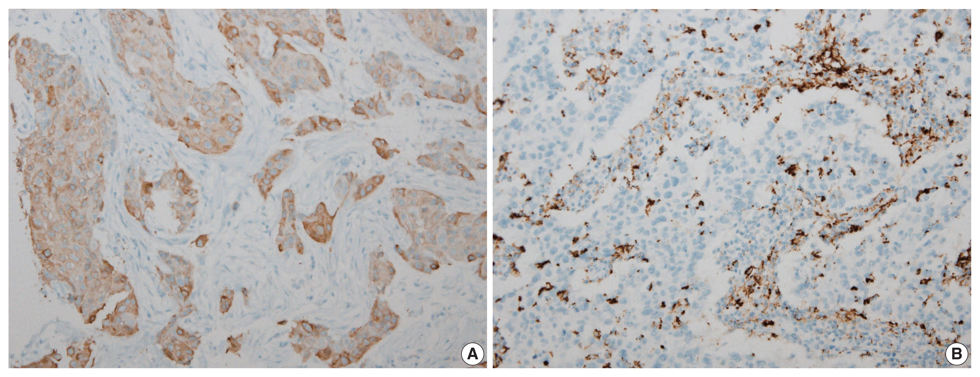
Two pathologists independently observed and agreed to the final score values, and in some cases, IHC study was performed up to three times. The FASN expression levels in breast cancer patients were classified as grade 0, 1, 2, or 3 according to staining intensity. FASN expression was observed in 47.1% (95/166) of TNBC patients. Intensity grades of 1, 2, or 3 were seen in 33.1% (55/166), 13.3% (22/166), and 10.8% (18/166) of TNBC patients, respectively. Grades 0 and 1 were classified as the low-FASN group and grades 2 and 3 as the high-FASN group (Fig. 1). Low and high expression of FASN was identified in 75.9% (126/166) and 24.1% (40/166) of TNBC patients, respectively. Table 2 shows a comparison between the clinicopathological data of 40 patients with high FASN expression compared with that of 126 patients with low FASN expression. In the high-FASN expression group, more patients were younger than 50 years, while in the low-FASN group, more patients were older than 50 years old. The major cancer type was invasive ductal carcinoma and most were histologic grade 3; therefore, there was no association with high and low FASN expression. No statistically significant differences in T category, N category, AJCC stage, and recurrence rate were found between the high- and low-FASN expression groups.
Correlation with immunohistochemical study of Ki-67
The Ki-67 proliferation level was statistically different between the FASN low and high expression groups, with median Ki-67 levels of 60% (range, 40% to 80%) and 73% (range, 55% to 87%), respectively, using the Mann-Whitney U test (p = .003) (Table 3). The Spearman correlation coefficient was 0.257, indicating weak correlation between Ki-67 proliferation level and FASN expression level (p = .001).
Correlation with PD-L1 SP142 IHC assay
Two pathologists independently observed and agreed to the final score values. PD-L1 expression was assessed using the PD-L1 SP142 assay. Of the 166 cases, 94 available cases were used to perform IHC for PD-L1 SP142. At the 1% cutoff value, 52.6% (50/94) of cases were positive for PD-L1 SP142 in infiltrating immune cells (ICs). We analyzed the correlation between FASN expression level and percentage of PD-L1 SP142 positive infiltrating ICs, but no correlation was identified. There was no statistically significant PD-L1 SP142 positivity between the low- and high-FASN expression groups. A representative case of SP142 expression in the expression of high FASN was confirmed (Fig. 2).
DISCUSSION
TNBC has no available special treatment other than chemotherapy after surgery, so early detection of recurrence through short-term follow-up observations will help patients with treatment and prognosis [22]. The development of interval breast cancer within the time interval between screening examinations has more adverse biological features, poorer survival outcomes, and is more highly associated with TNBC [23]. TNBC has high prevalence in young women under 50 and is more common in African Americans [24]. TNBC is known to have relatively poor prognosis compared to non-triple-negative breast cancer, most likely resulting from lack of special treatment methods such as hormone treatment or targeted treatment other than non-specific chemotherapy [25].
The TNBC subtype was classified into four distinct types by Burstein et al. [26] and is being actively studied because of different prognoses and target therapies. Clinical results and treatment responses have changed depending on classification [27]. These inherent differences related to TNBC sub-classification have resulted in a renewed effort to identify driver mutations and more appropriate targeted treatment [28]. For potential therapy, activated progesterone receptor, platinum, tyrosine kinase inhibitor, PI3K/mTOR inhibitor, anti–PD-L1 inhibitors, and/or androgen receptor targeted PI3K inhibitors are thought to be possible [29]. Therefore, these treatments are being attempted in TNBC patients to identify whether they can benefit from a more appropriate and targeted treatment. We performed IHC for FASN, a representative lipid metabolism–related protein, as a candidate for a potential specific target in TNBC.
FASN is a key lipogenic enzyme and is known to be overexpressed in various human cancers [30]. FASN is a lipid-producing enzyme that is expressed at low levels in normal human tissues but is reported to be highly expressed in breast, colon, prostate, ovarian, and endometrial cancers [11–14]. It has been hypothesized that useful indicators of prognosis can be found based on differences in the degree of expression between normal and cancer tissues, and patients with high FASN expression level have been reported to show lower disease-free survival and decreased survival rate [31]. High FASN expression was identified in 45% of TNBC cases [15] and high-FASN groups have been significantly associated with positive nodal status [15,16]. Therefore, FASN has emerged as a potential target, and FASN inhibitors are being evaluated in clinical trials [32]. First-generation (e.g., orlistat and cerulenin) and next-generation (TVB-3166 and TVB-2640) FASN-targeting drugs have been developed [33]. Adipophilin (ADP) and FASN are two lipid metabolism–related proteins of clinicopathological relevance for IHC expression in salivary duct carcinoma. ADP-positive expression was associated with the presence of prominent nuclear pleomorphism, high Ki-67, and poor prognosis [30].
In this study, we evaluated the association of FASN expression between clinicopathological features, including PD-L1 SP142 expression, and further evaluated its usefulness as a biomarker in TNBC. FASN expression was observed in 47.1% (95/166) of TNBC patients in our study. Staining intensity grades of 1, 2, and 3 were found in 33.1% (55/166), 13.3% (22/166), and 10.8% (18/166) of TNBC patients, respectively. Low (grade 0,1) and high (grade 2,3) expression of FASN was identified in 75.9% (126/166) and 24.1% (40/166) of TNBC, respectively. However, we could not identify an association with lymph node stage or TNM stage [15].
Ki-67 IHC is used as a proliferation marker in breast cancer and is a low-cost measurement method suitable for extensive use in clinical practice [34]. Higher Ki-67 level is associated with increased early recurrence and aggression, resulting in a lower patient survival rate [35]. In a Japanese study, when Ki-67 was greater than 10%, it was related with a high recurrence rate and low survival rate [36]. The Breast Cancer Working Group proposed guidelines for Ki-67 assessment in breast cancer and use of this potentially important marker based on current evidence [37]. Less than 5% of patients do not require chemotherapy, and more than 30% show chemotherapy indication. The ER negative case is referred to as insufficient evaluation for Ki-67 [37]. Another group reported that high Ki-67 expression (cutoff value of 30%) was correlated with early recurrence in luminal B/HER-2 negative breast cancer [38]. TNBC patients in the high Ki-67 group seem to benefit more from treatment with carboplatin [39]. The majority of TNBC cases has high Ki-67 expression level (≥ 20%), which is used as a proliferation factor [40]. In our study, 92.8% of cases had Ki-67 expression ≥ 20%. However, the Ki-67 proliferation level was statistically different between the FASN low and high expression groups. The median Ki-67 value of the low- and high-FASN expression groups was 60% (range, 40% to 80%) and 73% (range, 55% to 87%), respectively.
In 2019, the FDA-approved atezolizumab (TECENTRIQ, Genentech Inc.) treatment in combination with paclitaxel was approved for TNBC patients whose PD-L1 stained tumor-infiltrating ICs were over 1% intensity [41]. PD-L1 is a cell membrane protein expressed in tumor cells and ICs [42]. PD-L1 expression is increased in TNBC and is a positive predictor of immunotherapy [43]. PD-L1 IHC using the VENTANA SP142 assay, the current FDA-approved assay, improved clinical outcome in atezolizumab plus nab-paclitaxel-treated patients [44]. In our study, we investigated the association between PD-L1 and FASN expression level and unfortunately identified no association.
TNBC represents a small proportion of breast cancer subtypes but has the worst outcome [45]. Although this study was based on data from a small number of TNBC cases and short-term follow-up surveys, we included 166 TNBC patients in the study. More research on the expression of FASN on TNBC and its potential inhibitors will help identify new target treatments. Further research is needed on how FASN correlates with other factors in TNBC. In addition, further studies are required with a larger number of TNBC patients to investigate the specific role of FASN in TNBC and the possibility for FASN inhibition as a target treatment for TNBC.
In conclusion, we showed FASN expression in TNBC patients and Ki-67 proliferation index to be positively correlated with FASN level, indicating higher proliferation activity as FASN increases. However, there was no statistical association with PD-L1 SP142, the currently FDA-approved assay, or FASN expression level.

 XML Download