INTRODUCTION
Clinical Menifestation
Technical Approach
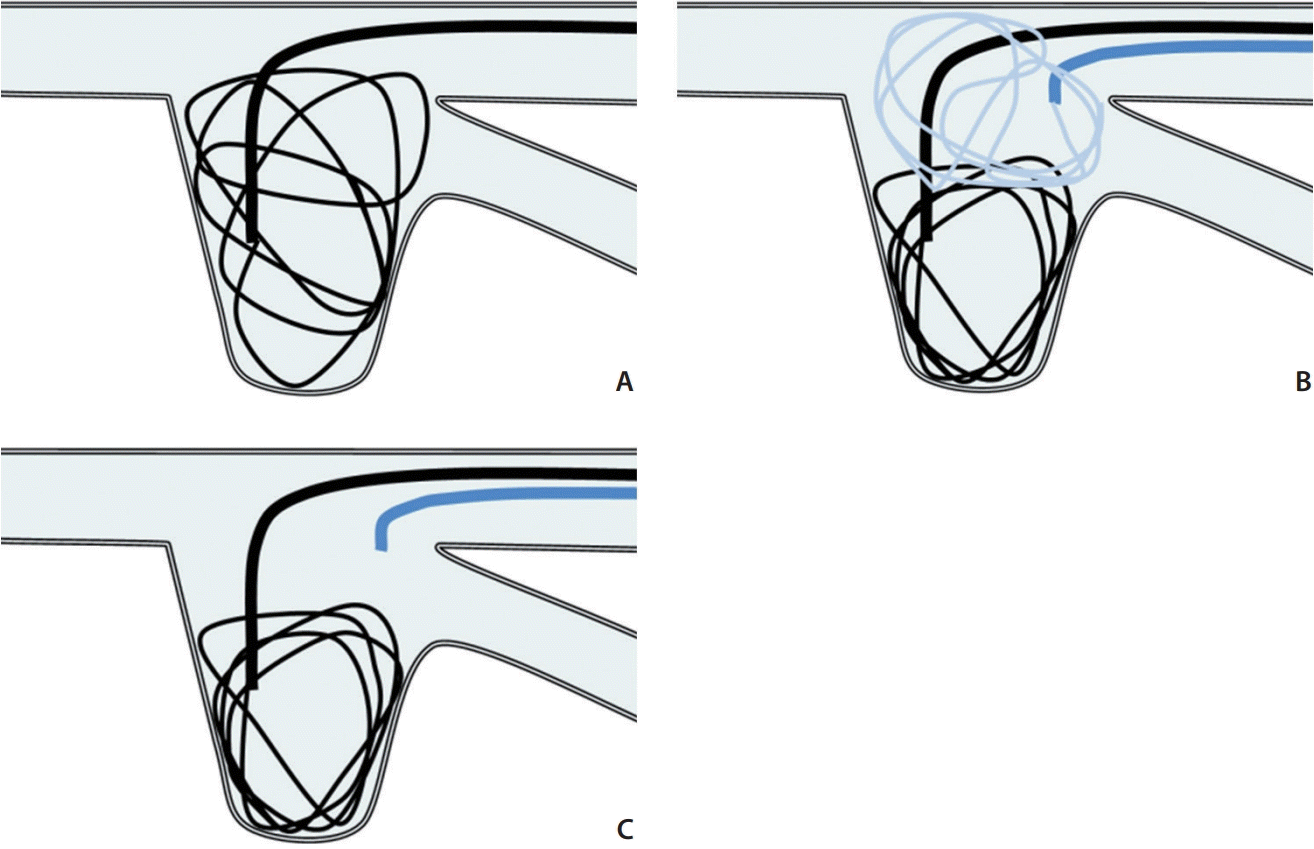
 | Fig. 1.Schematic of the parent artery coil protection technique for the side-branched aneurysm. (A) Because of the wide neck, the first framing coil protrudes into the side branch and parent artery. (B) The first framing coil is inserted into the aneurysm sac and is stable with a protective coil from the second microcatheter. (C) After withdrawing the protective coil, the stability of the framing coil should be confirmed. |
CASE REPORT
Case No. 1
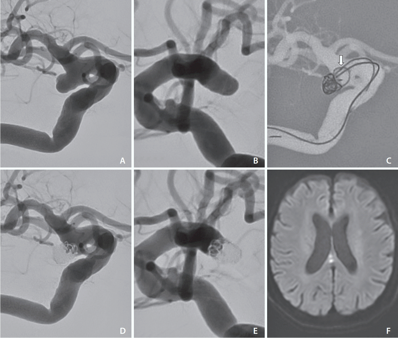
 | Fig. 2.(A, B) Working views of the right internal carotid angiograms show a wide neck IC-PC aneurysm. (C) First framing coil was inserted with the parent artery coil protection from the second microcatheter (white arrow). (D, E) Right internal carotid angiograms after the procedure show good preservation of the posterior communicating artery. (F) Postoperative magnetic resonance DWI shows spotty hyperintense areas, but there were no symptoms. IC-PC, internal carotid-posterior communicating artery; DWI, diffusion-weighted imaging. |
Case No. 2
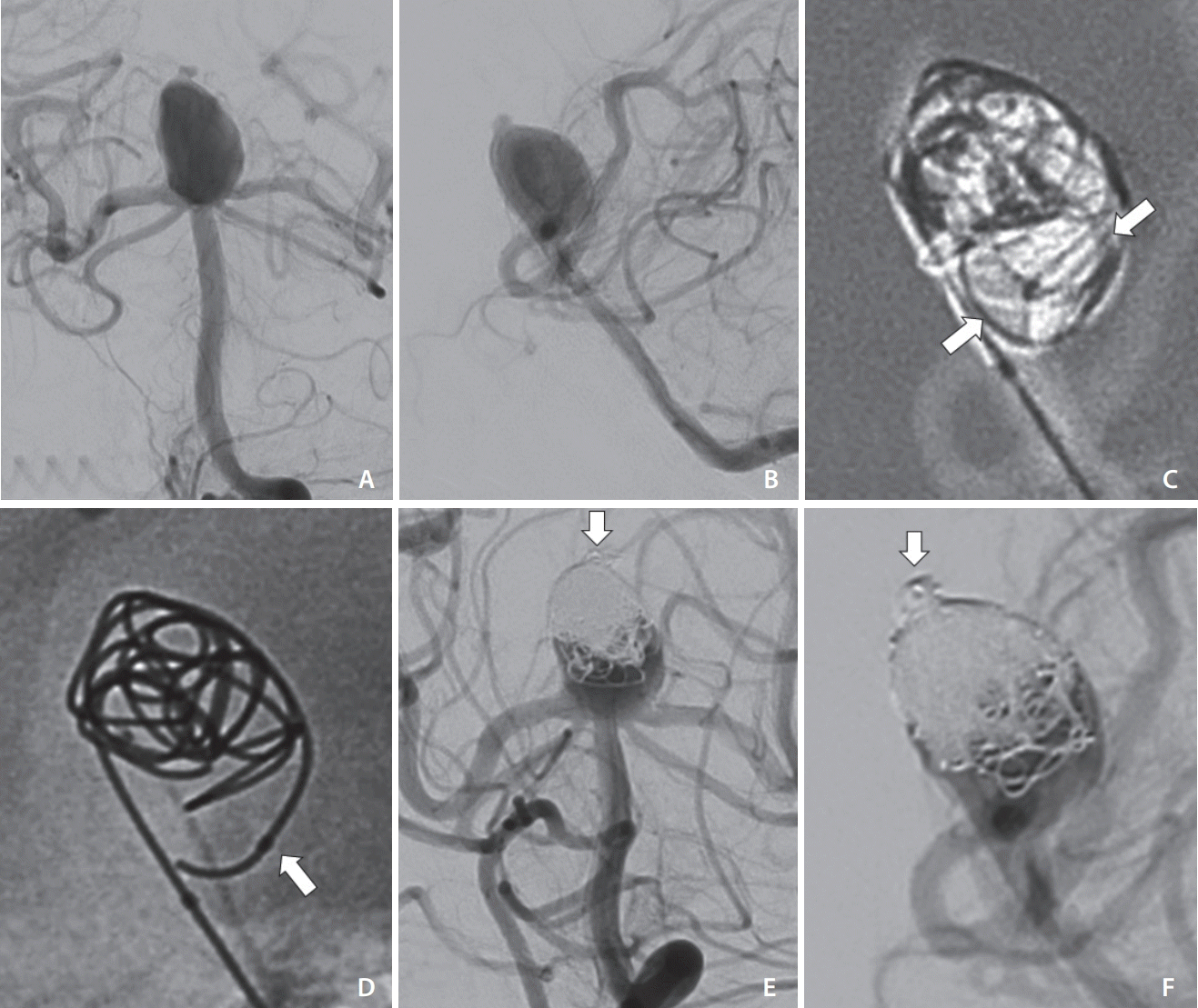
 | Fig. 3.(A, B) Working views for the left vertebral angiograms show a very wide-neck BA tip aneurysm. (C) First framing coil was inserted with the protection of the coil loops (between white arrows) from the second microcatheter. (D) After withdrawing the protection coil, the stability of the framing coil was confirmed. The white arrow shows the second microcatheter tip. (E, F) Left vertebral angiograms after the procedure show good preservation of the bilateral PCA and SCA. The white arrows show good obliteration of the ruptured bleb. BA, basilar artery; PCA, posterior cerebral artery; SCA, superior cerebellar artery. |


 PDF
PDF Citation
 Print
Print


 XML Download