

 PDF
PDF Citation
 Print
Print


INTRODUCTION
Vertebral artery dissecting aneurysm (VADA) is a special subtype of intracranial aneurysms, which can cause subarachnoid hemorrhage (SAH) or acute ischemic stroke, particularly in middle-aged adults [1,24]. Stent-assisted coiling as a reconstructive treatment of VADAs has been proven effective in maintaining the parent artery patency. However, single stent-assisted coiling seems to be insufficient with a high recurrence rate [11,17,20,22,24], which may cause an aneurysm rupture or rebleeding. Multiple stents in an overlapping manner can increase the metal coverage rate and flow-diverting effect, and have shown superior clinical results compared with traditional stent-assisted coiling [14,21,24].
In the reported studies, the number and variety of stents selected and deployment order were usually based on the preference of the operators, essentially considering the anatomy of the aneurysm and parent artery [5,6]. However, the use of more stents increases the operating steps and may cause more complications [4,9]. In addition, the ideal stents’ properties, and the interactions between stents and the aneurysm need to be further elucidated.
In this study, we used an Low-profile Visualized Intraluminal Support (LVIS) stent-within-Neuroform EZ stent manner combined with coiling for the treatment of VADAs. The Neuroform EZ stent is an open-cell designed stent with a strong radial force, and no flared ends. The LVIS stent is a braided stent with a high metal coverage rate. The safety and efficacy of this combination for the treatment of VADA were evaluated.
Go to : 
MATERIALS AND METHODS
This study has been performed in accordance with the Helsinki Declaration and its later amendments. Ethics approval from Institutional Review Board of the First Affiliated Hospital of Nanjing Medical University was obtained for this study (ethics number : 2020-SR-451).
Patient characteristics
From January 2017 to June 2019, a total of 766 consecutive patients with intracranial aneurysm(s) received endovascular treatment in our department. Among them, 18 consecutive cases (male : female=13 : 5; mean age, 52.8±12.3 years) received a reconstructive technique using a Neuroform EZ plus LVIS stent-assisted coiling for a single VADA (ruptured : unruptured=5 : 13). The diagnosis of VADAs was demonstrated by computed tomography angiography (CTA) or magnetic resonance angiography (MRA), and confirmed with digital subtraction angiography (DSA), which was based on typical features including the irregular dilation from the original lumen or a double-lumen sign of the affected segment of VA [5]. For ruptured aneurysms, all patients encountered moderate to severe headache (n=5). All unruptured aneurysms were symptomatic, the main complain of which may resulted from slight or moderate ischemia of posterior circulation, including dizziness (n=6), numbness (n=2), and hemiplegia (n=1), or act as a sign of rupture, including sentinel headache (n=4).
Antiplatelet protocols
For unruptured aneurysms, a dual antiplatelet solution (100 mg of aspirin and 75 mg of clopidogrel) was routinely loaded at least 7 days before procedure. As for ruptured aneurysms, a loading dose of dual antiplatelet medication (300 mg of aspirin and 300 mg of clopidogrel) was given orally at least 2 hours before treatment. All patients were recommended to continue on a dual antiplatelet therapy consisting of aspirin (100 mg) and clopidogrel (75 mg) for at least 8 weeks and mono antiplatelet therapy with aspirin (100 mg) indefinitely. Notably, after 7 days of dual antiplatelet therapy, all patients received a thrombelastogram (TEG) analysis [2] to test drug sensitivity. The drug dosage or category was changed based on the TEG analysis if necessary.
Endovascular treatment procedure
Informed patient consents were obtained before procedure. All procedures were performed under general anesthesia. A 6-F guiding catheter (Envoy, Coidis Neurovascular, Miami, FL, USA) was introduced through a femoral sheath into the distal vertebral artery. Systemic heparinization was initiated to maintain the activated clotting time between 250–300 seconds. Three-dimensional reconstruction angiography was performed to determine a proper working projection for coiling and stent placement. Considering the high radial force of Neuroform EZ stent, the ‘semi-jailing’ technique was used for all cases. Briefly, an XT27 microcatheter (Stryker Neurovascular, Cork, Ireland) was advanced across the aneurysm neck to a distal branch of the parent artery. A second microcatheter was inserted into the aneurysm sac. A Neuroform EZ stent (Stryker Neurovascular) was semi-deployed to partially bridge the aneurysm neck with the central portion and jail the second microcatheter. Followed by the coil (MicroVention, Tustin, CA, USA; ev3, Irvine, CA, USA) embolization, the Neuroform EZ stent was totally deployed. After pulling out the XT27 microcatheter, a headway 21 microcatheter (MicroVention) was advanced to the distal parent artery. An LVIS stent (MicroVention) was then advanced and deployed within the previous Neuroform EZ stent. The size of the stent was determined according to the vessel diameter and length of the affected vertebral artery segment. Usually both sides of the aneurysm neck were covered by at least 5 mm by the Neuroform EZ stent. To keep both sides of the LVIS stent within the Neuroform EZ stent, a shorter LVIS stent was preferred. Notably, to give a radial strength to the aneurysm neck part of the Neuroform stent, the LVIS stent was compressed before deployment at the aneurysm neck. This procedure reshaped the Neuroform EZ stent as a ‘lantern’ and could obtain a better packing density of the aneurysm.
The immediate angiographic outcomes were evaluated based on Raymond classification [18]. The degree of occlusion was divided into three categories : complete occlusion (Raymond class I), aneurysm neck residual (Raymond class II), and aneurysm sac residual (Raymond class III). Technical success was defined as correct stent placement with total aneurysmal neck coverage and patent parent or perforator arteries despite the occlusion degree for unruptured VADA, and successful coiling (Raymond I or II) for ruptured VADA [22]. Raymond class I and II were considered successful occlusion.
Clinical and angiographic follow-up
Follow-up angiography was scheduled 3 to 6 months after embolization. An MRA or CTA was recommended 1 year after the procedure and every 2 years thereafter. The MR and DSA were assessed by two independent observers. Clinical outcomes at discharge and follow-up were evaluated with a modified Rankin Scale (mRS) score via physical examination or a telephone interview. The most recent clinical follow-up records were used as the clinical results in this study.
Statistical analysis
Continuous variables are reported as the mean±standard deviation. Categorical variables are reported as a frequency/percentage. Statistical analysis was performed with IBM SPSS Statistic 20 (IBM-Armonk, New York, NY, USA)
Go to :
RESULTS
As shown in Table 1, 13 (72.2%) of the treated aneurysms were unruptured. The Hunt-Hess score of five ruptured patients ranged from class II to class IV. All dissecting aneurysms were located in V4 segment of vertebral artery. The mean aneurysm size was 6.4±2.3 mm in diameter and 10.6±4.7 mm in length. Posterior inferior cerebral artery (PICA) was involved in nine patients (50.0%).
Table 1.
Patient Manifestations and endovascular procedure-related data
![]()
All but one overlapping stents were successfully deployed achieving a 94.4% technical success rate. The immediate angiography after procedure confirmed Raymond class I in 12 cases (66.7%, including five ruptured VADAs), Raymond class II in two case (11.1%), and Raymond class III in four cases (22.2%). Accordingly, the successful occlusion (Raymond I and II) rate reached 14 cases (77.8%) after procedure.
There was one procedure-related complication in this series. A patient (case 8) with an unruptured aneurysm suffered acute in-stent thrombosis during procedure. After a venous tirofiban bolus injection followed by an immediate arterial infusion was given, the parent artery was recanalized. The MR scan confirmed a small brainstem infarction the second day after the procedure. The symptoms of this patient, which including dizziness, nausea and vomiting, were resolved in 3 days post-procedure with symptomatic treatments. All patients reached the antiplatelet inhibition rates tested by TEG, and the antiplatelet therapy was maintained as previously scheduled.
An angiographic follow-up at 3–9 months (mean, 5.7±1.6 months) showed (Table 1) complete occlusion in 15 cases (83.3%, Fig. 1). All patients elucidated successful occlusion (Raymond I and II) at last angiographic follow-up. Five cases (27.8%) were improved with progressive occlusion of the aneurysm (Fig. 2). One case (case 11) with a residual aneurysm sac at first angiographic follow-up 3 month postoperatively, and received another angiography at 9 months, which improved to Raymond class II. No decreased Raymond class was found. In the clinical follow-up period (mean, 21.3 months; range, 15–42 months), 17 patients (94.4%) had mRS scores of 0–2. The mRS of 16 patients (88.9%) improved and none were decreased. The MRA or CTA follow-up was conducted with clinical follow-up, which showed satisfied patency. No rebleeding occurred during the follow-up.
 | Fig. 1.It shows the treatment procedure and follow-up angiography of a 41-year-old female (case 9) with a ruptured vertebral artery dissecting aneurysm (VADA). A : Angiography before procedure shows the VADA (white arrow). B and C : Angiography after the first stent deployment. C : White arrowheads show that the coils at the neck of the VADA were relatively loose before the second stent insertion. D and E : Angiography after the second stent deployment. E : White arrowheads show that the coils become more compacted. F : Follow-up angiography at 3 months shows the complete occlusion of the aneurysm.
|
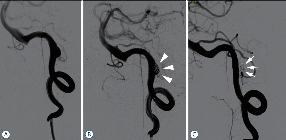 | Fig. 2.It shows that one patient (case 6) with an initial Raymond class III improved to class I during a 3-month angiographic follow-up. A : The initial angiography showed the vertebral artery dissecting aneurysm. B : Angiography after the procedure showed the aneurysm sac filling (arrowheads). C : Angiography after 3 months elucidated that no contrast filled the aneurysm (arrows).
|
Go to :
DISCUSSION
In this study, we used LVIS stent-within-Neuroform EZ stent combined with coiling for the treatment of VADAs and demonstrated the safety and efficacy of this endovascular therapy. Except for one patient who had a residual aneurysmal sac and one patient who had a perioperative ischemic complication, all patients showed satisfactory technical and clinical results during a mean follow-up period of 21.3 months.
VADA is a common cause of SAH or posterior ischemic stroke and is challenging to treat. Ruptured VADAs are associated with a high incidence of re-bleeding, necessitating early treatment [10]. For unruptured VADAs, aggressive treatment is sometimes suggested, especially for patients with symptoms, though the treatment indication and timing remain controversia [5]. Due to its special etiology and anatomy, surgical managements including trapping, clipping, and by-pass were preliminarily used but with relatively high procedure-related complications, especially when the posterior inferior cerebellar artery was involved [19]. Recently, the technique with multiple overlapping stent-assisted coiling for VADAs has been proven useful in maintaining the parent artery patent and preventing aneurysm rupture, and has become the preferred way as opposed to parent artery occlusion.
The manner of overlapping stents varied significantly in previous studies, and its standard strategy has not been defined. Most reported case series used multiple stents with a different combination manner of Enterprise, Neuroform or Solitaire stent. Although the immediate complete embolization rates and procedure-related complications were acceptable with two or more overlapped stents, high recurrence and recanalization rates were observed during the follow-up imaging [5,23,24]. Notably, in our case series, the successful occlusion increased from 77.8% to 100%. The progressive occlusion may be due to the higher metal coverage degree with our stent combination manner via the LVIS stent-within-Neuroform EZ stent. As far as we know, the metal coverage of the LVIS stent was reported as 23% on average [22], which was much higher than the other three types of stents used (ranging from 5.4% to 13.3% based on the parent artery diameter) [13].
To further improve the metal cover rate and flow diversion effect, flow-diverting stents (FDS) with a high metal surface area (30–35%) have been attempted for the treatment of VADAs with or without an overlapping manner. A multicenter study of 131 posterior circulation aneurysms showed that a pipeline embolization device (PED) was effective with an 89.7% complete or near complete occlusion rate for dissecting aneurysms [8]. In most recent, Kim et al. [12] demonstrated a satisfied angiographic result within 3–6 months follow-up, which showed with 77.8% (7/9) complete or near complete VADA obliteration using FDS. Although one patient showed with segmental occlusion, the clinical outcome was mRS 0 in all patients. However, a meta-analysis elucidated that dissecting aneurysms could be one of the risk factors for flow diverter-related complications and showed a trend of morbidity and mortality [16]. The safety of this off-label used FDS for the treatment of VADAs still needs to be furtherly verified. Zhu et al. [25] reported two overlapped LVIS stents combined with coiling for the treatment of blood-blister-like aneurysms (BBAs), and the double-LVIS stent improved metal coverage and resulted in a superior flow-diverting effect to a PED [7]. However, in our previous experiences with double LVIS stents for the treatment of VADAs or BBAs (unpublished data), we found that it was difficult to obtain a good position of the second stent because of the stent migration during the catheter passing through the first stent and placement. This may be a common situation for braided stents due to its relative lower wall adherence. Additionally, it seems to increase the ischemic complications because of the high coverage rate with the two LVIS stents. Based on these, we used Neuroform EZ as the first stent to enhance the radial force, followed by an LVIS stent to improve the metal coverage. Our results preliminarily confirmed that this combination is a reasonable manner not only on the properties and interactions between stents, but also on the metal coverage rate.
Most recently, Lim et al. [15] reported one case using an LIVS blue stent within-an-Enterprise stent for the treatment of VADA. In their theory, the Enterprise stent was first introduced to offer a scaffold to reduce the unstrained segment size across the aneurysm neck and prevent an outward expansion of the LVIS blue stent. This could maintain the flow-diverting effect with a high metal coverage distribution as well as the porosity of the aneurysm neck area. By overlapping the Enterprise stent with an LVIS blue stent, the metal coverage rate was similar to our overlapping manner with Neuroform EZ stent and LVIS stent. Notably, compared with other types of intracranial stents used in most previous studies, the Neuroform EZ stent has the highest radial force, no flared ends, and open cell structure [3,13]. The high radial force make it more stable in the parent artery during the second stent deployment, and no flared ends could decrease the vascular damage caused by stent migration compared with other stent types when pushing the LVIS stent at the aneurysm neck area. In this way, the LVIS stent gives internal support to the Neuroform EZ stent, and ‘destroys’ the continuity of the Neuroform EZ stent surface. This leads to the stent protruding into the aneurysm sac with the help of open cell structure and consequently improves the local coil compaction. Unfortunately, Neuroform EZ is still the only available open-cell designed stent with relative big delivery system (0.027 inch) in China [3]. The disadvantage of this overlapping manner is that the two different stent types require different navigation systems; an extra step of microcatheter exchanging is needed during the procedure. This may prolong the procedure time and lead to ischemic events, as case 8 who was encountered with in-stent thrombosis though with adequate antiplatelet drugs. Early recognition by angiography and the usage of tirofiban may play equally important role for reversing in-stent thrombosis as in our study. Although it may potentially increase the complication rate during the second microcatheter navigation, we found it is technically feasible considering the straight structure of the V4 segment of the vertebral artery.
This study has some limitations that should be mentioned. First, this is a retrospective study. Second, the technique was newly used in the latest years, which results in a relatively small case series and limited follow-up duration. A prospective study with a large case series and longtime observation are needed to confirm our results in the future.
Go to :
CONCLUSION
Based on the characteristics of Neuroform EZ and LVIS stents, we combined these two commonly used stents for the treatment of VADAs. The technical and clinical results were inspiring and proved the feasibility and efficacy of this modification to some extent. However, a prospective study with a large case series is still required.
Go to :
 XML Download