

 PDF
PDF Citation
 Print
Print


Introduction
The anatomical structures of the aortic arch branches (supra-aortic branches) should be considered during percutaneous dilatational tracheostomy (PDT) to prevent accidental vascular injury and associated hemorrhage [1]. Clinicians need to be aware of arterial deviations such as a high aortic arch, wide brachiocephalic trunk (BCT) that overlaps with the cervical trachea, the pretracheal course of the right subclavian artery (SCA) and common carotid artery (CCA), and unusual origins of branches of thyroid arteries [2].
Case
Ethical statements: This study was approved by the Institutional Ethics Committee of Eulji University (EMC 2021-08-005). The informed consent is waived for this case study.
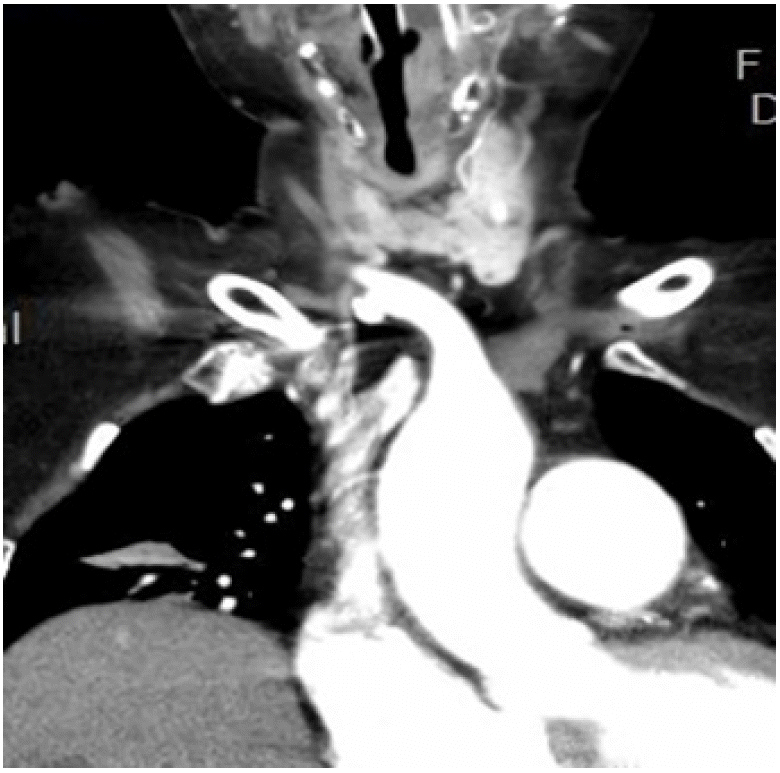
A 72-year-old woman presented as a consult with our team for a tracheostomy. She had a history of prolonged mechanical ventilator support for 1 month because ventilator weaning was not available. The patient had a recent percutaneous coronary intervention for a non-ST elevated myocardial infarction via the left circumflex artery culprit and heart failure. Her past history included right-sided weakness after a cerebrovascular accident with the right middle cerebral artery infarction that had persisted for 7 years, myotonic dystrophy for 3 years and bronchial asthma. Prior to the tracheostomy, we detected a pulsating portion on the anterior surface of the neck, which was targeted for tracheostomy, and a computed tomography (CT) scan showed a high riding anomalous BCT that crossed the tracheostomy target in the midline on the trachea (Fig. 1).
A literature review indicated that a transverse course of BCT or aberrant right SCA precluded longer tracheostomy cannulation and could result in tracheo-brachiocephalic fistula. Therefore, aortic debranching followed by PDT can present as a challenging case for an aortic surgeon.
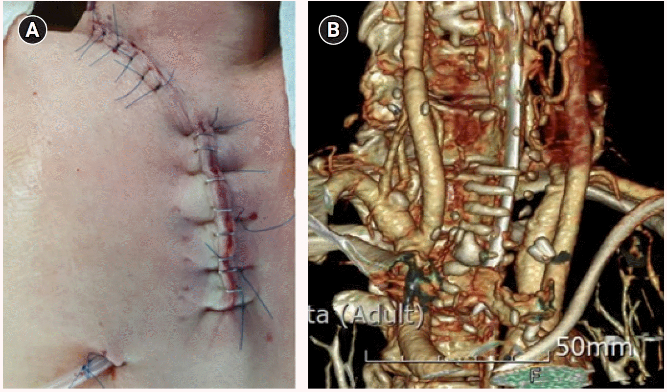
Under general endotracheal anesthesia, we applied a reversed T-shaped partial upper sternotomy from the manubrium to the second intercostal body of the sternum and separated the right supraclavicular incision to optimize exposure of the ascending aorta, aortic arch, and bifurcation of the BCT (Fig. 2A). After systemic heparinization, both sides of BCT were clamped and then divided after bifurcation. The proximal origin of BCT was oversewn and then the distal portion of the SCA and CCA were anastomosed with 10-mm and 8-mm straight Hemashield grafts, which were collagen-impregnated woven double velour polyester grafts (Hemashield Platinum; Maquet Cardiovascular, LLC, Wayne, NJ, USA). The graft from the CCA was anastomosed with the other graft from the SCA as a Y graft using an end-to-side approach. A Y graft produced at our institution was anastomosed with the ascending aorta with an end-to-side approach under a side bite for partial clamping (Fig. 2B). The postoperative wound (Fig. 3A) was examined and determined to be clean and the CT scan indicated that no additional vasculature crossed over the PDT target (Fig. 3B).
At postoperative day 12, we performed percutaneous dilational tracheostomy, which was uneventful and without wound infection, after the debranching of the anomalous BCT had healed. Unfortunately, the patient succumbed to sepsis with pneumonia after three months of prolonged mechanical ventilation via PDT.
Discussion
The anatomical structures of the aortic arch branches (supra-aortic branches) should be considered during PDT to prevent accidental vascular injury and associated hemorrhage PDT [1]. Clinicians need to be aware of arterial deviations such as a high aortic arch, wide BCT that overlaps with the cervical trachea, the pretracheal course of the right SCA and CCA, and unusual origins of branches of thyroid arteries [2].
To avoid injuries of vessels in the low neck, it is important for physicians to consider anatomic variations that can present in individual patients. Vascular variations such as a cervical aortic arch, an accessory inferior thyroidal artery, and high riding BCT in the pretracheal position could be detected by handheld Doppler before tracheostomy [2]. In addition to anatomic variations in the level and size of the arch vessels, maximal neck extension can elevate a normal BCT or arch [3]. Reduced neck extension can reduce injury to vessels that are lower in the neck [4].
In patients with aortic anomalies, a “debranching technique” can be applied as an alternative approach for tracheostomy without any vascular complications. This approach can be considered as an alternative method to prevent vascular complications during tracheostomy as well as for applying long-term mechanical ventilation for patients that cannot be weaned from mechanical ventilation.
In practice, patients are frequently identified with a portion of a major vessel that is anterior to the trachea at sites where a tracheostomy might be performed. Therefore, physicians must be cautious when performing a percutaneous dilational tracheostomy. Debranching techniques can be used for aortic surgeries and also for tracheostomy in patients with any major vessels that overlap with the trachea at the suprasternal notch.

 XML Download