Journal List > Diabetes Metab J > v.46(3) > 1516078002

Kim, Hur, Choi, Oh, and Roh: Influence of Pre-Pregnancy Underweight Body Mass Index on Fetal Abdominal Circumference, Estimated Weight, and Pregnancy Outcomes in Gestational Diabetes Mellitus

Abstract

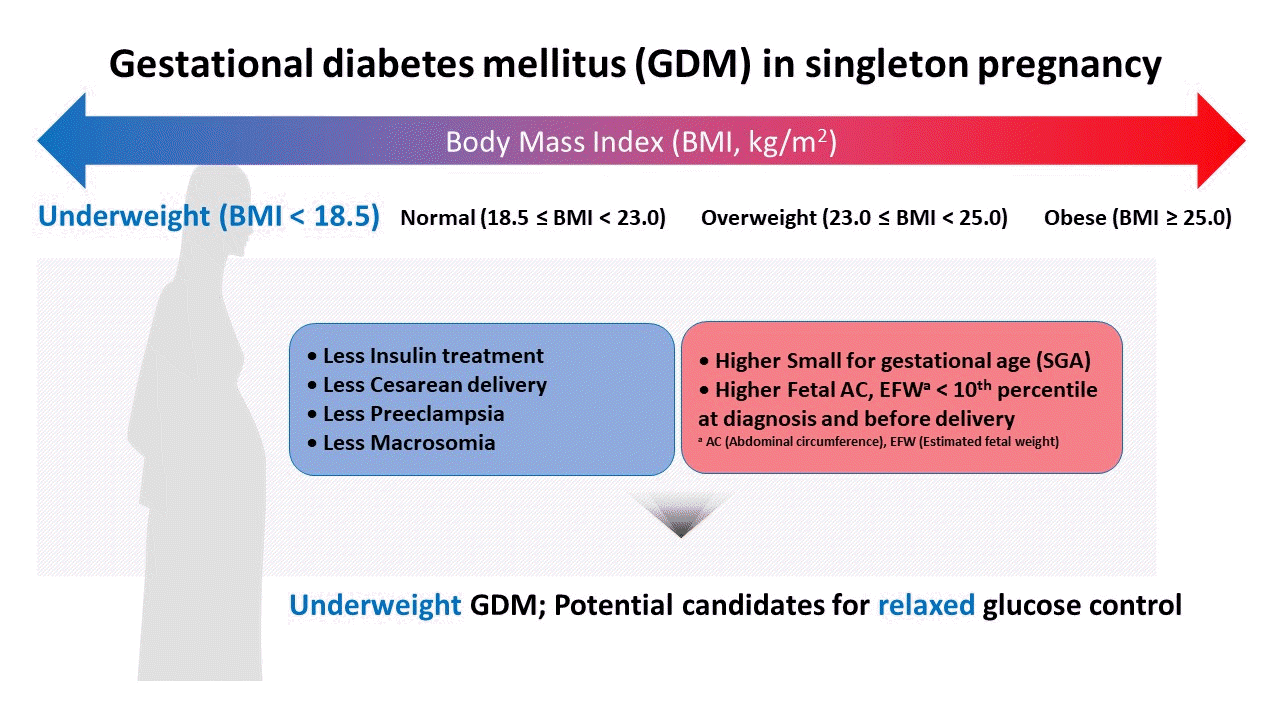
This study aimed to determine the influence of pre-pregnancy body mass index on pregnancy outcomes in gestational diabetes mellitus (GDM), comparing underweight patients with GDM with normal weight patients with GDM. Maternal baseline characteristics, ultrasonographic results, and pregnancy and neonatal outcomes were reviewed in 946 women with GDM with singleton pregnancies. Underweight patients with GDM showed a benign course in most aspects during pregnancy, except for developing a higher risk of giving birth to small for gestational age neonates. Underweight women with GDM required less insulin treatment, had a higher rate of vaginal delivery, and had a lower rate of cesarean delivery. In addition, their neonates were more likely to have fetal abdominal circumference and estimated fetal weight below the 10th percentile both at the time of GDM diagnosis and before delivery. Notably, their risk for preeclampsia and macrosomia were lower. Collectively, our data suggest that underweight women with GDM may require a different approach in terms of diagnosis and management throughout their pregnancy.

Graphical abstract

INTRODUCTION

Generally, conventional therapy for gestational diabetes mellitus (GDM) is based on maternal blood glucose levels [1]; strict control of blood glucose is recommended to prevent adverse pregnancy outcomes in patients with GDM, including macrosomia, increased cesarean section rate, and neonatal complications [2]. However, studies have shown that tight glycemic control of GDM, regardless of fetal growth, may increase the risk of giving birth to small for gestational age (SGA) neonates [3,4]. These data collectively suggest that glucose control in patients with GDM needs to be tailored according to individual growth potential or preexisting risk factors for SGA.
Given these, the purpose of this study was to characterize GDM in underweight women as potential candidates for more relaxed glucose control based on maternal characteristics, prenatal ultrasonographic findings, and pregnancy and neonatal outcomes.

METHODS

Study design

This was a retrospective cohort study of 946 consecutive women with GDM who gave birth between January 2006 and December 2015 at a single tertiary center located in Seoul, Republic of Korea. Based on electronic medical records, women with preexisting diabetes or multiple pregnancies were excluded [5,6]. The study was approved by the Institutional Review Board in Samsung Medical Center (reference number: 2018-02-041). Informed consent was waived by the board.
In this study, the last body weight recorded before confirmation of pregnancy was defined as the pre-pregnancy weight. Body weight at delivery was also obtained upon admission of the patient for delivery. According to pre-pregnancy body mass index (BMI) standards, the following definitions were used: underweight (BMI <18.5 kg/m2), normal weight (BMI ≥18.5 and <23.0 kg/m2), overweight (BMI ≥23.0 and <25.0 kg/m2), and obese (BMI ≥25.0 kg/m2) [7]. Ultrasonographic examinations were performed at the time of GDM diagnosis and within 3 weeks before delivery; we defined delta fetal abdominal circumference (AC) as the change in AC between the two sonographic evaluation periods (cm/week), which we used to determine whether change in fetal AC throughout gestation differs according to pre-pregnancy BMI. Delta weight was defined as the weight change of the mother from GDM diagnosis to delivery. Neonatal hypoglycemia was defined as an initial blood glucose level of <35 mg/dL.

Statistical analyses

Before performing statistical analyses, we evaluated the normality of the data gathered. Non-parametric data were reported as medians and ranges (minimum to maximum) for continuous variables, and parametric variables were reported as means and standard deviations. Linear-by-linear associations in the χ2 independence test were analyzed for categorical variables. The Jonckheere-Terpstra test was performed to evaluate the trend of continuous variables among the BMI categories. Each BMI group was compared with the normal weight group, and odds ratios of pregnancy outcomes were evaluated using multivariable analysis adjusted for age, parity, insulin treatment, and delta weight. Weight change after GDM diagnosis to delivery was included as a covariate due to previous research suggesting that it may impact pregnancy outcomes [810]. In addition, we compared pregnancy and neonatal outcomes between the underweight group and other groups. The underweight and normal weight, underweight and overweight, underweight and obese groups were analyzed separately to determine whether underweight women had discriminatory outcomes compared with the other pre-pregnancy BMI groups. Statistical analyses were conducted using SPSS version 21.0 (IBM Co., Armonk, NY, USA); statistical significance was set at a P<0.016 using the Bonferroni method due to the multiple comparisons made. A probability value of P<0.05 was considered statistically significant except for inter-group analysis.

RESULTS

Maternal characteristics

We analyzed 946 consecutive women with GDM who delivered at our institution between January 2006 and December 2015. Since the total number of deliveries in our institution was 17,804 during the study period, the incidence of singleton GDM was estimated to be 5.3%. The distribution of GDM according to the pre-pregnancy BMI categories was as follows: underweight (9.1%), normal weight (48.3%), overweight (16.0%), obese (22.2%). As shown in Table 1, the median maternal age was the youngest in the underweight group and was increased in the higher BMI groups (P=0.005). There was a significant difference observed in underweight women when age was compared with each pre-pregnancy BMI group. The proportion of primiparous women was the highest in the underweight group and appeared to decrease as BMI increased (P=0.001). When compared with each BMI category, parity showed more significant differences in underweight women than in overweight or obese women. Additionally, women treated with insulin also showed a significant decrease in BMI (P<0.001). Specifically, underweight women required less insulin treatment, and overweight women required more insulin treatment. Moreover, glycosylated hemoglobin (HbA1c) results at the time of diagnosis also showed a significant difference (P<0.001) among the groups and showed a significant difference in underweight women compared with overweight or obese women. Although insignificant, the difference in HbA1c level from diagnosis to delivery was also lower in underweight women than in normal weight women. In addition, correlation analysis between neonatal weight and HbA1c level at delivery in the total study population showed that neonatal weight was significantly correlated with the glycemic control status (r=0.259, P=0.004). Weight change from GDM diagnosis to delivery (delta weight) was the highest in underweight women (P<0.001).

Sonographic findings

Table 1 shows the sonographic findings of the study population. Among the sonographic results obtained at the time of GDM diagnosis, both AC and estimated fetal weight (EFW) showed significant differences among the groups. Underweight women had the highest proportion of AC and EFW percentiles below 10% (P<0.001 and P=0.015, respectively). Similar results were obtained using sonography before delivery. AC percentile at the time of GDM diagnosis was significantly different in underweight women compared with obese women. AC percentile before delivery showed a significant difference in underweight women compared with overweight women. Both EFW percentile before delivery and delta AC were significantly different in underweight women compared with overweight or obese women. In addition, correlation analysis between fetal AC before delivery and HbA1c level at delivery in the total study population showed that fetal AC before delivery was significantly correlated with the glycemic control status (r=0.293, P=0.001). Delta AC also showed statistically significant differences among the BMI groups (P=0.001). Underweight women had the lowest change in AC (delta AC) with an increasing trend observed as BMI increased (P=0.001).

Pregnancy and neonatal outcomes

Table 2 describes the pregnancy and neonatal outcomes. When stratified using the four pre-pregnancy BMI categories, the proportion of women delivered by cesarean section showed an increasing trend across the BMI categories (P<0.001). Interestingly, spontaneous labor showed the highest incidence in underweight women with a decreasing trend observed as BMI increased (P<0.001). Neonatal weight at birth and incidence of macrosomia appeared to be the lowest in the underweight group (P<0.001 and P=0.003, respectively). However, there was a trend toward an increased prevalence of SGA observed in the underweight group (P=0.173). Cesarean delivery and neonatal weight showed significant differences in underweight women compared with overweight or obese women. Neonatal glucose was significantly different in underweight women compared with obese women.

Odds ratios for adverse pregnancy and neonatal outcomes

To assess the relative risk of adverse pregnancy outcomes in underweight women with GDM compared with normal weight patients with GDM, we performed multivariable analysis adjusted for age, parity, insulin treatment, and delta weight, with normal weight used as a reference group (Supplementary Table 1). Although the underweight group did not have a significant increase in the odds for adverse outcome variables such as cesarean delivery (adjusted odds ratio [aOR], 0.738; 95% confidence interval [CI], 0.429 to 1.268), preeclampsia (aOR, 2.910; 95% CI, 0.663 to 12.762), macrosomia (aOR, 0.616; 95% CI, 0.071 to 5.332), and admission to the neonatal intensive care unit (aOR, 1.491; 95% CI, 0.575 to 3.865), the odds of SGA were five times higher in the underweight group than in the normal weight group (aOR, 4.958; 95% CI, 1.172 to 20.971).

DISCUSSION

This study was motivated by a relatively common situation in our clinical practice in which lean women are diagnosed with GDM with the measurements of their neonates by ultrasound already falling under SGA. Considering that the main purpose of GDM management is to prevent macrosomia, it is unclear whether strict glucose control is mandatory for underweight women with GDM.
The general population of patients with diabetes shows that the fewest complications occur among patients with normal weight, with a slight increase in adverse outcomes in the underweight group [11]. However, this may not be applicable to complications of GDM. With exemption to SGA, our data showed that pre-pregnancy underweight women with GDM had the lowest incidence of adverse pregnancy outcomes, covering both cesarean section rate and neonatal respiratory distress syndrome. With regard to macrosomia and increased cesarean section rates, which are important complications of uncontrolled GDM, underweight women manifested the most benign course among women grouped by BMI category. These results are in line with another study showing that increased maternal BMI was significantly associated with the risk of adverse pregnancy outcomes such as the requirement for insulin, development of preeclampsia, and need for cesarean delivery [12].
Normalizing glucose levels in all patients with GDM may result in unnecessary insulin treatment in pregnancies not at increased risk for fetal complications and may result in intrauterine growth restriction [3,4]. Studies have suggested that limiting insulin therapy to fetuses with increased AC for gestational age is associated with good outcomes, without increasing the risks for both macrosomia and SGA [6,13,14]. Despite having research results and evidence-based recommendations, ultrasonography-based therapy for GDM is still not commonly practiced [13,15]. In our study, the proportion of women with fetal AC below the 10th percentile in underweight women with GDM increased from 12% at the time of GDM diagnosis to 18% at delivery. This change is in contrast with that of the normal weight group, which decreased from 9.5% to 8.9%. Thus, it is presumable that current management, which is uniformly applied to patients with GDM regardless of pre-pregnancy BMI, may negatively affect fetal growth. This observation is in concordance with previous studies [3,16], arguing that initiating insulin treatment to strictly control blood glucose levels in low-risk women is unnecessary. This may be true, considering that the results of our study show that SGA in underweight women is increased more than that in the normal weight group, even after adjusting for age, parity, and management after GDM diagnosis (insulin treatment and weight change from diagnosis to delivery) in multivariable analysis. Our finding of a lower increase in the fetal AC (delta AC) in underweight individuals with GDM also raises the need for strict glucose control in the underweight group.
Our study is unique in that we focused on the characterization of women with underweight GDM. Notably, this is the first study to use the concept of delta AC, which was smaller in underweight women with GDM than in overweight women with GDM. This study had a relatively large sample size, and all patients received concurrent uniform screening and diagnosis, antenatal care, and perinatal care in a single tertiary hospital. In fact, our study appears to have the largest sample size among the most recent studies evaluating fetal AC as well as maternal blood glucose levels (n=98 [4], n=199 [17], and n=299 [18]). However, this study had some limitations. Although the total study population was relatively large, the small sample size of underweight women compared with the other pre-pregnancy BMI groups may have decreased the power of this study. Additionally, this study does not contain data on the long-term outcomes of the offspring. Finally, since our study population only included women from a single tertiary center, our data may not represent the entire population of the Republic of Korea.
In conclusion, underweight women with GDM have a benign course in most aspects except for a higher risk of their neonates becoming SGA, indicating that underweight women with GDM may require a different approach in terms of diagnosis and management throughout pregnancy. Therefore, we agree on a “relaxed maternal glycemic target” in the management of underweight women with GDM as previously suggested [14].

SUPPLEMENTARY MATERIALS

Supplementary materials related to this article can be found online at https://doi.org/10.4093/dmj.2021.0059.

Notes

CONFLICTS OF INTEREST

Kyu-Yeon Hur was editorial board member of the Diabetes & Metabolism Journal from 2020 to 2021. She was not involved in the review process of this article. Otherwise, there was no conflict of interest.

AUTHOR CONTRIBUTIONS

Conception or design: M.K., S.Y.O.

Acquisition, analysis, or interpretation of data: M.K., K.Y.H., S.J.C., S.Y.O., C.R.R.

Drafting the work or revising: M.K., K.Y.H., S.J.C., S.Y.O., C.R.R.

Final approval of the manuscript: S.Y.O

FUNDING

None

ACKNOWLEDGMENTS

We would like to thank the staff of the Department of Obstetrics and Gynecology of Samsung Medical Center, Seoul, Republic of Korea and editorial support of Sungkyunkwan University, Seoul, Republic of Korea.

REFERENCES

1. Hyperglycemia and Adverse Pregnancy Outcome (HAPO) Study Cooperative Research Group. Hyperglycemia and Adverse Pregnancy Outcome (HAPO) study: preeclampsia. Am J Obstet Gynecol. 2010; 202:255.
2. Vaarasmaki M. Is it worth treating gestational diabetes: if so, when and how? Diabetologia. 2016; 59:1391–5.
crossref
3. Langer O, Levy J, Brustman L, Anyaegbunam A, Merkatz R, Divon M. Glycemic control in gestational diabetes mellitus: how tight is tight enough: small for gestational age versus large for gestational age? Am J Obstet Gynecol. 1989; 161:646–53.
4. Kjos SL, Schaefer-Graf U, Sardesi S, Peters RK, Buley A, Xiang AH, et al. A randomized controlled trial using glycemic plus fetal ultrasound parameters versus glycemic parameters to determine insulin therapy in gestational diabetes with fasting hyperglycemia. Diabetes Care. 2001; 24:1904–10.
crossref
5. Yogev Y, Langer O. Pregnancy outcome in obese and morbidly obese gestational diabetic women. Eur J Obstet Gynecol Reprod Biol. 2008; 137:21–6.
crossref
6. Buchanan TA, Kjos SL, Schafer U, Peters RK, Xiang A, Byrne J, et al. Utility of fetal measurements in the management of gestational diabetes mellitus. Diabetes Care. 1998; 21(Suppl 2):B99–106.
7. Kim M, Park J, Kim SH, Kim YM, Yee C, Choi SJ, et al. The trends and risk factors to predict adverse outcomes in gestational diabetes mellitus: a 10-year experience from 2006 to 2015 in a single tertiary center. Obstet Gynecol Sci. 2018; 61:309–18.
crossref
8. Katon J, Reiber G, Williams MA, Yanez D, Miller E. Weight loss after diagnosis with gestational diabetes and birth weight among overweight and obese women. Matern Child Health J. 2013; 17:374–83.
crossref
9. Kurtzhals LL, Norgaard SK, Secher AL, Nichum VL, Ronneby H, Tabor A, et al. The impact of restricted gestational weight gain by dietary intervention on fetal growth in women with gestational diabetes mellitus. Diabetologia. 2018; 61:2528–38.
crossref
10. Zheng W, Huang W, Liu C, Yan Q, Zhang L, Tian Z, et al. Weight gain after diagnosis of gestational diabetes mellitus and its association with adverse pregnancy outcomes: a cohort study. BMC Pregnancy Childbirth. 2021; 21:216.
crossref
11. Zheng W, McLerran DF, Rolland B, Zhang X, Inoue M, Matsuo K, et al. Association between body-mass index and risk of death in more than 1 million Asians. N Engl J Med. 2011; 364:719–29.
12. Roman AS, Rebarber A, Fox NS, Klauser CK, Istwan N, Rhea D, et al. The effect of maternal obesity on pregnancy outcomes in women with gestational diabetes. J Matern Fetal Neonatal Med. 2011; 24:723–7.
crossref
13. Quevedo SF, Bovbjerg ML, Kington RL. Translation of fetal abdominal circumference-guided therapy of gestational diabetes complicated by maternal obesity to a clinical outpatient setting. J Matern Fetal Neonatal Med. 2017; 30:1450–5.
crossref
14. Kjos SL, Schaefer-Graf UM. Modified therapy for gestational diabetes using high-risk and low-risk fetal abdominal circumference growth to select strict versus relaxed maternal glycemic targets. Diabetes Care. 2007; 30(Suppl 2):S200–5.
crossref
15. Metzger BE, Buchanan TA, Coustan DR, de Leiva A, Dunger DB, Hadden DR, et al. Summary and recommendations of the Fifth International Workshop-Conference on Gestational Diabetes Mellitus. Diabetes Care. 2007; 30(Suppl 2):S251–60.
crossref
16. Dooley SL, Metzger BE, Cho NH. Gestational diabetes mellitus: influence of race on disease prevalence and perinatal outcome in a U.S. population. Diabetes. 1991; 40(Suppl 2):25–9.
crossref
17. Schaefer-Graf UM, Kjos SL, Fauzan OH, Buhling KJ, Siebert G, Buhrer C, et al. A randomized trial evaluating a predominantly fetal growth-based strategy to guide management of gestational diabetes in Caucasian women. Diabetes Care. 2004; 27:297–302.
crossref
18. Bonomo M, Cetin I, Pisoni MP, Faden D, Mion E, Taricco E, et al. Flexible treatment of gestational diabetes modulated on ultrasound evaluation of intrauterine growth: a controlled randomized clinical trial. Diabetes Metab. 2004; 30:237–44.
crossref

dmj-2021-0059f1.tif
Table 1
Maternal characteristics and sonographic findings according to pre-pregnancy BMI
Characteristic Total (n=946) Underweight (n=86) Normal (n=457) Overweight (n=151) Obese (n=210) P value
Baseline characteristics
 Age, yr 34 (20–52) 32 (25–45)e,f,g 34 (20–46) 35 (25–46) 34 (22–52) 0.005c
 Primiparous 461 (48.7) 52 (60.5)f,g 231 (50.5) 66 (43.7) 86 (41.0) 0.001d
 Insulin treatment 262 (27.7) 19 (22.1) 97 (21.2) 52 (34.4) 75 (35.7) <0.001d
 HbA1c at diagnosis, % 5.3 (3.2–6.5) 5.2 (4.5–6.3)f,g 5.2 (3.2–6.5) 5.4 (4.5–6.4) 5.6 (4.7–6.4) <0.001c
 HbA1c before delivery, % 5.5±0.4 5.2±0.4 5.5±0.5 5.5±0.5 5.6±0.3 0.065c

Weight variable
 Pre-pregnancy BMI, kg/m2 22.0 (14.5–44.6) 17.9 (14.5–18.5) 20.7 (18.5–23.0) 24.0 (23.0–25.0) 27.5 (25.0–44.6) <0.001c
 Diagnosis BMI, kg/m2 25.0 (16.6–47.9) 20.4 (16.9–24.9) 23.6 (18.3–32.5) 26.9 (21.7–32.6) 30.2 (23.7–47.9) <0.001c
 Delta weight, kg 2.7±3.7 3.4±3.4g 2.8±3.5 2.5±3.8 2.4±4.0 0.003c
 Delivery BMI, kg/m2 26.2 (16.7–47.6) 21.6 (16.7–27.4) 24.6 (18.4–32.5) 27.9 (22.7–35.1) 31.2 (25.3–47.6) <0.001c

Sonography at diagnosisa 601 50 308 96 137
 AC percentile at diagnosis, % g <0.001d
  <10 48 (8.0) 6 (12.0) 29 (9.5) 6 (6.3) 6 (4.4)
  10–50 342 (57.1) 27 (54.0) 183 (59.8) 60 (62.5) 69 (50.4)
  50–90 184 (30.7) 15 (30.0) 89 (29.1) 28 (29.2) 47 (34.3)
  >90 25 (4.2) 2 (4.0) 5 (1.6) 2 (2.1) 15 (11.0)
 EFW percentile at diagnosis, % 0.015d
  <10 9 (1.5) 2 (4.0) 5 (1.6) 0 2 (1.5)
  10–25 68 (11.3) 5 (10.0) 35 (11.4) 13 (13.5) 11 (8.0)
  25–50 237 (39.4) 19 (38.0) 133 (43.2) 35 (36.5) 50 (36.5)
  50–75 170 (28.3) 15 (30.0) 85 (27.6) 29 (30.2) 38 (27.7)
  75–90 84 (14.0) 5 (10.0) 41 (13.3) 13 (13.5) 24 (17.5)
  >90 33 (5.5) 4 (8.0) 9 (2.9) 6 (6.3) 12 (8.8)

Sonography before delivery 863 80 417 133 197
 AC percentile before deliveryb, % f <0.001d
  <10 77 (9.5) 13 (17.8) 35 (8.9) 7 (5.6) 18 (9.5)
  10–50 411 (50.4) 39 (53.4) 220 (56.0) 54 (42.9) 81 (42.6)
  50–90 291 (35.7) 19 (26.0) 124 (31.6) 59 (46.8) 77 (40.5)
  >90 37 (4.6) 2 (2.7) 14 (3.6) 6 (4.8) 14 (7.4)
 EFW percentile before deliveryb, % f,g <0.001d
  <10 61 (7.1) 6 (7.5) 35 (8.4) 4 (3.0) 13 (6.6)
  10–25 138 (16.0) 22 (27.5) 66 (15.9) 18 (13.5) 28 (14.3)
  25–50 250 (29.1) 24 (30.0) 120 (28.9) 37 (27.8) 51 (26.0)
  50–75 250 (29.1) 18 (22.5) 124 (29.9) 46 (34.6) 53 (27.0)
  75–90 109 (12.7) 9 (11.3) 51 (12.3) 19 (14.3) 28 (14.3)
  >90 52 (6.0) 1 (1.3) 19 (4.6) 9 (6.8) 23 (11.7)

Delta AC, cm/week 0.90 (−0.50 to 4.08) 0.87 (0.00 to 1.36)f,g 0.89 (−0.04 to 4.08) 0.93 (0.47 to 1.70) 0.91 (−0.50 to 1.42) 0.001c

Values are presented as median (range), number (%), or mean±standard deviation. Delta weight: weight change from gestational diabetes mellitus diagnosis to delivery (n=794). Significance was set at P<0.016 by Bonferroni method due to multiple comparison.

BMI, body mass index; HbA1c, glycosylated hemoglobin; AC, abdominal circumference; EFW, estimated fetal weight.

a Ten cases were excluded from the analysis according to pre-pregnancy BMI due to unavailability of data,

b Thirty-four (AC percentile) and 36 (EFW percentile) cases were excluded from analysis according to pre-pregnancy BMI due to unavailability of data,

c Jonckheere-Terpstra test,

d Linear-by-linear association,

e Inter-group difference between the underweight and normal weight groups,

f Inter-group difference between the underweight and overweight groups,

g Inter-group difference between the underweight and obese groups.

Table 2
Pregnancy and neonatal outcome according to pre-pregnancy body mass index
Variable Total (n=946) Underweight (n=86) Normal (n=457) Overweight (n=151) Obese (n=210) P value
Gestational age at delivery, day 272 (163–294) 272 (204–288) 273 (163–294) 271 (188–289) 270 (176–288) 0.006b
Neonate weight, kg 3.16 (0.46–4.77) 3.10 (1.12–4.29)d,e 3.16 (0.56–4.41) 3.20 (0.78–4.77) 3.28 (0.46–4.62) <0.001b
Male sex 509 (53.8) 49 (57.0) 251 (54.9) 82 (54.3) 103 (49.0) 0.137c
Neonate glucose, g/dL 67 (19–190) 70 (37–128)e 68 (25–185) 68 (33–190) 63 (19–177) 0.004b
Preterm delivery 165 (17.4) 13 (15.1) 52 (11.4) 26 (17.2) 43 (20.5) 0.008c
Cesarean delivery 439 (46.4) 25 (29.1)d,e 185 (40.5) 85 (56.3) 119 (56.7) <0.001c
Preeclampsia 30 (3.2) 3 (3.5) 6 (1.3) 3 (2.0) 14 (6.7) 0.003c
SGA 21 (2.2) 4 (4.7) 11 (2.4) 1 (0.7) 4 (1.9) 0.173c
Fetal anomaly 18 (1.9) 2 (2.3) 4 (0.9) 5 (3.3) 6 (2.9) 0.130c
Macrosomia 33 (3.5) 1 (1.2) 10 (2.2) 7 (4.6) 13 (6.2) 0.003c
NICU 110 (11.6) 9 (10.5) 33 (7.2) 15 (9.9) 30 (14.3) 0.022c
Shoulder dystociaa 3 (0.3) 0 2 (0.4) 0 1 (0.5) 0.812c
Hypoglycemia 25 (2.7) 0 10 (2.2) 2 (1.4) 11 (5.4) 0.009c
RDS 52 (5.5) 1 (1.2) 11 (2.4) 6 (4.0) 17 (8.1) <0.001c

Values are presented as median (range) or number (%). Significance was set at P<0.016 by Bonferroni method due to multiple comparison.

SGA, small for gestational age; NICU, neonatal intensive care unit; RDS, respiratory distress syndrome.

a Number of vaginal deliveries was used as the denominator,

b Jonckheere-Terpstra test,

c Linear-by-linear association,

d Inter-group difference between the underweight and overweight groups,

e Inter-group difference between the underweight and obese groups.

TOOLS
Similar articles