Journal List > J Korean Med Sci > v.37(22) > 1162234

Kim, Yang, Paik, Jeong, Kim, Park, and Lee: The Assessment and Outcomes of Crossmatching in Lung Transplantation in Korean Patients

Abstract

Background

In lung transplantation, human leukocyte antigen (HLA) compatibility is not included in the lung allocation score system or considered when placing donor allografts. However, HLA matching may affect the outcomes of lung transplantation. This study evaluated the current assessment status, prevalence, and effects of HLA crossmatching in lung transplantation in Korean patients using nationwide multicenter registry data.

Methods

Two hundred and twenty patients who received lung transplantation at six tertiary hospitals in South Korea between March 2015 and December 2019 were retrospectively reviewed. Clinical data, including general demographic characteristics, primary diagnosis, and pretransplant status of the recipients and donors registered by the Korean Organ Transplant Registry, were retrospectively analyzed. Survival analysis was performed using the Kaplan-Meier method with log-rank tests.

Results

Complement-dependent cytotoxic crossmatch (CDC-XM) was performed in 208 patients (94.5%) and flow cytometric crossmatch (flow-XM) was performed in 125 patients (56.8%). Among them, nine patients (4.1%) showed T cell- and/or B cell-positive crossmatches. The incidences of postoperative complications, including primary graft dysfunction, acute rejection, and chronic allograft dysfunction in positively crossmatched patients, were not significant compared with those in patients without mismatches. Moreover, Kaplan-Meier analyses showed poorer 1-year survival in patients with positive crossmatch according to CDC-XM (P < 0.001) and T lymphocyte XM (P = 0.002) than in patients without mismatches.

Conclusion

Positive CDC and T lymphocyte crossmatching results should be considered in the allocation of donor lungs. If unavailable, the result should be considered for postoperative management in lung transplantation.

Graphical Abstract

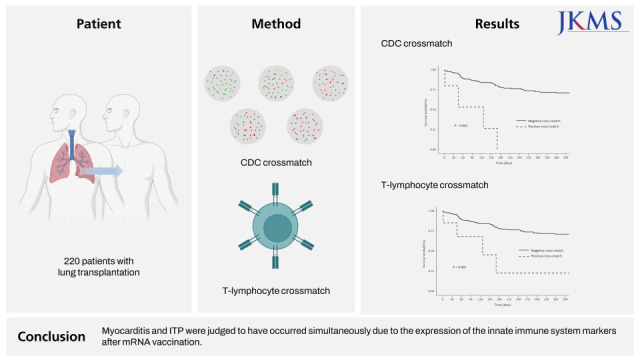
jkms-37-e177-abf001.jpg

INTRODUCTION

Crossmatching is the assessment of the immune compatibility of a particular donor and the recipient. The association between positive crossmatches and hyperacute rejection was first demonstrated in the 1960s with renal transplantation.1 Over the following decades, a positive crossmatch result began to be considered a contraindication to transplantation because of its devastating postoperative effects.23 In particular, complement-dependent cytotoxicity (CDC) results against T lymphocytes are considered an absolute contraindication.34 However, the meaning of positive crossmatches has changed with the development of crossmatching methodology and immunosuppression strategies.56 Early research suggested that it was crucial to avoid the impact of positive T cell crossmatches on renal transplantation; however, because of their low specificity and sensitivity, recent analyses have found that avoidance is not mandatory.2 A combination of different crossmatching techniques has recently been recommended for solid organ transplantations.7
In lung transplantation, considering the limited number of available donors, persistent demand for required organs, and risk of increasing incidences of wait-list mortality and morbidity, immunological “mismatches” have to be accepted despite the potential for the development of donor-specific antibodies (DSAs), which can trigger antibody-mediated rejection (AMR).8910 A pretransplant crossmatch is not mandatory in the lung allocation system suggested by the International Society of Heart and Lung Transplantation (ISHLT); interpretation and decisions are left to local protocols of each center and no definitive guidelines are available.7810
According to the Korea donor allocation system, pretransplant crossmatching is only considered mandatory for renal and pancreas transplantation; the system does not play a role in nonrenal transplantation, including lung transplantation. According to a report from Korean Network for Organ Sharing, there was no lung transplantation with positive crossmatching between March 2014 to February 2015.11 However, the total number of nationwide lung transplantations has almost tripled since 2014 (55 cases in 2014; 64 cases in 2015; 89 cases in 2016; 93 cases in 2017; 92 cases in 2018; 157 cases in 2019)12; thus, the incidences and outcomes of positive crossmatching are worthy of analysis. In this study, we aimed to investigate the positive crossmatch rate in lung transplantation and their posttransplant outcomes using a multicenter nationwide cohort. In addition, we analyzed the impact of each crossmatch technique on the crossmatching results to clarify the meaning of positive crossmatching.

METHODS

Lung transplant cohort

Clinical lung transplantation data were derived from patients who received lung transplantation from deceased donors at one of five tertiary centers which performed more than 10 cases annually between 2010 to 2012 in South Korea via the Korean Organ Transplant Registry (KOTRY). The KOTRY was established in 2014 and began to organize the lung transplantation registry in 2015.13 We analyzed the data on registered lung transplantations performed between March 2015 and December 2019. Among these patients, ten patients who did not undergo crossmatching were excluded. Finally, 210 patients were included in the study.
Clinical data, including the general demographic characteristics, primary diagnosis, and pretransplant status of the recipients and donors, were prospectively collected. The details of desensitization protocols, transplant operations, and postoperative follow-up results were also prospectively collected. All clinical data were collected and registered using a web-based report form by the attending physician.
Because of donor shortage, transplantation was performed regardless of the status of DSA screening. Moreover, according to a medical urgency-based allocation system in Korea, the results of human leukocyte antigen (HLA) crossmatching were not regarded as mandatory considerations.14 Most patients received induction therapy with high-dose steroids (methylprednisolone, 500 mg) or interleukin-2 antagonist followed by a standard triple immunosuppressant regimen consisting of prednisolone, mycophenolate, and tacrolimus, except when there were contraindications to these medications. Pretransplant immunological results did not affect the choice of immunosuppressant regimen. Desensitization protocols, including plasma exchange and intravenous infusion of immunoglobulin, were considered after transplantation in patients with pretransplant DSA and high mean fluorescence intensity (MFI).

HLA crossmatching and other immunologic evaluation

The Korean Organ Donation Agency (KODA) laboratory performed crossmatching of registered lung transplantations. Both CDC crossmatch (CDC-XM) and flow cytometric crossmatch (flow-XM) were performed. Although virtual crossmatch techniques have gained influence in many countries, they are currently unavailable in Korea.
For the CDC crossmatch, both T and B lymphocytes were isolated by negative selection methods using the EasySep HLA Total Lymphocyte Enrichment kit (STEMCELL Technologies Inc., Tukwila, WA, USA). Both the National Institutes of Health and antihuman globulin augmented methods were performed using standard protocols with some minor modifications.11 Cells and duplicate serum dilutions of 1:1 to 1:4, respectively, were incubated at 25°C for 30 minutes for complement reaction. Cells were stained with commercial staining reagent (FluoroQuench Stain; One Lambda, Canoga Park, CA, USA) and observed under an inverted fluorescent microscope. The positive crossmatch results were recorded when the cytotoxic reaction resulted in more than 11% cell lysis.
For flow-XM, both T and B lymphocytes were stained using three-color immunofluorescence staining in a single tube as previously described,1415 with minor modifications. After the incubation of cells and serum at 25°C for 15 minutes, a fluorescent conjugate reaction was performed at 25°C for 20 minutes using titrated goat F(ab’)2 antihuman immunoglobulin G fluorescein isothiocyanate (Jackson Immunoresearch Laboratories, West Grove, PA, USA), anti-CD3 PerCP (Becton Dickinson, San Jose, CA, USA), and anti-CD19 allophycocyanin conjugates (Becton Dickinson). Fluorescence was analyzed using a FACSCalibur Flow Cytometer with an HTS microplate acquisition system (Becton Dickinson). Fluorescence was considered positive when the MFI ratio to negative reference test was over 2.0 for both T and B lymphocytes.
Panel reactive antibody (PRA) class I and II identifications were performed before transplantation with the identification kit (One Lambda, Inc., West Hills, CA, USA or Gen-Probe Inc., San Diego, CA, USA). PRA over 10% was considered positive, and over 50% was considered highly sensitized.16 Antibodies against donor HLA-A, B, DR, and DQ were defined DSAs, and the strength of each DSA was quantified based on MFI.

Clinical outcomes

Clinical outcomes, including acute rejection, primary graft dysfunction (PGD), chronic lung allograft dysfunction (CLAD), and mortality, were analyzed. Acute cellular rejection (ACR) was diagnosed according to the ISHLT grading system with a transbronchial biopsy specimen.17 However, not all treated recipients with acute rejection were available for transbronchial biopsies for histopathologic confirmation. As such, clinical diagnosis of acute rejection was also assumed when allograft dysfunction without definite entities was responsive to steroid pulse therapy. Though there is no distinct definition of AMR in lung transplantation, AMR was diagnosed as per a proposal published by the ISHLT in 2016, including allograft dysfunction, lung histology, C4d+, and DSA without other explainable causes.818
PGD in lung transplantation was defined as an allograft dysfunction of the transplanted lung within the first 72 hours after the procedure. PGD was diagnosed and graded according to the ISHLT criteria.19 Bronchiolitis obliterans syndrome (BOS) and restrictive allograft syndrome (RAS) were the two main phenotypes of CLAD, which represented the irreversible loss of lung function and a major cause of limiting long-term survival. BOS was identified by the sustained and irreversible reduction in forced expiratory volume in 1 second (FEV1) compared with the post-lung transplant baseline FEV1 in the absence of any other etiologies.20 RAS is defined by restrictive physiology according to a pulmonary function test, positive findings of a radiologic study, the presence of ground-glass opacity, and interstitial fibrosis.21

Statistical analysis

Descriptive statistics were used to demonstrate baseline clinical characteristics of the study cohort. For categorical variables, data are shown as numbers and percentages, and chi-squared or Fisher’s exact tests were used where appropriate. For continuous variables, data were analyzed using Student’s t-test or Mann-Whitney U test and presented as the mean ± standard deviation or the median, interquartile range (IQR). Univariable and multivariable regression analysis was used to evaluate the factors associated with clinical outcomes. To confirm the independent association between variables, linear regression was applied for confounders. Survival analysis was performed by the Kaplan-Meier method with log-rank tests. All statistical analysis was performed with SPSS (version 25.0; SPSS Inc., Chicago, IL, USA), and a P value < 0.05 was considered statistically significant.

Ethics statement

This study was approved by the Institutional Review Board (IRB) of the Severance Hospital of Yonsei University (IRB No. 4-2021-0673). The IRB waived the requirement for obtaining informed consent from the patients.

RESULTS

Baseline characteristics

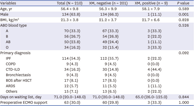
The baseline characteristics of the patients according to the results of crossmatches are shown in Table 1. Among the included 210 recipients, nine patients showed positive crossmatches. The median age was 56.4 years, and 134 patients (63.8%) were male. Only one patient was male in the positive crossmatch group. There was no RH− blood type within the study cohort. The most common primary diagnosis was idiopathic pulmonary fibrosis (114 patients, 54.3%), followed by connective tissue-related interstitial lung disease (CTD-ILD) (34 patients, 16.2%). The median number of days on the waiting list was 71.0, and 63 patients (30.0%) had undergone pretransplant extracorporeal membrane oxygenation support. The baseline characteristics were not significantly different between the positive and negative crossmatch groups.
Table 1

Baseline characteristics

jkms-37-e177-i001
Variables Total (N = 210) XM, negative (n = 201) XM, positive (n = 9) P value
Age, yr 56.4 ± 9.8 56.3 ± 9.9 58.1 ± 7.9 0.589
Male 134 (63.8) 134 (66.2) 1 (11.1) 0.001
BMI, kg/m2 21.3 ± 3.8 21.2 ± 3.7 21.7 ± 6.6 0.828
ABO blood type 0.526
A 70 (33.3) 67 (33.3) 3 (33.3)
B 56 (26.7) 54 (26.9) 2 (22.2)
AB 50 (23.8) 49 (24.4) 1 (11.1)
O 34 (16.2) 32 (15.4) 3 (33.3)
Primary diagnosis 0.092
IPF 114 (54.3) 112 (55.7) 2 (22.2)
COPD 9 (4.3) 9 (4.5) 0 (0.0)
CTD-ILD 34 (16.2) 30 (14.9) 4 (44.4)
Bronchiectasis 9 (4.3) 9 (4.5) 0 (0.0)
BOS after HSCT 17 (8.1) 17 (8.5) 0 (0.0)
ARDS 12 (5.7) 11 (5.5) 1 (11.1)
Others 15 (7.1) 13 (6.5) 2 (22.2)
Days on waiting list, day 71.0 (20.0–148.0) 71.0 (20.0–148.0) 65.0 (26.0–125.0) 0.844
Preoperative ECMO support 63 (30.0) 60 (29.9) 3 (33.3) 1.000
Values are presented as mean ± standard deviation or number (%).
XM = crossmatch, BMI = body mass index, COPD = chronic obstructive pulmonary disease, CTD-ILD = connective tissue-related interstitial lung disease, BOS = bronchiolitis obliterans syndrome, ECMO = extracorporeal membrane oxygenation, ARDS = acute respiratory distress syndrome, IPF = idiopathic pulmonary fibrosis, HSCT = hematopoietic stem cell transplantation.
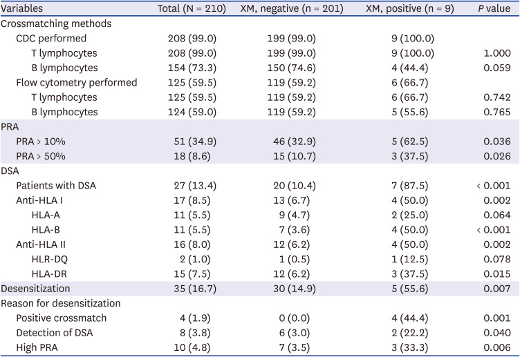
Table 2 shows the results of immunological evaluations, including crossmatching. CDC-XM was performed in 208 patients (99.0%) and flow-XM was performed in 125 patients (59.5%). PRA was identified in 208 patients (99.0%). Among these patients, high levels of class I or class II PRA (> 50%) were detected in 18 patients (8.7%). The proportion of patients with high PRA was higher in the positive crossmatch group than in the negative crossmatch group (37.5% vs. 10.7%, P = 0.026).
Table 2

Immunologic evaluation for transplantation

jkms-37-e177-i002
Variables Total (N = 210) XM, negative (n = 201) XM, positive (n = 9) P value
Crossmatching methods
CDC performed 208 (99.0) 199 (99.0) 9 (100.0)
T lymphocytes 208 (99.0) 199 (99.0) 9 (100.0) 1.000
B lymphocytes 154 (73.3) 150 (74.6) 4 (44.4) 0.059
Flow cytometry performed 125 (59.5) 119 (59.2) 6 (66.7)
T lymphocytes 125 (59.5) 119 (59.2) 6 (66.7) 0.742
B lymphocytes 124 (59.0) 119 (59.2) 5 (55.6) 0.765
PRA
PRA > 10% 51 (34.9) 46 (32.9) 5 (62.5) 0.036
PRA > 50% 18 (8.6) 15 (10.7) 3 (37.5) 0.026
DSA
Patients with DSA 27 (13.4) 20 (10.4) 7 (87.5) < 0.001
Anti-HLA I 17 (8.5) 13 (6.7) 4 (50.0) 0.002
HLA-A 11 (5.5) 9 (4.7) 2 (25.0) 0.064
HLA-B 11 (5.5) 7 (3.6) 4 (50.0) < 0.001
Anti-HLA II 16 (8.0) 12 (6.2) 4 (50.0) 0.002
HLR-DQ 2 (1.0) 1 (0.5) 1 (12.5) 0.078
HLA-DR 15 (7.5) 12 (6.2) 3 (37.5) 0.015
Desensitization 35 (16.7) 30 (14.9) 5 (55.6) 0.007
Reason for desensitization
Positive crossmatch 4 (1.9) 0 (0.0) 4 (44.4) 0.001
Detection of DSA 8 (3.8) 6 (3.0) 2 (22.2) 0.040
High PRA 10 (4.8) 7 (3.5) 3 (33.3) 0.006
Values are presented as number (%).
XM = crossmatch, CDC = complement-dependent cytotoxicity, PRA = panel-reactive antibody, DSA = donor specific antibody, HLA = human leukocyte antigen.
At the time of lung transplantation, most patients (201 patients, 95.7%) had undergone the DSA screening test. Twenty-seven patients (13.4%) were revealed to have DSA. A higher proportion of the patients in the positive crossmatch group had DSA (7 patients, 87.5%, vs. 20 patients, 10.4%, P < 0.001) compared with that in the negative crossmatch group. Desensitization was performed in 35 patients (16.7%), and four patients in the positive crossmatch group underwent desensitization.
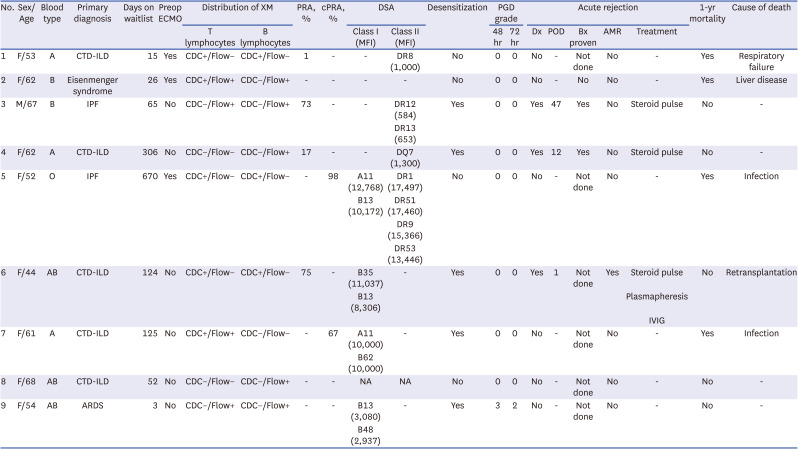
The characteristics of nine patients with positive crossmatch are shown in Table 3. Among these patients, the most common etiology for lung transplantation was CTD-ILD. CDC-XM and flow-XM revealed positive crossmatches in five and six patients, respectively. DSA was detected in six patients, and three patients (patient numbers 5, 6, and 7) had an MFI higher than 5,000. Five patients underwent desensitization before lung transplantation. Acute rejection was diagnosed in three patients (patient numbers 3, 4, and 6), and two patients underwent transbronchial lung biopsy (patient numbers 3 and 4). Patient number 6 was positively crossmatched to the donor as observed using CDC-XM with both T land B lymphocytes, and class I DSA was detected with MFI over 5,000. The patient was clinically diagnosed with AMR and received aggressive treatments for rejection, including steroid pulse, plasmapheresis, and intravenous infusion of immunoglobulin. However, the patient received retransplantation on postoperative day 15 due to rejection.
Table 3

Characteristics of patients with positive crossmatch

jkms-37-e177-i003
No. Sex/Age Blood type Primary diagnosis Days on waitlist Preop ECMO Distribution of XM PRA, % cPRA, % DSA Desensitization PGD grade Acute rejection 1-yr mortality Cause of death
T lymphocytes B lymphocytes Class I (MFI) Class II (MFI) 48 hr 72 hr Dx POD Bx proven AMR Treatment
1 F/53 A CTD-ILD 15 Yes CDC+/Flow− CDC+/Flow− 1 - - DR8 (1,000) No 0 0 No - Not done No - Yes Respiratory failure
2 F/62 B Eisenmenger syndrome 26 Yes CDC+/Flow+ CDC−/Flow+ - - - - No 0 0 No - No No - Yes Liver disease
3 M/67 B IPF 65 No CDC−/Flow+ CDC−/Flow+ 73 - - DR12 (584) Yes 0 0 Yes 47 Yes No Steroid pulse No -
DR13 (653)
4 F/62 A CTD-ILD 306 No CDC−/Flow− CDC−/Flow+ 17 - - DQ7 (1,300) Yes 0 0 Yes 12 Yes No Steroid pulse No -
5 F/52 O IPF 670 Yes CDC+/Flow− CDC+/Flow− - 98 A11 (12,768) DR1 (17,497) No 0 0 No - Not done No - Yes Infection
B13 (10,172) DR51 (17,460)
DR9 (15,366)
DR53 (13,446)
6 F/44 AB CTD-ILD 124 No CDC+/Flow− CDC+/Flow− 75 - B35 (11,037) - Yes 0 0 Yes 1 Not done Yes Steroid pulse No Retransplantation
B13 (8,306) Plasmapheresis
IVIG
7 F/61 A CTD-ILD 125 No CDC+/Flow+ CDC−/Flow− - 67 A11 (10,000) - Yes 0 0 No - Not done No - Yes Infection
B62 (10,000)
8 F/68 AB CTD-ILD 52 No CDC−/Flow− CDC−/Flow+ - - NA NA No 0 0 No - Not done No - No -
9 F/54 AB ARDS 3 No CDC−/Flow+ CDC−/Flow+ - - B13 (3,080) - Yes 3 2 No - Not done No - No -
B48 (2,937)
XM = crossmatch, CTD-ILD = connective tissue-related interstitial lung disease, ECMO = extracorporeal membrane oxygenation, CDC = complement-dependent cytotoxicity, PRA = panel-reactive antibody, DSA = donor specific antibodies, PGD = primary graft dysfunction, MFI = mean fluorescence intensity, AMR = antibody-mediated rejection, ARDS = acute respiratory distress syndrome, IPF = idiopathic pulmonary fibrosis, IVIG = intravenous immunoglobulin.

Operative details and postoperative outcomes

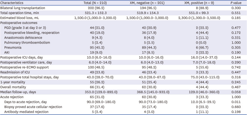
Most patients underwent bilateral lung transplantation (202 patients, 96.2%). According to the PGD grade, patients presenting with grade 3 at postoperative 48 hours or 72 hours were regarded as high-grade PGD. Forty-four patients showed high-grade PGD, and no statistical difference was observed between the groups. The incidences of other postoperative complications, including postoperative bleeding, anastomosis dehiscence, pulmonary thromboembolism, pneumonia, and acute kidney injury, were not statistically distinguishable between the groups (Table 4).
Table 4

Operative and postoperative outcomes

jkms-37-e177-i004
Characteristics Total (N = 210) XM, negative (n = 201) XM, positive (n = 9) P value
Bilateral lung transplantation 202 (96.2) 194 (96.5) 8 (88.9) 0.300
Total operation time, min 521.3 ± 152.3 519.9 ± 154.3 551.0 ± 99.3 0.551
Estimated blood loss, mL 1,500.0 (1,000.0–3,200.0) 1,500.0 (1,000.0–3,000.0) 2,300.0 (1,300.0–3,500.0) 0.185
Postoperative outcomes
PGD (grade 3 at day 2 or 3) 44 (21.0) 42 (20.9) 2 (22.2) 0.477
Postoperative bleeding, reoperation 40 (19.0) 36 (17.9) 4 (44.4) 0.172
Anastomosis dehiscence 9 (4.3) 8 (4.0) 1 (11.1) 0.331
Pulmonary thromboembolism 5 (2.4) 5 (2.5) 0 (0.0) 1.000
Pneumonia 95 (45.2) 89 (44.3) 6 (66.7) 0.305
AKI 19 (9.0) 17 (8.5) 2 (22.2) 0.190
Postoperative ICU days, day 10.0 (6.0–16.0) 10.0 (6.0–16.0) 16.0 (14.0–37.0) 0.144
Postoperative ventilator care, day 6.0 (4.0–14.0) 6.0 (4.0–13.0) 7.0 (7.0–18.0) 0.290
Postoperative re-ECMO support 100 (48.5) 95 (48.2) 5 (55.6) 0.742
Readmission of ICU 49 (23.8) 46 (23.4) 3 (33.3) 0.447
Postoperative total hospital stays, day 43.0 (28.0–70.0) 43.0 (28.0–67.0) 75.0 (43.0–115.0) 0.316
1-year mortality 55 (26.2) 51 (25.4) 4 (44.4) 0.245
Overall mortality 66 (31.4) 62 (30.8) 4 (44.4) 0.467
Median follow-up, days 353.0 (129.0–892.0) 368.5 (145.0–932.0) 129.0 (46.0–360.0) 0.058
Acute rejection 65 (31.0) 62 (30.8) 3 (33.3) 1.000
Days to acute rejection, day 90.0 (69.0–180.0) 90.0 (73.0–180.0) 12.0 (6.5–29.5) 0.011
Biopsy proved acute cellular rejection 37 (17.6) 35 (17.4) 2 (22.2) 0.660
Antibody-mediated rejection 5 (2.4) 4 (2.0) 1 (11.1) 0.198
Values are presented as number (%), mean ± standard deviation or number (interquartile range).
XM = crossmatch, PGD = primary graft dysfunction, ECMO = extracorporeal membrane oxygenation, ICU = intensive care unit, AKI = acute kidney injury.
Acute rejection was diagnosed in 65 patients. Transbronchial lung biopsy-proven ACR was diagnosed in 37 patients, and other patients were diagnosed clinically. Though the incidence of acute rejection with regard to crossmatching results was not statistically different between the groups, the positive crossmatch group showed shorter intervals between transplantation and diagnosis of rejection. Five patients had clinical presentations that were compatible with AMR. However, none of these patients were diagnosed using the biopsy specimen or capillary C4d composition. Seven patients (3.3%) showed physiologic changes that were considered as CLAD. BOS was diagnosed in five patients (2.4%), and RAS was diagnosed in three patients (1.4%). The median number of days from transplant to CLAD diagnosis was 648 (IQR, 488.0–759.0). All of the patients showed CLAD were in the negative crossmatch group.

Survival analysis

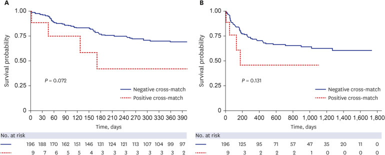
As shown in Table 4, 55 patients (26.2%) showed 1-year mortality. The 1-year and overall mortality rates were not statistically different between the groups (Table 4). Fig. 1 shows the Kaplan-Meier curves of 1-year and overall mortality rates of the patients. According to Kaplan-Meier analysis, there were no significant differences in the survival rates, regardless of crossmatching status.
Fig. 1

Impact of positive crossmatching on survival of lung transplant patients. (A) The 1-year and (B) overall survival of lung transplant patients.

jkms-37-e177-g001
We also performed a regression analysis to identify the risk factors for poor 1-year survival. Positive CDC crossmatching was the only significant risk factor for poor 1-year survival in the univariable analysis (odds ratio, 11.922, 95% confidence interval, 1.302–19.125, P = 0.018).
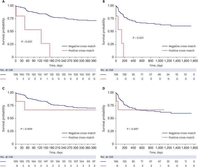
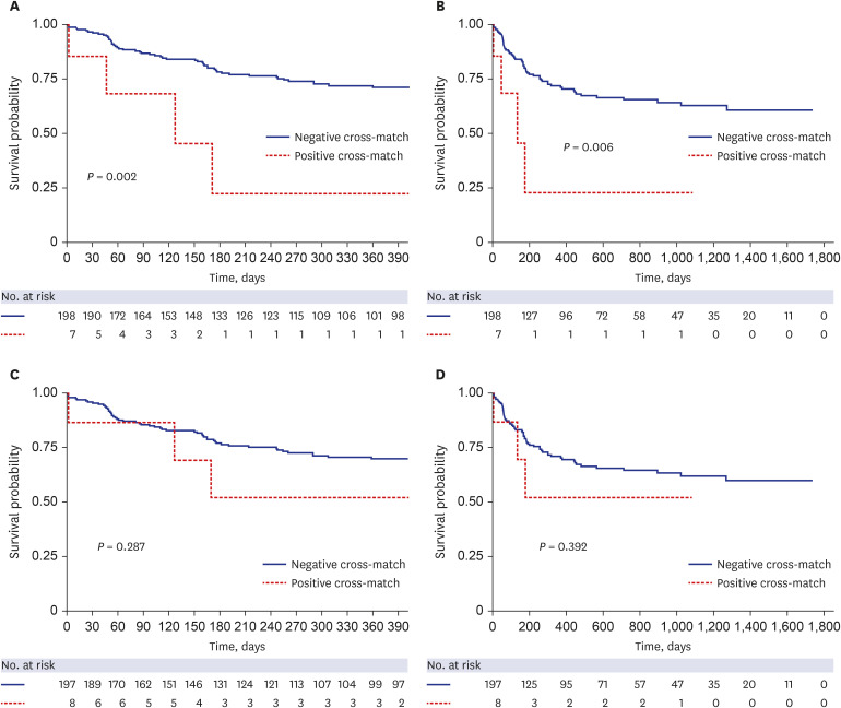
We further divided the patients based on crossmatching methods and performed survival analysis for the subgroups. When positive crossmatching was confirmed by CDC-XM, the post-transplant 1-year and overall mortality rates were poorer than the negative crossmatching results (P < 0.001 and P < 0.001, respectively) (Fig. 2A and B). According to the Kaplan-Meier method, positive crossmatching by the flow-XM method did not significantly differ in the 1-year and overall morality (Fig. 2C and D). When considering the detection of T and B lymphocytes individually, only positive crossmatching by T lymphocytes was related to mortality within 1-year after transplantation (P = 0.002) and overall mortality (P = 0.006) (Fig. 3).
Fig. 2

Impact of positive CDC and flow crossmatches on survival of lung transplant patients. (A) The 1-year survival according to the results of CDC crossmatch. (B) Overall survival according to the results of CDC crossmatch. (C) The 1-year survival according to the results of flow cytometric crossmatch. (D) Overall survival according to the results of flow cytometric crossmatch.

CDC = complement-dependent cytotoxicity.
jkms-37-e177-g002
Fig. 3

Survival analysis of positive T and B lymphocyte crossmatches in lung transplantation. (A) The 1-year survival according to the results of T lymphocyte crossmatching. (B) Overall survival according to the results of T lymphocyte crossmatching. (C) The 1-year survival according to the results of B lymphocyte crossmatching. (D) Overall survival according to the results of B lymphocyte crossmatching.

jkms-37-e177-g003

DISCUSSION

We conducted a multicenter nationwide study to analyze the prevalence of positive crossmatching and the outcomes of positively crossmatched patients in lung transplantation. Although the incidence of positive crossmatching was low (4.3%), positive CDC-XM and T lymphocyte crossmatching results were related to poor 1-year and overall survival.
It is controversial to consider HLA matching as an allocation factor. ISHLT introduced the lung allocation score (LAS) in 2005 and revised it in 2015 and 2020.22 The LAS includes factors related to waiting list mortality and post-transplant 1-year survival. HLA matching is not considered in the LAS system for several reasons. First, as lung transplantation is mostly performed under emergency settings, it is not always possible to conduct donor HLA typing prior to allocation. Furthermore, there is also a possibility of prolonging allograft ischemic time while waiting for crossmatching results.810 Brugière et al.23 analyzed the relative impact of mismatching and graft ischemic time; an allograft ischemic time of over 330 minutes worsened patient survival, even with well-matched donor and recipient pairs. Second, because of the shortage of possible donor organs and the extensive complexity of the HLA system, the probability of finding a matched recipient is extremely low.10 Furthermore, nonimmunologic factors are considered more important for 1-year survival. Early mortality resulting from the ischemia and reperfusion injury has been shown to decrease after introducing low-potassium dextran lung preservation solution.24 Advances in immunosuppressive regimens and enhanced combinations of antibiotics may also be responsible for improved unstable hemodynamics during the early post-transplant period.25 After alleviating these nonimmunological factors for early graft dysfunction and mortality in lung transplantation, the effect of HLA compatibility between the donor and recipient becomes more important for early outcomes. In addition, an improved methodology for HLA crossmatching has facilitated more accurate and rapid access to crossmatching results, leading to increased awareness of the importance of HLA crossmatching in lung transplantation.
Several studies analyzed the impact of HLA mismatching on lung transplantation. It is well known that HLA mismatching can trigger graft damage or even mortality.10 Donor HLA molecules are recognized by the immune system of the recipient. Immunogenetic discrepancies can lead to ACR and AMR, which play major roles in graft dysfunction and loss.
In 2003, the Eurotransplant Foundation analyzed 590 patients who underwent cadaveric lung transplantation between January 1997 and December 1999. In this study, patients with more than four mismatches of HLA loci showed poorer 1-year survival than the others.26 A retrospective study by the United Network for Organ Sharing/ISHLT also confirmed HLA mismatches as a risk factor for 1-year mortality in lung transplantation.27 As routine pretransplant crossmatching is not considered feasible in lung transplantation; these studies did not analyze prospective crossmatching; however, the results confirmed that HLA mismatching is related to graft dysfunction and survival.
In our study, the incidence of rejection or 1-year survival was not statistically different according to the crossmatching results. This result may be because of the low incidence of positive crossmatching in the cohort (4.3%) and the relatively short-term follow-up period of the positive group (median 129.0 vs. 386.5 days). Moreover, most centers involved in the registry tended to decline the allocation when positive crossmatching had been verified not only by the KODA lab but also by the center’s study. However, the relationship between 1-year survival and positive crossmatching was observed when the cohort was subdivided according to the crossmatching method.
Flow-XM is reportedly more sensitive for detecting anti-HLA antibodies than CDC-XM.28 Several studies have shown the relationship between positive flow-XM results and a higher risk of graft dysfunction and poor survival.237 However, as flow-XM is sometimes too sensitive, denying the allocation because of a positive flow crossmatch is still controversial.29 Moreover, flow-XM is not a functional test, and the binding of antibodies to lymphocytes may not always reflect complement system activation.6 Thus, transplantation across a positive flow crossmatch and negative CDC crossmatch is acceptable, whereas transplantation across a positive CDC result is not typically recommended. In our study, the Kaplan-Meier analysis also supported this principle. The patients with positive CDC crossmatch showed significantly lower 1-year and overall survival, whereas positive flow crossmatch did not significantly affect the outcome.
The impact of positive T cell crossmatches on poor graft outcomes was apparent in renal, cardiac, and liver transplantation.230 A negative lymphocytotoxic T cell result is generally considered as grounds to proceed with transplantation. Here, the results of Kaplan-Meier analysis also demonstrated significantly poor 1-year and overall survival in positive T-cell crossmatched patients, whereas positive B-cell crossmatching did not show a significant difference in graft survival. Positive T-cell crossmatching indicates the presence of DSAs against class I antigens, which can lead to antibody-mediated damages to the graft.378 This damage can cause hyperacute rejection or graft loss.
This study has certain limitations. First, although this study analyzed a nationwide cohort, this is a retrospective study with a relatively small number of patients and a short follow-up period. CLAD was not observed in the positively crossmatched patients, probably because of the short follow-up time (median 129.0 days). Thus, further follow-up for the CLAD is needed for the positive crossmath group. Second, the incidence of positive crossmatching was too low to perform further statistical analyses. As mentioned above, we performed regression analysis to identify the risk factors of poor survival, and positive CDC crossmatching was the only meaningful risk factor for poor 1-year survival. Third, a nationwide multicenter study with registry data does not reflect detailed information on each case. In particular, it is difficult to determine the actual immunologic consequences on graft survival.
In conclusion, this study analyzed the outcomes of positively crossmatched patients in lung transplantation and revealed the importance of crossmatching methods in lung transplantation using nationwide data. Positive results of CDC and T lymphocyte crossmatching may lead to devastating outcomes in lung transplantation. Although crossmatching is not part of the allocation system, the results of crossmatching should be considered with caution during postoperative management after lung transplantation.

Notes

Presentation: Presented at the Plenary Session of the Asian Transplantation Week 2020 (ATW 2020), Seoul, Korea, December 3–5, 2020.

Funding: This research was supported by Research of Korea Centers for Disease Control and Prevention (grant No. 2020-ER7201-01).

Disclosure: The authors have no potential conflicts of interest to disclose.

Author Contributions:

  • Conceptualization: Kim H.

  • Data curation: Kim H.

  • Formal analysis: Kim H, Lee JG.

  • Funding acquisition: Kim H.

  • Investigation: Kim H.

  • Methodology: Kim H.

  • Project administration: Kim H, Lee JG.

  • Resources: Kim H.

  • Software: Kim H.

  • Supervision: Paik HC, Jeong SJ, Kim SY, Park MS, Lee JG.

  • Validation: Kim H.

  • Visualization: Kim H.

  • Writing - original draft: Kim H, Lee JG.

  • Writing - review & editing: Kim H, Yang YH, Paik HC, Jeong SJ, Kim SY, Park MS, Lee JG.

References

1. Patel R, Terasaki PI. Significance of the positive crossmatch test in kidney transplantation. N Engl J Med. 1969; 280(14):735–739. PMID: 4886455.
2. Opelz G, Döhler B, Süsal C. Analysis of positive kidney, heart, and liver transplant crossmatches reported to the Collaborative Transplant Study. Hum Immunol. 2009; 70(8):627–630. PMID: 19375469.
3. Tinckam KJ, Chandraker A. Mechanisms and role of HLA and non-HLA alloantibodies. Clin J Am Soc Nephrol. 2006; 1(3):404–414. PMID: 17699239.
4. Fidler SJ, Irish AB, Lim W, Ferrari P, Witt CS, Christiansen FT. Pre-transplant donor specific anti-HLA antibody is associated with antibody-mediated rejection, progressive graft dysfunction and patient death. Transpl Immunol. 2013; 28(4):148–153. PMID: 23665534.
5. Leech SH, Rubin S, Eisen HJ, Mather PJ, Goldman BI, McClurken JB, et al. Cardiac transplantation across a positive prospective lymphocyte cross-match in sensitized recipients. Clin Transplant. 2003; 17(Suppl 9):17–26. PMID: 12795663.
6. Colvin MM, Cook JL, Chang PP, Hsu DT, Kiernan MS, Kobashigawa JA, et al. Sensitization in heart transplantation: emerging knowledge: a scientific statement from the American Heart Association. Circulation. 2019; 139(12):e553–e578. PMID: 30776902.
7. Tait BD, Süsal C, Gebel HM, Nickerson PW, Zachary AA, Claas FH, et al. Consensus guidelines on the testing and clinical management issues associated with HLA and non-HLA antibodies in transplantation. Transplantation. 2013; 95(1):19–47. PMID: 23238534.
8. Dick A, Humpe A, Kauke T. Impact, screening, and therapy of HLA antibodies in patients before and after lung transplantation. Transfus Med Hemother. 2019; 46(5):337–347. PMID: 31832059.
9. Lobo LJ, Aris RM, Schmitz J, Neuringer IP. Donor-specific antibodies are associated with antibody-mediated rejection, acute cellular rejection, bronchiolitis obliterans syndrome, and cystic fibrosis after lung transplantation. J Heart Lung Transplant. 2013; 32(1):70–77. PMID: 23260706.
10. Ju L, Suberbielle C, Li X, Mooney N, Charron D. HLA and lung transplantation. Front Med. 2019; 13(3):298–313. PMID: 29974325.
11. Park MH, Kim S, Hwang H, Park H, Kwak J, Kwon EK, et al. Positive rates of preliminary crossmatches among transplantation candidates waitlisted for different organs in the Korean Network for Organ Sharing. Transplant Proc. 2016; 48(7):2464–2466. PMID: 27742323.
12. Jeong YH, Kim DK, Choi S. Current status of lung transplantation in Korea. Curr Chall Thorac Surg. 2022; 4:4.
13. Moon SW, Park MS, Lee JG, Paik HC, Kim YT, Lee HJ, et al. Panel-reactive and donor-specific antibodies before lung transplantation can affect outcomes in Korean patients receiving lung transplantation. Yonsei Med J. 2020; 61(7):606–613. PMID: 32608204.
14. Yu WS, Kim SY, Kim YT, Lee HJ, Park S, Choi SM, et al. Characteristics of lung allocation and outcomes of lung transplant according to the Korean urgency status. Yonsei Med J. 2019; 60(10):992–997. PMID: 31538435.
15. Hamrick CW, Lebeck LK. Flow cytometric T and B cell crossmatching. Hahn AB, Land GA, Strothman RM, editors. ASHI Laboratory Manual. 4th ed. Philadelphia, PA, USA: American Society for Histocompatibility and Immunogenetics;2000. p. 41–45.
16. Smith JD, Danskine AJ, Laylor RM, Rose ML, Yacoub MH. The effect of panel reactive antibodies and the donor specific crossmatch on graft survival after heart and heart-lung transplantation. Transpl Immunol. 1993; 1(1):60–65. PMID: 8081763.
17. Stewart S, Fishbein MC, Snell GI, Berry GJ, Boehler A, Burke MM, et al. Revision of the 1996 working formulation for the standardization of nomenclature in the diagnosis of lung rejection. J Heart Lung Transplant. 2007; 26(12):1229–1242. PMID: 18096473.
18. Otani S, Davis AK, Cantwell L, Ivulich S, Pham A, Paraskeva MA, et al. Evolving experience of treating antibody-mediated rejection following lung transplantation. Transpl Immunol. 2014; 31(2):75–80. PMID: 25004453.
19. Christie JD, Edwards LB, Kucheryavaya AY, Benden C, Dipchand AI, Dobbels F, et al. The registry of the International Society for Heart and Lung Transplantation: 29th adult lung and heart-lung transplant report-2012. J Heart Lung Transplant. 2012; 31(10):1073–1086. PMID: 22975097.
20. Estenne M, Maurer JR, Boehler A, Egan JJ, Frost A, Hertz M, et al. Bronchiolitis obliterans syndrome 2001: an update of the diagnostic criteria. J Heart Lung Transplant. 2002; 21(3):297–310. PMID: 11897517.
21. Sato M, Waddell TK, Wagnetz U, Roberts HC, Hwang DM, Haroon A, et al. Restrictive allograft syndrome (RAS): a novel form of chronic lung allograft dysfunction. J Heart Lung Transplant. 2011; 30(7):735–742. PMID: 21419659.
22. Lyu DM, Goff RR, Chan KM. The lung allocation score and its relevance. Semin Respir Crit Care Med. 2021; 42(3):346–356. PMID: 34030198.
23. Brugière O, Thabut G, Suberbielle C, Reynaud-Gaubert M, Thomas P, Pison C, et al. Relative impact of human leukocyte antigen mismatching and graft ischemic time after lung transplantation. J Heart Lung Transplant. 2008; 27(6):628–634. PMID: 18503962.
24. Strüber M, Wilhelmi M, Harringer W, Niedermeyer J, Anssar M, Künsebeck A, et al. Flush perfusion with low potassium dextran solution improves early graft function in clinical lung transplantation. Eur J Cardiothorac Surg. 2001; 19(2):190–194. PMID: 11167111.
25. Hopkins PM, McNeil K. Evidence for immunosuppression in lung transplantation. Curr Opin Organ Transplant. 2008; 13(5):477–483. PMID: 19060529.
26. Smits JM, Mertens BJ, Van Houwelingen HC, Haverich A, Persijn GG, Laufer G. Predictors of lung transplant survival in Eurotransplant. Am J Transplant. 2003; 3(11):1400–1406. PMID: 14525601.
27. Quantz MA, Bennett LE, Meyer DM, Novick RJ. Does human leukocyte antigen matching influence the outcome of lung transplantation? An analysis of 3,549 lung transplantations. J Heart Lung Transplant. 2000; 19(5):473–479. PMID: 10808155.
28. Sutton PM, Harmer AH, Bayne AM, Welsh KI. The flow cytometric detection of alloantibodies in screening for renal transplantation. Transpl Int. 1995; 8(5):360–365. PMID: 7576017.
29. Cervelli C, Pisani F, Aureli A, Azzarone R, Scimitarra M, Battistoni C, et al. For anti-HLA-specific donor antibodies detection by flow cytometry cytotoxic crossmatches comparison of methods. Transplant Proc. 2013; 45(7):2761–2764. PMID: 24034042.
30. Mulley WR, Kanellis J. Understanding crossmatch testing in organ transplantation: a case-based guide for the general nephrologist. Nephrology (Carlton). 2011; 16(2):125–133. PMID: 21272123.
TOOLS
Similar articles