Journal List > J Korean Neurosurg Soc > v.61(1) > 1162035

Lee, Chang, Lee, Hwang, Bae, and Doh: Morphometric Study of the Lumbar Posterior Longitudinal Ligament

Abstract

Objective

Morphometric data for the lumbar posterior longitudinal ligament (PLL) was investigated to identify whether there is a difference in the morphometry of the PLL of the lumbar spine at each level with respect to the pattern of intervertebral disc displacement.

Methods

In 14 formalin-fixed adult cadavers (12 males and 2 females), from L1 to L5, the authors measured the width and height of the PLL and compared them with other landmarks such as the disc and the pedicle.

Results

Horizontally, at the upper margin of the disc, the central portion of the superficial PLL covered 17.8–36.9% of the disc width and the fan-like portion of the PLL covered 63.9–76.7% of the disc width. At the level of the median portion of the disc, the PLL covered 69.1–74.5% of the disc width. Vertically, at the level of the medial margin of the pedicle, the fan-like portion of the PLL covered 23.5–29.9% of the disc height. In general, a significant difference in length was not found in the right-left and male-female comparisons.

Conclusion

This study presents the morphometric data on the pattern of intervertebral disc displacement and helps to improve the knowledge of the surgical anatomy of the lumbar PLL.

INTRODUCTION

Gray8) reported that the posterior longitudinal ligament (PLL) is situated within the vertebral canal, and extends along the posterior surfaces of the bodies of the vertebrae, from the body of the axis, where it is continuous with the tectorial membrane, to the sacrum. It is broader above than below, and thicker in the thoracic region than in the cervical and lumbar regions.
Among the 379 cases reviewed by MRI scans10), there were 244 patients (64.4%) with disc pathologies involving the L4–L5 and/or L5–S1 levels, 67 patients (17.7%) with disc pathologies involving the L3–L4 level, and 42 patients (11.1%) with disc pathologies involving the T12–L1, L1–L2, and/or L2–L3 levels. Out of the 279 cases of surgically proven disc herniation5), 166 cases (59.4%) of disc herniation were found at the L5–S1 level, 103 cases (37%) of disc herniation were found at the L4–L5 level, 8 cases (2.9%) of disc herniation were found at the L3–L4 level, and 2 cases (0.7%) of disc herniation were found at the L2–3 level.
On the other hand, Rosner and Campbell19) explained that posterolateral disc herniation is common, perhaps because of thinning of the PLL at the periphery. The most frequent site of rupture of the disc is the mediolateral portion (lateral border of the dural sac)6) because the lateral portion of the PLL does not have a firm connection with the annulus fibrosus17). However, in their papers, there was no proper morphometric reference supporting their assumption. Also, there is no clear information in the literature regarding the actual length, thickness or cross-sectional area of this ligament1,9,19).
Combining the above three individual studies, a question may arise whether there is a difference in the morphometry of the PLL of the lumbar spine at each level with respect to the pattern of intervertebral disc displacement. However, a morphometric study of the PLL is not common and a study of the lumbar PLL in Korean people has rarely been published to date. To solve this problem, the authors measured the width and height of the PLL at each lumbar spine level in 14 adult cadavers.

MATERIALS AND METHODS

The authors used 14 adult cadavers (12 males and 2 females) perfused with a fixative solution containing formalin, phenol, alcohol and glycerin. The age of the cadavers ranged from 25 to 95 years (mean, 61.6 years), and they did not have a history of spinal trauma, tumor, severe spondylosis. Each cadaver was placed in a prone position so that the first through fifth lumbar spines (L1–L5) and the sacrum could be observed, while the PLL, disc, and pedicle were exposed by removing the soft and bony structure in the posterior lumbar spine (Fig. 1).
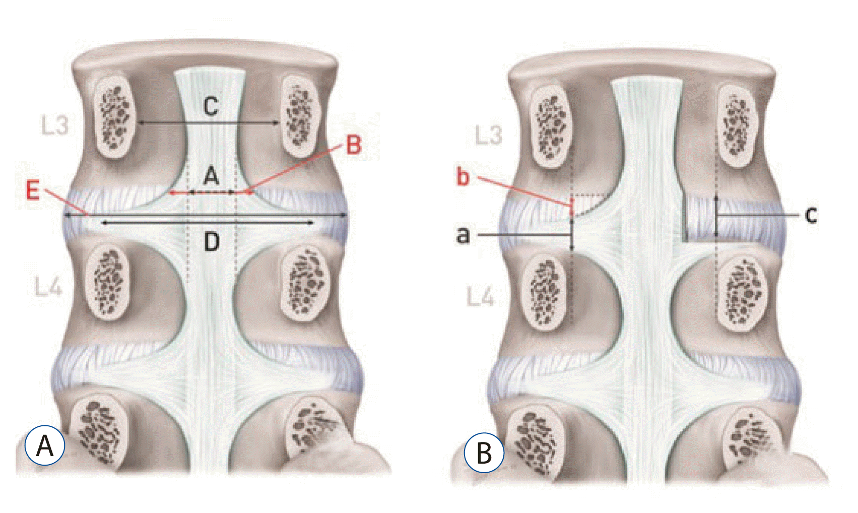
In 14 formalin-fixed adult cadavers, from L1 to L5, the authors performed measurements by using digital calipers: 1) horizontally, the distance between both pedicles, the width of the central portion of the superficial PLL at the level of the upper margin of the disc, the width of the PLL at the level of the upper margin of the disc, the width of the PLL, and the disc at the level of the median portion of the disc; and 2) vertically, at the virtual line of the medial margin of the pedicle, the height of the PLL, the disc height not covered by the PLL and the disc height on both left and right sides.
A standard instrument was used for performing all measurements. Every measurement was performed by one operator in order to minimize possible errors. The t-test and the Mann-Whitney test were conducted using SPSS software version 20.0 (SPSS Inc., Chicago, IL, USA) to analyze the results. Values were considered statistically significant if they had a p<0.05.

RESULTS

The gross anatomical properties of the PLL (Fig. 2)

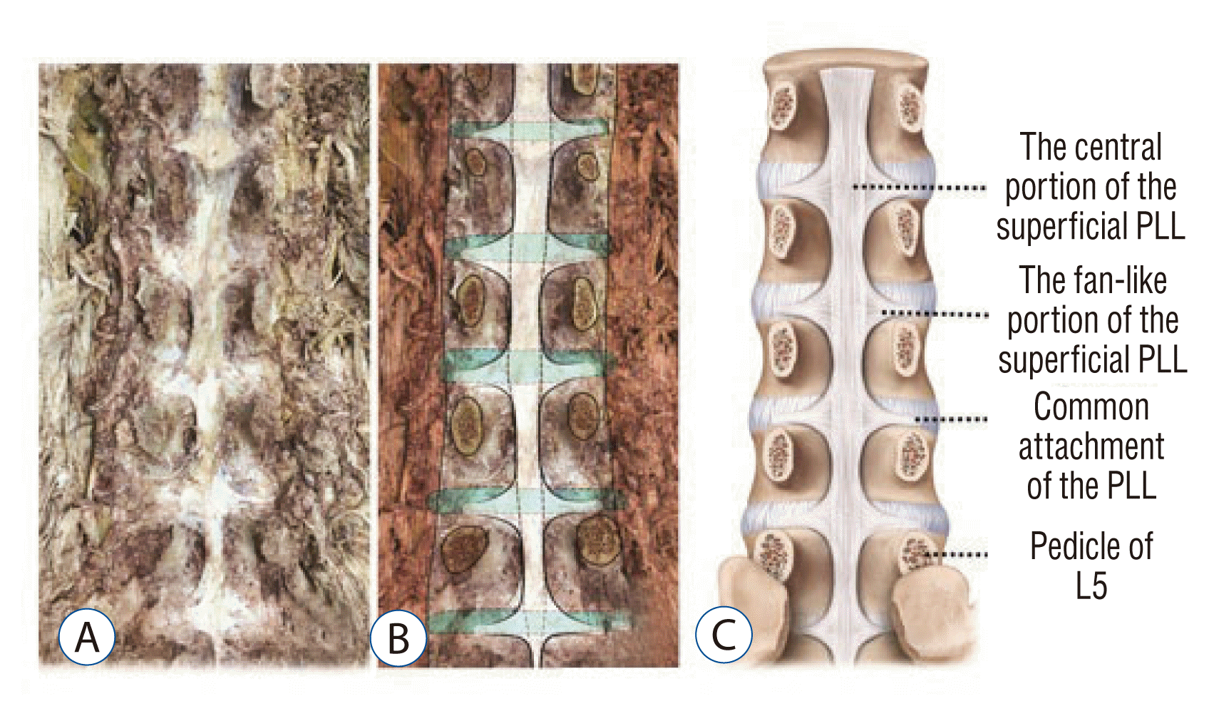
1) The width of the PLL at a low lumbar level was lesser than that at the upper lumbar level; 2) Common attachment of the PLL had an asymmetric shape. The upper border had a more obtuse angle but the lower border had a more concave angle; 3) Common attachment of the PLL covered almost all of the lower disc area, but some of the upper lateral disc area was not covered by the PLL; and 4) Common attachment had an end-point within the pedicle width and it did not extend to the extraforaminal area.

The width of the central portion of the superficial PLL at the level of the upper margin of the disc (A)

A was 8.5±0.5 mm at L1/2, 8.1±1.2 mm at L2/3, 7.9±1.6 mm at L3/4, 7.0±1.6 mm at L4/5, and 4.8±1.3 mm at L5/S1 (Table 1). There was a decreasing pattern from L1/2 to L5/S1. At the L4/5 level, A was 8.5±0.0 mm in the female group but it was 6.8±1.6 mm in the male group. In a male-female comparison, we found a statistically significant difference at L4/5 (p=0.003) (Table 2).

The width of the PLL at the level of the upper margin of the disc (B)

B was 14.7±2.7 mm at L1/2, 17.5±2.7 mm at L2/3, 17.7±3.8 mm at L3/4, 20.5±3.4 mm at L4/5, and 18.2±3.9 mm at L5/S1 (Table 1). B showed an increasing pattern from L1/2 to L4/5. In a male-female comparison, we found no statistically significant difference (Table 2).

The distance between both pedicles (C)

C was 23.0±3.0 mm at L1, 23.3±2.3 mm at L2, 24.8±1.7 mm at L3, 26.7±2.8 mm at L4, and 27.0±2.7 mm at L5 (Table 1). C showed a consistently increasing tendency from L1 to L5. In a male-female comparison, we found no statistically significant difference (Table 2).

The width of the PLL at the level of the median portion of the disc (D)

D was 28.6±3.0 mm at L1/2, 28.2±2.3 mm at L2/3, 29.9±3.5 mm at L3/4, 31.0±3.5 mm at L4/5, and 32.5±5.6 mm at L5/S1 (Table 1). D showed an increasing tendency from L2/3 to L5/S1 level. In a male-female comparison, we found no statistically significant difference (Table 2).

The width of the disc at the level of the median portion of the disc (E)

E was 38.4±2.5 mm at L1/2, 40.4±3.1 mm at L2/3, 43.1±4.4 mm at L3/4, 44.1±5.9 mm at L4/5, and 44.0±3.5 mm at L5/S1 (Table 1). E showed an increasing tendency from L1/2 to L4/5. In a male-female comparison, we found no statistically significant difference (Table 2).

The height of the PLL at the level of the medial margin of the pedicle (a), left (aL) and right (aR) sides

aL was 6.2±2.4 mm at L1/2, 5.5±1.8 mm at L2/3, 7.0±1.2 mm at L3/4, 7.5±1.4 mm at L4/5, and 6.6±1.4 mm at L5/S1. aR was 6.4±2.3 mm at L1/2, 5.7±1.8 mm at L2/3, 7.0±1.4 mm at L3/4, 7.4±1.1 mm at L4/5, and 7.1±2.0 mm at L5/S1 (Table 1). In a male-female comparison and a left-right comparison, we found no statistically significant difference (Tables 2 and 3).

The height of the disc not covered by the PLL at the level of the medial margin of the pedicle (b), left (bL) and right (bR) sides

bL was 2.6±1.3 mm at L1/2, 2.8±1.1 mm at L2/3, 3.8±2.1 mm at L3/4, 3.5±0.9 mm at L4/5, and 3.0±1.1 mm at L5/S1. bR was 2.4±1.1 mm at L1/2, 2.8±1.5 mm at L2/3, 3.3±1.8 mm at L3/4, 3.1±0.7 mm at L4/5, and 3.1±1.1 mm at L5/S1 (Table 1). In a male-female comparison and a left-right comparison, we found no statistically significant difference (Tables 2 and 3).

The disc height at the level of the medial margin of the pedicle (c), left (cL) and right pedicles (cR)

cL was 10.4±3.5 mm at L1/2, 10.8±3.3 mm at L2/3, 12.7±3.5 mm at L3/4, 13.1±2.6 mm at L4/5, and 12.0±3.1 mm at L5/S1. cR was 10.2±3.4 mm at L1/2, 11.0±3.5 mm at L2/3, 12.7±4.1 mm at L3/4, 13.1±2.5 mm at L4/5, and 11.8±2.5 mm at L5/S1 (Table 1). Both cL and cR showed the lowest disc height at L1/2 and the highest disc height at L4/5. In a male-female comparison and a left-right comparison, we found no statistically significant difference (Tables 2 and 3).

DISCUSSION

The PLL shows a broad, uniform shape in the cervical and upper thoracic regions1,8). On the contrary, in the thoracic and lumbar regions, it presents a series of concave2), oblique15) dentations (Fig. 2). However, it is narrow and thick over the centers of the bodies, from which it is separated by the basivertebral veins, and the paravertebral venous plexus8). The PLL attaches to the posterior aspect of the intervertebral discs and to the adjacent margins of the vertebral bodies8,15). On gross observation, each of the PLL fibers does not connect merely one segment but extends far through at least two levels of segments2). This ligament is composed of smooth, shining, longitudinal fibers which are more compact than those of the anterior longitudinal ligament (ALL), and it consists of superficial layers occupying the interval between 3rd and 4th vertebrae, and deeper layers which extend between the adjacent 1 or 2 vertebræ2).
The PLL resists hyperflexion of the spine, attaches primarily to the annulus fibrosis of the vertebral discs, and lacks the mechanical strength of the ALL19). According to Loughenbury et al.12), the clinical relevance of the PLL and associated membranes tends to be based on a protective rather than a supportive role. The observed patterns of migration of foreign material into the vertebral canal confirm that the PLL and its extensions act to protect the spinal cord from displaced disc material, invading masses and bony fragments following a vertebral burst fracture. With a dorsocentral protrusion of a semifluid mass, the strong midline strap of posterior longitudinal fibers tends to restrain the herniation15).
The PLL is made of superficial and deep layers12). The superficial layer is located on the posterior surface than the deep layer such that its distinction from the dura mater is often is difficult18). Due to its wide attachment to the intervertebral disc, it is described as a ‘fan-like’14), structure presenting a denticulate appearance over each vertebral body2,21). This denticulate appearance is more apparent in the lower thoracic and lumbar regions, where the superficial layer is divided into the fan-like portion and the thick central portion12).
The central portion of the superficial PLL is made of a central band of fibers 8–10 mm wide21). However, in our study, it had a more extensive range of 4.8–8.5 mm on average, which was different from the above-mentioned study. In the lumbar region, the thick central portion appears to decrease in width from L1 to L514). This finding suggests that the vulnerable area for disc herniation is broader in the lower lumbar region than in the upper lumbar region, horizontally.
As observed in the above result, the shape of each lumbar vertebra is slightly different among the levels. In order to determine whether it contributes to the occurrence of herniation or not, we calculated the relative ratios among the parameters, as shown in Table 4.
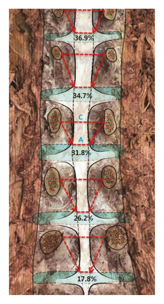
As per Table 4, horizontally, at the upper margin of the disc, the central portion of the superficial PLL covered 17.8–36.9% of interpedicular distance and the fan-like portion of the PLL covered 63.9–76.7% of interpedicular distance, and the PLL covered 69.1–74.5% of the disc width. Specifically, the relative ratio of the width of the central portion of the superficial PLL (A) to the inter-pedicular distance (C) was highest at a percentage of 36.9% at L1/2 but it was lowest at a percentage of 17.8% at L5/S1 (Fig. 3). As there are diverse causes and mechanisms of disc herniation, the incidence of disc herniation shows a large difference between the upper and lower lumbar regions3,4,16), and this suggests the possibility that the difference in A/C (%) may be another anatomical reason why the incidence of disc herniation is different among lumbar levels. The aspect ratio of the horizontal distance which is not covered by the central portion of the PLL becomes larger consecutively at the lower lumbar level. Based on this point, there is a possibility that a correlation exists between the morphometry of the PLL and the pattern of the intervertebral disc displacement.
In addition, the width of the PLL at the level of upper margin of the disc (B), which includes the fan-like portion in the transverse direction, did not show any tendency in connection to the level, in consideration of its relative ratio to the shortest distance between both pedicles (C) (Table 4). Different from the thick central portion of the superficial PLL, the fan-like portion showed a thinner transparent membranous pattern even on gross observation. The author conjectures that the structural protection role of the fan-like portion may be less significant than that of the central portion in the pattern of the intervertebral disc displacement.
Kapoor11) reported that C was 18.51 mm at L1, 21.47 mm at L2–L4, and 21.50 mm at L5, and El-Rakhawy et al.7) reported that it was 21.6 mm, 22.6 mm, 21.4 mm, 23.5 mm, and 25.1 mm at L1 through L5. In our study also, a similar range was obtained for L1–L5 (Table 1). The increasing pattern of C reported in these two studies was similar to the result of our study. Clinically, C is the transverse diameter of the spinal canal, and the patients with canal stenosis showed lower figures for all diameters of the central canal20). This is a typical finding when degenerative changes occur7,13).
When the aspect ratio of disc width covered by the PLL (D) to the width of the discs (E) was calculated, the ratio (D/E) was 73.1% at L1/2, 70.4% at L2/3, 69.1% at L3/4, 70.0% at L4/5, and 74.5% at L5/S1, showing that the PLL covers about 70% of the transverse width of the lumbar discs (Table 4). In addition, if only the leftmost and rightmost 15% of the disc area are invaded by surgery, it is possible to preserve the fan-like portion of the superficial PLL at the upper margin of the disc. Also, the leftmost and rightmost 15% of the disc area may be found to be not covered by the PLL during surgery through the transforaminal and extraforaminal approaches but the disc area would be completely covered by the PLL through the posterolateral approach.
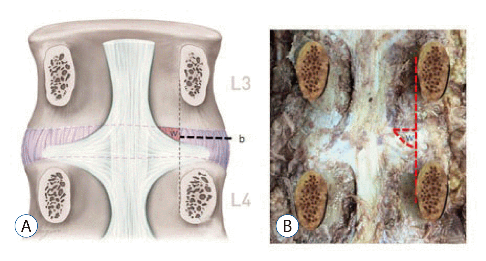
The height of the disc not covered by the PLL at the level of the medial margin of the pedicle (b), which is related to the longitudinal axis of the weak point (Fig. 4), was about 21–33 mm at all the levels, and its ratio to the full disc height at the level of the medial margin of the pedicle (c) was not significantly different among the levels either (Table 4). As b/c (%) was about 25% on an average among the levels, if the upper 25% of the disc height at the level of the medial margin of the pedicle is invaded during a disc-related procedure, it is highly likely that injury to the PLL is avoided. Anatomically, this portion is regarded as the most vulnerable region for herniation within the spinal canal. If there is herniation of the subligamentous type in the lower 3/4th of the disc height, the likelihood of a remnant disc fragment should be considered, and a more careful probe is recommended.
According to this result, anatomical weak point (W) not covered by any portion of the PLL is on upper-lateral disc area (Fig. 4). However, we know empirically that the downward displacement of the disc is more common than upward. Ebeling and Reulen6) reported the medio-lateral disc herniations(lateral border of the dural sac or the exit zone of the nerve root) were displaced more often in a caudal direction (63%) and only rarely in a cranial direction (8%), lateral disc herniation (compressing exclusively the nerve root, for instance in the lateral recess inside the spinal canal) were displaced with same frequency in both direction: caudal direction (34%), cranial direction (39%). Authors estimate that the disc displacement occurs basically to caudal direction rather than cranial direction in spinal canal by some factors which cannot be explained by only a morphometry of the PLL but W contributes to making the difference of the cranio-caudal direction of the disc displacement between medio-lateral and lateral area.
This study has limitations such as absence of exclusion criteria related to disc pathology, body height and the high average age of a small number of cadavers. However, if the relative length ratios for individual variables are determined and used, it helps to compensate for variations in the absolute values of measurement results with the body size of specimens. In order to exclude biases related to the reduced disc height resulting from disc degeneration due to aging, future researches should use younger cadavers. Also, it is less meaningful to compare female with male due to a small number of cadavers.
CONCLUSION

In this cadaveric study, the authors confirmed that the relatively upper-lateral disc area in the spinal canal is an anatomical weak point for lumbar intervertebral disc displacement because there is no covering by any portion of the PLL and lumbar intervertebral disc displacement may be more common in the lower lumbar region because the PLL at the lower level has a more narrow shape. Finally, we believe that this study will be a representative morphometric study on the pattern of the intervertebral disc displacement and it will help gain a known anatomical understanding of the lumbar PLL.

PATIENT CONSENT

The patient provided written informed consent for the publication and the use of their images.

Acknowledgements

This work was supported by the Soonchunhyang Research Fund.

References

1. Behrsin JF, Briggs CA. Ligaments of the lumbar spine: a review. Surg Radiol Anat. 10:211–219. 1988.
crossref
2. Bogduk N. Clinical anatomy of the lumbar spine and sacrum. ed 4. Edinburgh: Elsevier Health Sciences;2005. p. 41–42.
3. Bosacco SJ, Berman AT. Surgical management of lumbar disc disease. Radiol Clin North Am. 21:377–393. 1983.
4. Bosacco SJ, Berman AT, Raisis LW, Zamarin RI. High lumbar disk herniations. Case reports Orthopedics. 12:275–278. 1989.
5. Decker HG, Shapiro SW. Herniated lumbar intervertebral disks; results of surgical treatment without the routine use of spinal fusion. AMA Arch Surg. 75:77–84. 1957.
6. Ebeling U, Reulen HJ. Are there typical localisations of lumbar disc herniations? A prospective study. Acta Neurochir (Wien). 117:143–148. 1992.
crossref
7. El-Rakhawy M, El-Shahat AER, Labib I, Abdulaziz E. Lumbar vertebral canal stenosis: concept of morphometric and radiometric study of the human lumbar vertebral canal. Anatomy. 4:51–62. 2010.
crossref
8. Gray H. Anatomy of the human body. ed 20. Philadelphia: Lea & Febiger;1918.
9. Hardy RW, Ball PA. Treatment of disk disease of the lumbar spine. Youmans Neurological Surgery. ed 5. Philadelphia: WB Saunders Company;2004.
10. Hsu K, Zucherman J, Shea W, Kaiser J, White A, Schofferman J, et al. High lumbar disc degeneration. Incidence and etiology. Spine (Phila Pa 1976). 15:679–682. 1990.
crossref
11. Kapoor Y. Morphometry of the lumbar vertebrae and its clinical significance. Sch J App Med Sci. 2:1045–1052. 2014.
12. Loughenbury PR, Wadhwani S, Soames RW. The posterior longitudinal ligament and peridural (epidural) membrane. Clin Anat. 19:487–492. 2006.
crossref
13. Modic MT, Ross JS. Lumbar degenerative disk disease. Radiology. 245:43–61. 2007.
crossref
14. Ohshima H, Hirano N, Osada R, Matsui H, Tsuji H. Morphologic variation of lumbar posterior longitudinal ligament and the modality of disc herniation. Spine (Phila Pa 1976). 18:2408–2411. 1993.
crossref
15. Parke WW, Bono CM, Garfin SR. Applied anatomy of the spine. Rothman-Simeone The Spine. ed 6. Philadelphia: Saunders;2011. p. 26–27.
16. Prasad R, Hoda MF, Dhakal MM, Singh K, Srivastava A, Sharma V. Epidemiological characteristics of lumbar disc prolapse in atertiary care hospital. Int J Neurosurg. 3:534–536. 2006.
17. Prestar FJ. Morphology and function of the interspinal ligaments and the supraspinal ligament of the lumbar portion of the spine. Morphol Med. 2:53–58. 1982.
18. Putz R. The detailed functional anatomy of the ligaments of the vertebral column. Ann Anat. 174:40–47. 1992.
crossref
19. Rosner MK, Campbell VA. Treatment of Disk Disease of the Lumbar Spine Youmans Neurological Surgery. ed 6. Philadelphia: WB Saunders Company;2011. p. 2919.
20. Santiago FR, Milena GL, Herrera RO, Romero PA, Plazas PG. Morphometry of the lower lumbar vertebrae in patients with and without low back pain. Eur Spine J. 10:228–233. 2001.
crossref
21. Wiltse LL. Anatomy of the extradural compartments of the lumbar spinal canal. Peridural membrane and circumneural sheath. Radiol Clin North Am. 38:1177–1206. 2000.

Fig. 1
Parameters. A: Horizontal direction. B: Vertical direction. (A) The width of the central portion of the superficial PLL at the level of the upper margin of the disc. (B) The width of the PLL at the level of the upper margin of the disc. (C) The distance between both pedicles. (D) The width of the PLL at the level of median portion of the disc. (E) The width of the disc at the level of median portion of the disc. (a) The height of the PLL at the level of the medial margin of the pedicle. (b) The height of the disc not covered by the PLL at the level of the medial margin of the pedicle. (c) The disc height at the level of the medial margin of the pedicle. PLL: posterior longitudinal ligament.
jkns-61-1-89f1.gif
Fig. 2
Photographic (A), drawing (B), and schematic (C) images of the lumbar spine after removing the posterior structure from L1 to L5.
jkns-61-1-89f2.gif
Fig. 3
A/C (%) decreased from L1 to L5. On the contrary, relative horizontal width which is not covered by the central portion of the PLL was increased. Note: most of the herniation of the lumbar intervertebral disc displacement occurs at L4/5, L5/S13,4,5). (A) The width of the central portion of the superficial PLL at the level of the upper margin of the disc. (C) The distance between both pedicles. PLL: posterior longitudinal ligament.
jkns-61-1-89f3.gif
Fig. 4
Schematic (A) and photographic drawing (B) images indicate the anatomical weak point (W) that is not covered by any portion of the PLL. (b) The height of the disc not covered by the PLL at the level of the medial margin of the pedicle. (W) The anatomical weak point regarding herniation of the disc in the spinal canal because it is not covered by any portion of the PLL. PLL: posterior longitudinal ligament.
jkns-61-1-89f4.gif
Table 1
Measured data at each vertebral level from 14 cadavers
Distance (mm)
L1/2 L2/3 L3/4 L4/5 L5/S1
A 8.5±0.5 8.1±1.2 7.9±1.6 7.0±1.6 4.8±1.3
B 14.7±2.7 17.5±2.7 17.7±3.8 20.5±3.4 18.2±3.9
C 23.0±3.0 23.3±2.3 24.8±1.7 26.7±2.8 27.0±2.7
D 28.6±3.0 28.2±2.3 29.9±3.5 31.0±3.5 32.5±5.6
E 38.4±2.5 40.4±3.1 43.1±4.4 44.1±5.9 44.0±3.5
aL 6.2±2.4 5.5±1.8 7.0±1.2 7.5±1.4 6.6±1.4
bL 2.6±1.3 2.8±1.1 3.8±2.1 3.5±0.9 3.0±1.1
cL 10.4±3.5 10.8±3.3 12.7±3.5 13.1±2.6 12.0±3.1
aR 6.4±2.3 5.7±1.8 7.0±1.4 7.4±1.1 7.1±2.0
bR 2.4±1.1 2.8±1.5 3.3±1.8 3.1±0.7 3.1±1.1
cR 10.2±3.4 11.0±3.5 12.7±4.1 13.1±2.5 11.8±2.5

Values are presented as mean±standard deviation. A: the width of the central portion of the superficial posterior longitudinal ligament (PLL) at the level of the upper margin of the disc, B: the width of the PLL at the level of the upper margin of the disc, C: the distance between both pedicles, D: the width of the PLL at the level of median portion of the disc, E: the width of the disc at the level of median portion of the disc, a: the height of the PLL at the level of the medial margin of the pedicle (a), left (aL), and right (aR) sides, b: the height of the disc not covered by the PLL at the level of the medial margin of the pedicle (b), left (bL), and right (bR) sides, c: the disc height at the level of the medial margin of the pedicle (c), left (cL), and right (cR) sides

Table 2
Sex difference in the biometric at each vertebral level from 14 cadavers (F=2, M=12)
Distance (mm)

L1/2 L2/3 L3/4 L4/5 L5/S1


F M p-value F M p-value F M p-value F M p-value F M p-value
A 8.7±0.4 8.5±0.5 0.581 9.3±0.3 8.0±1.2 0.154 9.2±0.8 7.7±1.6 0.224 8.5±0.0 6.8±1.6 0.003 5.7±0.4 4.7±1.4 0.378

B 13.0±1.7 15.0±2.8 0.354 19.4±0.1 17.2±2.8 0.024 20.7±0.1 17.7±3.8 0.306 21.6±0.9 20.3±3.7 0.619 17.9±0.1 18.2±4.3 0.819

C 25.9±1.5 22.5±3.0 0.155 23.9±3.4 23.2±2.2 0.715 24.7±3.1 24.8±1.6 0.92 27.8±2.5 26.6±2.9 0.598 29.5±2.0 26.6±2.6 0.166

D 30.8±3.4 28.2±2.9 0.284 28.5±3.4 28.2±2.3 0.859 33.0±3.3 29.4±3.3 0.189 34.0±3.3 30.5±3.4 0.197 39.8±0.3 31.3±5.0 0.039

E 41.1±4.1 38.0±2.1 0.104 42.1±6.1 40.1±2.7 0.419 46.8±0.9 42.5±4.5 0.218 44.9±3.4 44.0±6.3 0.852 43.0±0.2 44.2±3.7 0.666

cL 14.4±2.1 9.7±3.3 0.078 15.2±2.8 10.1±2.9 0.039 17.6±5.1 11.8±2.6 0.024 11.4±1.1 13.4±2.7 0.336 16.4±2.8 11.2±2.5 0.019

aL 8.4±0.3 5.9±2.4 0.181 8.3±1.7 5.0±1.3 0.008 8.7±1.2 6.7±1.0 0.029 8.7±0.0 7.3±1.5 0.008 8.9±0.1 6.2±1.1 0.006

bL 3.9±1.5 2.4±1.3 0.151 3.8±0.1 2.6±1.2 0.214 4.0±0.9 3.7±2.3 0.843 3.0±1.1 3.5±0.9 0.433 4.2±1.1 2.8±1.1 0.131

cR 13.6±1.9 9.6±3.3 0.132 16.0±2.6 10.2±2.9 0.02 19.1±5.8 11.6±2.8 0.01 11.4±0.3 13.4±2.6 0.318 14.3±1.6 11.4±2.4 0.13

aR 9.1±0.4 6.0±2.2 0.076 8.8±0.4 5.2±1.4 0.005 8.9±2.4 6.6±1.1 0.034 7.7±2.0 7.4±1.1 0.719 9.8±1.4 6.6±1.8 0.033

bR 3.4±1.9 2.2±0.9 0.181 3.3±1.7 2.7±1.5 0.628 3.6±0.7 3.2±1.9 0.806 3.2±0.1 3.1±0.8 0.819 2.9±0.2 3.1±1.2 0.752

Values are presented as mean±standard deviation. F: female, M: male, A: the width of the central portion of the superficial posterior longitudinal ligament (PLL) at the level of the upper margin of the disc, B: the width of the PLL at the level of the upper margin of the disc, C: the distance between both pedicles, D: the width of the PLL at the level of median portion of the disc, E: the width of the disc at the level of median portion of the disc, a: the height of the PLL at the level of the medial margin of the pedicle (a), left (aL), and right (aR) sides, b: the height of the disc not covered by the PLL at the level of the medial margin of the pedicle (b), left (bL), and right (bR) sides, c: the disc height at the level of the medial margin of the pedicle (c), left (cL), and right (cR) sides

Table 3
Left-Right differences in the biometric at each vertebral level from 14 cadavers
Distance (mm)

L1/2 L2/3 L3/4 L4/5 L5/S1


L R p-value L R p-value L R p-value L R p-value L R p-value
a 5.7±2.4 6.4±2.3 0.438 5.1±1.8 5.7±1.8 0.384 7.4±1.3 7.0±1.4 0.451 6.6±1.8 7.4±1.1 0.162 6.1±1.1 7.1±2.0 0.122

b 2.1±1.0 2.4±1.1 0.524 2.2±1.0 2.8±1.5 0.221 3.1±1.5 3.3±1.8 0.731 2.9±1.2 3.4±1.3 0.32 3.3±1.8 3.1±1.1 0.762

c 9.6±3.0 10.2±3.4 0.619 9.9±3.9 11.0±3.5 0.442 12.0±3.0 12.7±4.1 0.617 11.2±3.0 13.0±2.5 0.083 10.8±2.5 11.8±2.5 0.261

Values are presented as mean±standard deviation. L: left, R: right, a: the height of the PLL at the level of the medial margin of the pedicle, b: the height of the disc not covered by the posterior longitudinal ligament at the level of the medial margin of the pedicle, c: the disc height at the level of the medial margin of the pedicle

Table 4
The relative ratio (%) of the measured data
A/C B/C D/E bL/cL bR/cR
L1/2 36.9 63.9 73.1 25.0 23.5
L2/3 34.7 75.1 70.4 25.9 25.5
L3/4 31.8 71.3 69.1 29.9 26.0
L4/5 26.2 76.7 70.0 26.7 23.7
L5/S1 17.8 67.4 74.5 25.0 26.3

A: the width of the central portion of the superficial posterior longitudinal ligament (PLL) at the upper margin of the disc, C: the distance between both pedicles, B: the width of the PLL at the level of at the upper margin of the disc, D: the width of the PLL at the level of median portion of the disc, E: the width of the disc at the level of median portion of the disc, b: the height of the disc not covered by the PLL at the level of the medial margin of the pedicle (b), left (bL), and right (bR) sides, c: the disc height at the level of the medial margin of the pedicle (c), left (cL), and right pedicles (cR)

TOOLS
Similar articles