Abstract
Objective
Duplication of the vertebral artery (VA) is a rare vascular variant. This paper describes the anatomy and embryological development of duplicated VAs and reviews the clinical significance.
Methods
Computed tomography (CT) angiography was performed in 3386 patients (1880 females, 1506 males) between March 2014 and November 2015. We defined duplication of the VA as a condition in which the VA has two origins that fused at different levels of the neck.
Results
Ten of the 3386 patients (0.295%) who received CT angiography had a dual origin of the VA; three on the left side, and seven on the right side. In all seven with right dual origin of the VA, both limbs of the VA origin originated from the right subclavian artery. In all three patients with left dual origin of the VA, both limbs of the VA originated from the left subclavian artery and aortic arch. In all 10 patients, the medial limb of the duplicated VA was located posteriorly and medially to the common carotid artery (CCA) and anteriorly and laterally to the vertebral transverse foramen. In two patients, the medial limb of the duplicated VA was located in close proximity to the CCA. In another two patients, the medial limb of the duplicated VA was located in close proximity to the CCA, carotid bifurcation, and proximal internal carotid artery.
Duplication of the vertebral artery (VA) is defined as a VA that has two origins, a variable course, and a fusion level in the neck. Duplication of the VA is rarely reported in the literature and has been detected as an incidental finding in autopsy series, in angiographic studies, and in computed tomography (CT) angiography and magnetic resonance angiography5,10,14,17,18). Knowledge of these variants may be important, especially when choosing the right procedure to avoid misinterpretation or injury of a duplicated VA before vascular head and neck surgery, spinal surgery, or cerebral angiography.
Here, I report 10 cases of a duplicated VA that were identified incidentally by CT angiography. This paper describes the anatomy and embryological development of duplicated VAs and reviews the clinical significance of this anomaly.
Images obtained by CT angiography were analyzed for the presence of dual origin of the VA. Duplication of the VA was defined as the presence of two origins of the VA that had fused at different levels of the neck5). CT angiography performed on foreign patients or obtained outside a hospital was excluded. The CT angiographic studies were performed for a variety of clinical reasons, including symptoms of cerebral ischemia, hemorrhagic contusion, intracerebral hemorrhage, headache, dizziness, sensory changes, and routine check-up. All images were evaluated by one neurosurgeon.
CT angiography of the intracranial and extracranial vessels was performed in 3386 patients (1880 females, 1506 males; 58.73±15.57, age range 11–94 years) between March 3, 2014, and November 12, 2015, and the results were analyzed. An Aquilion Prime 160-slice CT scanner (Toshiba Medical Systems Corp., Otawara, Japan) was used in 2454 patients, and an Aquilion CXL edition 128-slice CT scanner (Toshiba Medical Systems Corp.) was used in 932 patients. After acquisition of the nonenhanced CT data, contrast-enhanced CT angiography was performed. The parameters for CT angiographic acquisition were as follows. For the Aquilion Prime : 100 kVp, 225 mAs, field of view 220 mm, detector collimation 80×0.5 mm, table speed 25.5 mm/rotation, gantry rotation speed 0.75 s/rotation, reconstructed section thickness 0.5 mm, and reconstruction increment 0.3 mm. For the Aquilion CXL : 120 kVp, 250 mAs, field of view 240 mm, detector collimation 64×0.5 mm, table speed 20.5 mm/rotation, gantry rotation speed 0.5 s/rotation, reconstructed section thickness 0.5 mm, and reconstruction increment 0.5 mm. The scan range extended from 2 cm below the aortic arch to a point 1 cm above the level of the lateral ventricles.
CT angiography was performed, and the results were analyzed using the following method. A total of 100 mL of iopamidol (Pamiray® 370; Dongkook Pharmaceuticals Co., Seoul, Korea) was administered intravenously using a power injector at a rate of 4.0 mL/s via an 18-gauge catheter positioned in a peripheral vein, and the scan delay was individually adapted using a bolus-tracking technique. For the bolus tracking, first, a single nonenhanced low-dose scan at the level of the upper neck was obtained. With the start of contrast material administration, repeated low-dose monitoring scans were obtained every second. When the first contrast was seen in the common carotid artery (CCA), the CT angiography was triggered automatically without any time delay. The data were transferred to a personal computer. Three dimensional reconstructions of the images were performed using commercially available software (Vitrea 2; Vital Images, Minnetonka, MN, USA). From the data, 3D CT angiography images were reconstructed using a volume-rendering technique. A series of 17 projection images at every 20° around the cephalocaudal axis were generated and then transferred to the picture archiving and communication system.
Ten of the 3386 patients (0.295%) who received CT angiography had a dual origin of the VA. Their age range was 34–75 years, and eight were females and two were males. Seven patients had a right dual origin of the VA, and three patients had a left dual origin of the VA (Table 1). In all seven patients with a right dual origin of the VA, both limbs of the VA origin originated from the right subclavian artery. In all three patients with a left dual origin of the VA, both limbs of the VA originated from the left subclavian artery and aortic arch. All 10 patients had no clinical symptoms associated with the dual origin of the VA. Associated vascular variations included one basilar artery fenestration and two left VA origins from the aortic arch.
In all 10 patients, axial CT demonstrated that the medial limb of the duplicated VA was located posteriorly and medially to the CCA and anteriorly and laterally to the vertebral transverse foramen. The medial limb of the duplicated VA was located between the anterior scalene muscle and longus coli muscle in all 10 patients.
In two patients, sagittal CT revealed that the medial limb of the duplicated VA was located in close proximity to the CCA. In another two patients, sagittal CT showed that the medial limb of the duplicated VA was located in close proximity to the CCA, carotid bifurcation, and proximal internal carotid artery (Table 1).
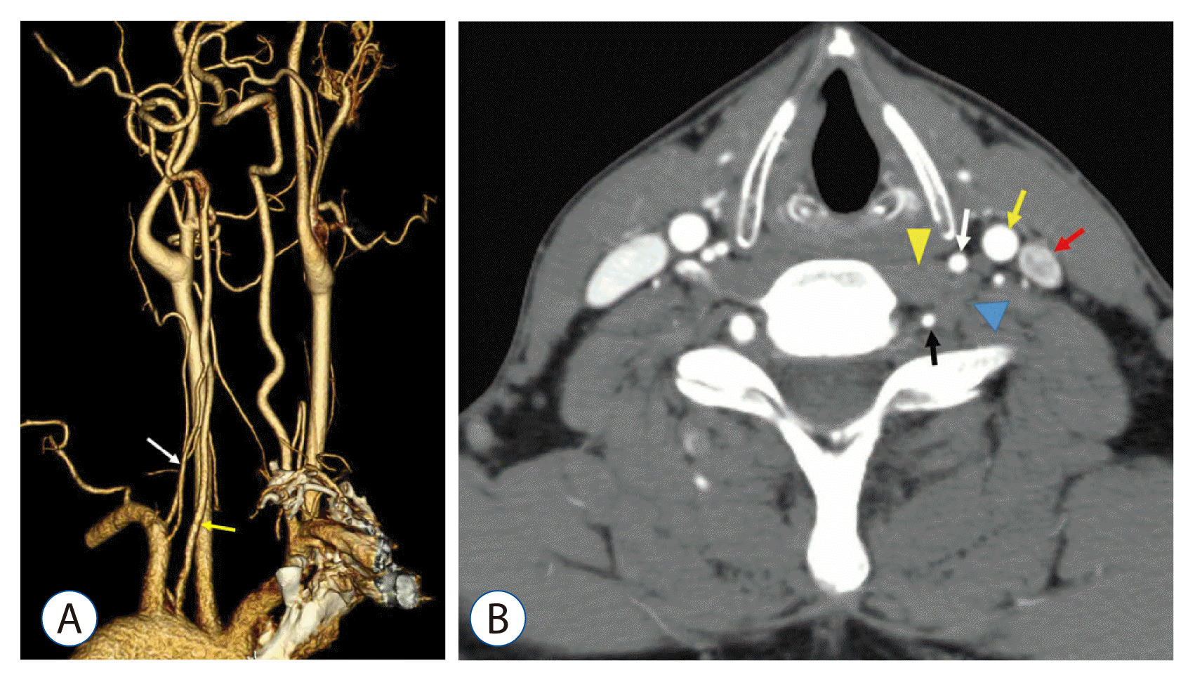
A 35-year-old man with left occipital pain underwent CT angiography. Reconstructed CT angiography showed a left duplicated VA originating from the left subclavian artery and aortic arch. Axial CT demonstrated that the medial limb of the duplicated VA was located anteriorly to the vertebral transverse foramen and medially to the CCA (Fig. 1). The patient’s symptom was relieved after conservative management.
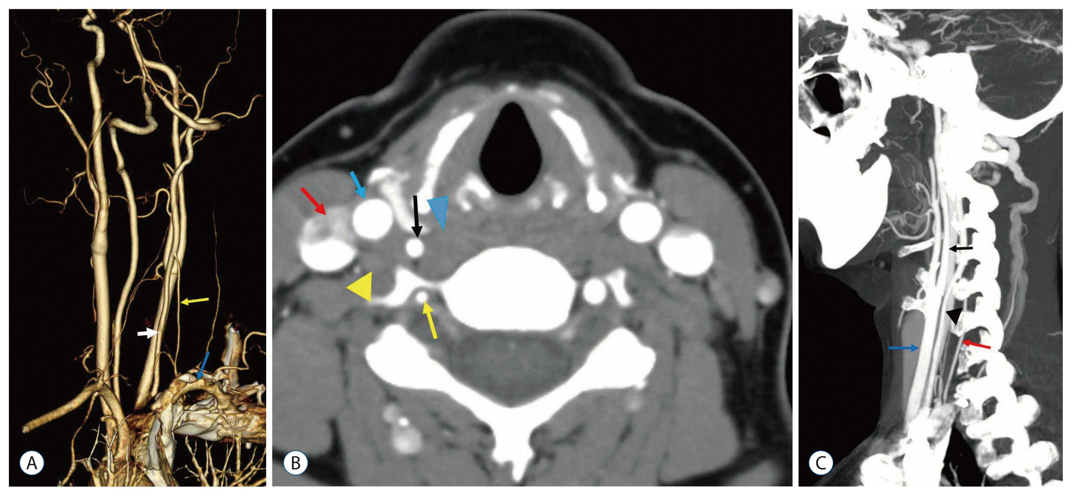
A 38-year-old woman was referred to our department because of dizziness. CT angiography demonstrated an incidental right duplicated VA originating from the right subclavian artery. Axial CT showed that the medial limb of the duplicated VA was located posteromedially to the right CCA and anteriorly to the vertebral transverse foramen. Sagittal CT revealed that the medial limb of the duplicated VA was located in close proximity to the CCA and internal carotid artery (Fig. 2). The patient was managed conservatively, and her dizziness improved.
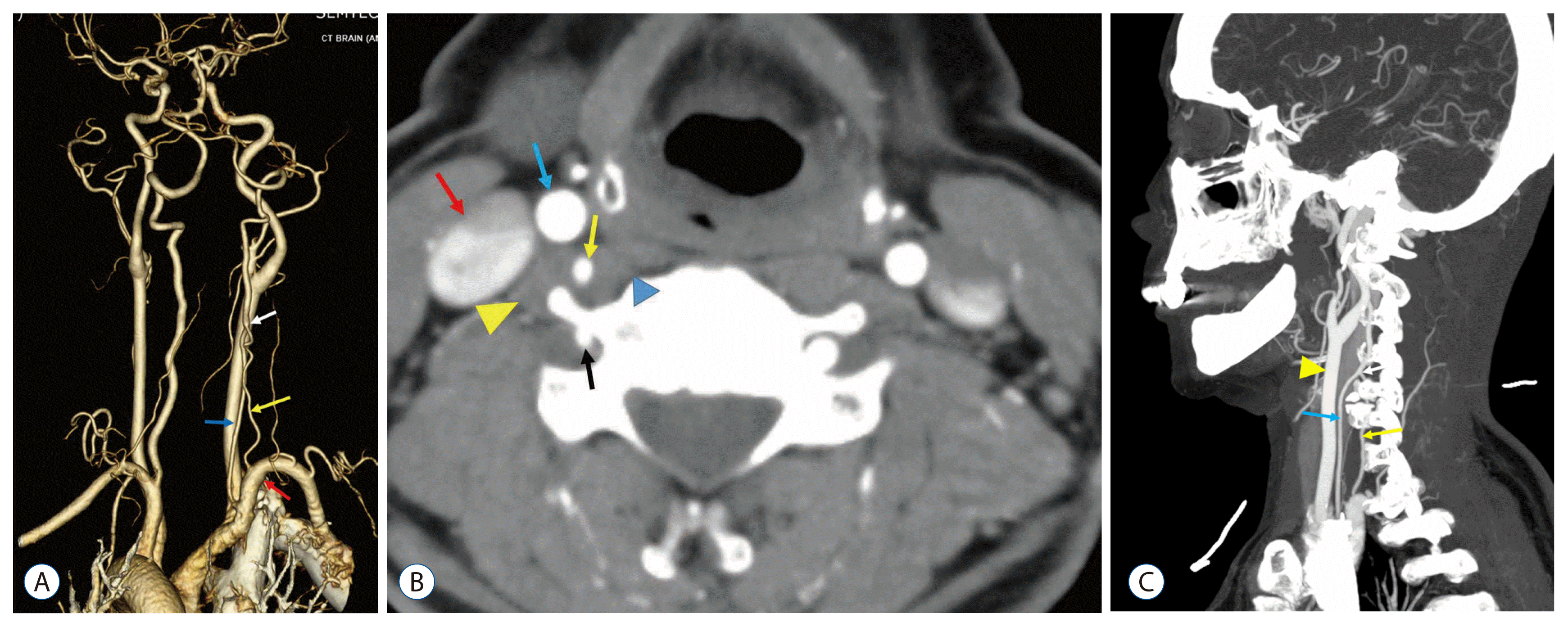
A 58-year-old woman visited our department for paresthesia of the left occipital area and right arm, and intermittent weakness in both legs. CT angiography showed a right duplicated VA originating from the right subclavian artery. Axial CT demonstrated that the medial limb of the duplicated VA was located posteriorly to the right CCA and anteriorly to the vertebral transverse foramen. Sagittal CT revealed that the medial limb of the duplicated VA was located in close proximity to the CCA (Fig. 3). The patient was managed conservatively, and her clinical symptoms improved.
During embryological development, the VA is formed by fusion of the longitudinal anastomoses of the cervical intersegmental arteries, which branch off the primitive paired dorsal aorta. In normal situations, almost all intersegmental arteries regress, with the exception of the seventh intersegmental artery (i.e., the sixth cervical intersegmental artery), which forms the proximal portion of the subclavian artery15). Several mechanisms that may give rise to a separated origin of the VA have been proposed. One explanation suggests that the primitive dorsal aorta does not regress together with the two intersegmental arteries that connect to the VA. This arrangement may give rise to duplication of the VA16,19). Another possibility is failure of the regression of the fourth, fifth, or sixth intersegmental arteries (i.e., the third, fourth, or fifth cervical intersegmental arteries)4–6). This adds an additional origin to the VA in addition to the normal seventh intersegmental artery. Persistence of the left fourth or fifth (or less frequently third) cervical intersegmental artery may result in an aortic origin or dual origin of the left VA.
During duplication of the VA, one limb can originate from the subclavian artery, whereas the second limb can originate from the aortic arch, subclavian artery, thyrocervical trunk, or innominate trunk4,12). Rarely, both limbs of a duplicated left VA originate from the aorta8). The left fourth and fifth intersegmental arteries rarely branch off from a stem artery, which originates from the aorta between the left CCA and left subclavian artery. Usually, the medial limb enters the higher vertebral transverse foramen, which is consistent with the theory of intersegmental vessel regression failure, and results from intersegmental arteries that accompany the cervical nerve roots21). In all 10 cases reported here, the medial limb of the duplicated VA entered the higher vertebral transverse foramen.
Duplication of the origin of the VA has no clinical expression in most cases. I reviewed the reports of symptomatic cases of a duplicated VA and their clinical implications for diagnosis and treatment.
Only two cases of duplication of the VA associated with dissection have been reported1,11). Dare et al.1) reported a 38-year-old man who had a duplication of the right proximal VA and who experienced extensive vertebrobasilar arterial dissection after sexual intercourse. Melki et al.11) reported a 51-year-old man who presented with acute infarction of the cerebellar vermis resulting from dissection of the medial limb of a duplicated right VA after minor neck trauma. Mahmutyazicioğlu et al.9) reported one case of symptomatic thrombosis in one limb of a left duplicated VA. In this patient, electrocardiography revealed paroxysmal atrial tachycardia, which was the most likely explanation for the thrombus. This thrombosis was identified using color Doppler sonography and was treated with anticoagulation, after which the patient’s symptoms, including vertigo, weakness, and nausea, ceased.
In a duplicated VA, the other limb may be collateral to the distal VA when the proximal segment of the subclavian artery or the VA originating from the subclavian artery exhibits stenosis2,20). Thrombosis or dissection of one limb of a duplicated VA may result in less severe symptoms compared with other types of VA thrombosis or dissection because of a collateral pathway via another limb of the duplicated VA.
Operative correction of a kinked duplicated origin of the VA, which was suspected to underlie disabling dizziness in a 70-year-old patient, was performed despite the absence of any other evidence of its benefits13). A dual origin of the VA mimicked a dissection on conventional angiography7,14). Polguj et al.15) reported that the lumen of the duplicated VA is smaller than normal; hence, this variant has clinical implications and should be considered when a VA needs catheterization. In such patients, interventional procedures should be performed from the normal side, if possible. Satti et al.18) suggested that a VA can be easily damaged during severe cervical spine injuries with rapid subluxation, deceleration, fracture through the transverse foramen, or flexion of the cervical spine. The additional point of attachment of the VA predisposes individuals with a duplicated origin of the VA to experience more severe outcomes from the kind of contusion that most commonly occurs during motor vehicle accidents.
Duplication of the VA may be encountered unexpectedly during lower anterior cervical spinal surgery because the entry of the medial limb of the duplicated VA into the vertebral transverse foramen is generally higher than usual3,4,8). In all 10 patients in this study, the medial limb of the dual-origin VA was located anteriorly and laterally to the cervical transverse foramen, and between the longus coli muscle and anterior scalene muscle. In the anterior spinal approach, the surgeon retracts the longus coli muscle laterally to expose the vertebral body; therefore, the location of the medial limb of the VA should be considered to avoid VA injury. Excessive retraction on the longus coli muscle may lead to injury of the medial limb of the VA. The additional point of attachment of the VA (junction point of both limbs of a duplicated VA) predisposes individuals with a duplicated origin of the VA to suffer from the kind of retraction injury that can occur during the lower cervical anterior approach. During anterior cervical spine operations, the surgeon might select the contralateral approach to avoid damaging the medial limb of a duplicated VA.
In six patients in this study, both limbs of the VA were located in distant position to the CCA and common carotid bifurcation. In the other four patients in this study, the medial limb of the duplicated VA was located in close proximity to the common carotid bifurcation or CCA. In carotid endarterectomy, the surgeon may injure the medial limb of the VA during CCA dissection and proximal clamping if the medial limb of the duplicated VA is located in close proximity to the common carotid bifurcation or CCA. Thus, surgeons should consider this anomaly when performing carotid endarterectomy4).
References
1. Dare AO, Chaloupka JC, Putman CM, Mayer PL, Schneck MJ, Fayad PB. Vertebrobasilar dissection in a duplicated cervical vertebral artery: a possible pathoetiologic association? A case report Vasc Endovascular Surg. 31:103–109. 1997.

2. Eisenberg RA, Vines FS, Taylor SB. Bifid origin of the left vertebral artery. Radiology. 159:429–430. 1986.

3. Giuffrè R, Sherkat S. Maldevelopmental pathology of the vertebral artery in infancy and childhood. Childs Nerv Syst. 16:627–632. 2000.

4. Goddard AJ, Annesley-Williams D, Guthrie JA, Weston M. Duplication of the vertebral artery: report of two cases and review of the literature. Neuroradiology. 43:477–480. 2001.

5. Ionete C, Omojola MF. MR angiographic demonstration of bilateral duplication of the extracranial vertebral artery: unusual course and review of the literature. AJNR Am J Neuroradiol. 27:1304–1306. 2006.
6. Kendi AT, Brace JR. Vertebral artery duplication and aneurysms: 64-slice multidetector CT findings. Br J Radiol. 82:e216–e218. 2009.

7. Kim DW. Concomitant dual origin and fenestration of the left vertebral artery resembling dissection. J Korean Neurosurg Soc. 46:498–500. 2009.

8. Komiyama M, Nakajima H, Yamanaka K, Iwai Y. Dual origin of the vertebral artery--case report. Neurol Med Chir (Tokyo). 39:932–937. 1999.
9. Mahmutyazicioğlu K, Sarac K, Bölük A, Kutlu R. Duplicate origin of left vertebral artery with thrombosis at the origin: color doppler sonography and CT angiography findings. J Clin Ultrasound. 26:323–325. 1998.

10. Meila D, Tysiac M, Petersen M, Theisen O, Wetter A, Mangold A, et al. Origin and course of the extracranial vertebral artery: CTA findings and embryologic considerations. Clin Neuroradiol. 22:327–333. 2012.

11. Melki E, Nasser G, Vandendries C, Adams D, Ducreux D, Denier C. Congenital vertebral duplication: a predisposing risk factor for dissection. J Neurol Sci. 314:161–162. 2012.

12. Mizukami M, Tomita T, Mine T, Mihara H. Bypass anomaly of the vertebral artery associated with cerebral aneurysm and arteriovenous malformation. J Neurosurg. 37:204–209. 1972.

13. Nishijima M, Harada J, Akai T, Endo S, Takaku A. Operative correction of a kinked duplicate origin of the vertebral artery in a patient with dizziness. Case report Surg Neurol. 32:356–359. 1989.

14. Nogueira TE, Chambers AA, Brueggemeyer MT, Miller TJ. Dual origin of the vertebral artery mimicking dissection. AJNR Am J Neuroradiol. 18:382–384. 1997.
15. Polguj M, Jędrzejewski K, Topol M, Wieczorek-Pastusiak J, Majos A. Duplication of the left vertebral artery in a patient with dissection of the right internal carotid artery and Ehlers-Danlos syndrome: case report and review of the literature. Anat Sci Int. 88:109–114. 2013.

16. Rameshbabu C, Gupta OP, Gupta KK, Qasim M. Bilateral asymmetrical duplicated origin of vertebral arteries: multidetector row CT angiographic study. Indian J Radiol Imaging. 24:61–65. 2014.

18. Satti SR, Cerniglia CA, Koenigsberg RA. Cervical vertebral artery variations: an anatomic study. AJNR Am J Neuroradiol. 28:976–980. 2007.
19. Sim E, Vaccaro AR, Berzlanovich A, Thaler H, Ullrich CG. Fenestration of the extracranial vertebral artery: review of the literature. Spine (Phila Pa 1976). 26:E139–E142. 2001.
Fig. 1
Reconstructed computed tomographic (CT) angiography shows a left duplicated vertebral artery (VA) originating from the left subclavian artery and aortic arch (A : yellow arrow, medial limb of the left duplicated VA; white arrow, lateral limb of the left duplicated VA). Axial neck CT at the sixth cervical vertebral body level after contrast enhancement shows that the medial limb of the left duplicated VA is located anteriorly to the vertebral transverse foramen. The medial limb of the duplicated VA is located between the longus coli muscle and anterior scalene muscle (B : black arrow, lateral limb of the left duplicated VA; white arrow, medial limb of left VA, red arrow, left internal jugular vein; yellow arrow, left common carotid artery; yellow arrowhead, longus coli muscle; blue arrowhead, anterior scalene muscle).

Fig. 2
Computed tomographic (CT) angiography of a 38-year-old woman with a right duplicated vertebral artery (VA) originating from the right subclavian artery. Reconstructed CT angiography shows a right duplicated VA originating from the right subclavian artery (A : blue arrow, right subclavian artery; yellow arrow, lateral limb of the right duplicated VA; white arrow, medial limb of the right duplicated VA). Axial neck CT at the fifth cervical vertebral body level after contrast enhancement shows that the medial limb of the duplicated right VA is located anteriorly to the vertebral transverse foramen and between the longus coli muscle and anterior scalene muscle (B : black arrow, medial limb of the right duplicated VA; yellow arrow, lateral limb of the right VA; blue arrow, right common carotid artery [CCA]; red arrow, right internal jugular vein; blue arrowhead, longus coli muscle; yellow arrowhead, anterior scalene muscle). Sagittal CT after contrast enhancement shows that the medial limb of the duplicated VA is located in close proximity to the CCA and proximal internal carotid artery (C : black arrowhead, medial limb of the duplicated VA; blue arrow, CCA; red arrow, lateral limb of the duplicated VA; black arrow, internal carotid artery).

Fig. 3
Computed tomographic (CT) angiography of a 58-year-old woman with a right dual-origin vertebral artery (VA). Reconstructed CT angiography shows a right duplicated VA originating from the right subclavian artery (A : red arrow, right subclavian artery; yellow arrow, lateral limb of the duplicated VA; blue arrow, medial limb of the duplicated VA; white arrow, junction of the two limbs of the duplicated VA). Axial neck CT at the fifth cervical vertebral body level after contrast enhancement shows that the medial limb of the duplicated VA is located posteriorly to the right common carotid artery (CCA) and anteriorly to the vertebral transverse foramen. The medial limb of the duplicated VA is located between the longus coli muscle and anterior scalene muscle (B : yellow arrow, medial limb of the right duplicated VA; black arrow, lateral limb of the right VA; blue arrow, right CCA; red arrow, right internal jugular vein; yellow arrowhead, anterior scalene muscle; blue arrowhead, longus coli muscle). Sagittal CT after contrast enhancement shows that the medial limb of the duplicated VA is located in close proximity to the CCA (C : blue arrow, medial limb of the duplicated VA; yellow arrowhead, CCA; yellow arrow, lateral limb of the duplicated VA; white arrow, junction of the two limbs of the duplicated VA).

Table 1
Characteristics of 10 patients with duplicated VA


 Citation
 Print
Print


 XML Download