INTRODUCTION
Bone cement augmentation procedures, such as percutaneous vertebroplasty (VP) or balloon kyphoplasty (KP), for osteoporotic compression fractures are simple and minimally invasive. Owing to their simplicity and rapid efficacy, they have been popular for the management of incapacitating pain related to osteoporotic compression fractures4,6).
Nonetheless, complications or delayed sequelae after VP or KP include leakage of cement into the spinal canal, cement dislodgement or fragmentation, and adjacent vertebral fractures. In such cases, patients usually require posterior screw fixation for structural stabilization of the augmented vertebrae10). Furthermore, spine surgeons occasionally encounter that posterior screw fixation is indicated in previously augmented vertebrae for degenerative spinal diseases.
Many clinical reports have examined the safety and efficacy of bone cement-augmented screw fixation for the treatment of osteoporotic spines to increase the pull-out strength3,5). However, to the best of the authors’ knowledge, no previous reports have evaluated the clinical results of posterior screw fixation in previously augmented vertebrae with bone cement for osteoporotic compression fractures.
Screw fixation in previously augmented vertebrae is considered difficult or even a contraindication owing to the excessive reinforcement of stiffness and strength after VP or KP. The purpose of this study was to evaluate the clinical results of screw fixation in previously augmented vertebrae for osteoporotic compression fractures and to determine whether this technique is feasible.
Go to : 
MATERIALS AND METHODS
From 2010 to 2015, 14 patients (3 men and 11 women) who underwent screw fixation in the vertebrae previously augmented for osteoporotic compression fractures were enrolled in this study. The following were the inclusion criteria: 1) previous VP or KP for an osteoporotic compression fracture, 2) symptom-free period after VP or KP for more than1month, and 3) newly developed mechanical back pain or neurologic deficits requiring posterior screw fixation for structural stabilization.
The study also included revision surgeries for delayed complications of VP or KP, such as cement dislodgement or neurologic deficits due to cement leakage. Degenerative spinal diseases requiring decompression and screw fixation at augmented levels were also included. However, patients with intraoperative complications and late spondylodiscitis requiring an anterior approach were excluded. All procedures were performed by a single surgeon. The augmented vertebral bodies were classified into two groups according to cement distribution pattern on simple radiographs. The criteria were as follows: 1) solid-type vertebral bodies with compact and 2) solid cement filling and trabecular-type vertebral bodies with sponge-like filling cement.
Surgical procedure
After explanation of the procedure, the patients were anesthetized and placed in the prone position on a radiolucent table. A standard open posterior approach with transpedicular screw fixation was performed in six patients; percutaneous screw fixation was confined to the augmented vertebrae in the other eight patients. All patients were allowed to ambulate in a thoracolumbosacral orthosis brace on postoperative day 2. Simple radiographs were taken preoperatively and serially and were used to investigate the pattern of previous cement distribution, fusion status, or screw loosening. All pre-VP, pre-KP, and preoperative magnetic resonance images were also obtained and assessed by the authors.
Safety and outcome evaluations
The patients were evaluated at the final follow-up using the visual analog scale (VAS) and a modified version of MacNab’s criteria for characterizing the clinical outcomes after spine surgery. Comparisons at time points were performed using paired t-tests, and differences were considered statistically significant when the p-values were <0.05.
Go to : 
RESULTS
A total of 14 patients (3 men and 11 women) underwent screw fixation in the previously augmented vertebrae for osteoporotic compression fractures. Twelve patients underwent VP, and two underwent KP. The mean age of the patients was 74.1 (68 –82) years, and the mean follow-up period was 11.5 (6–18) months. Table 1 shows the patient demographics in detail. It was possible to insert screws in the previously augmented vertebrae in all patients. However, the screw trajectory was blocked by the cement in the three patients who showed a solid cleft cement distribution pattern; this necessitated a change from the optimal direction or insertion of a short screw.
Table 1
Demographics of the patients
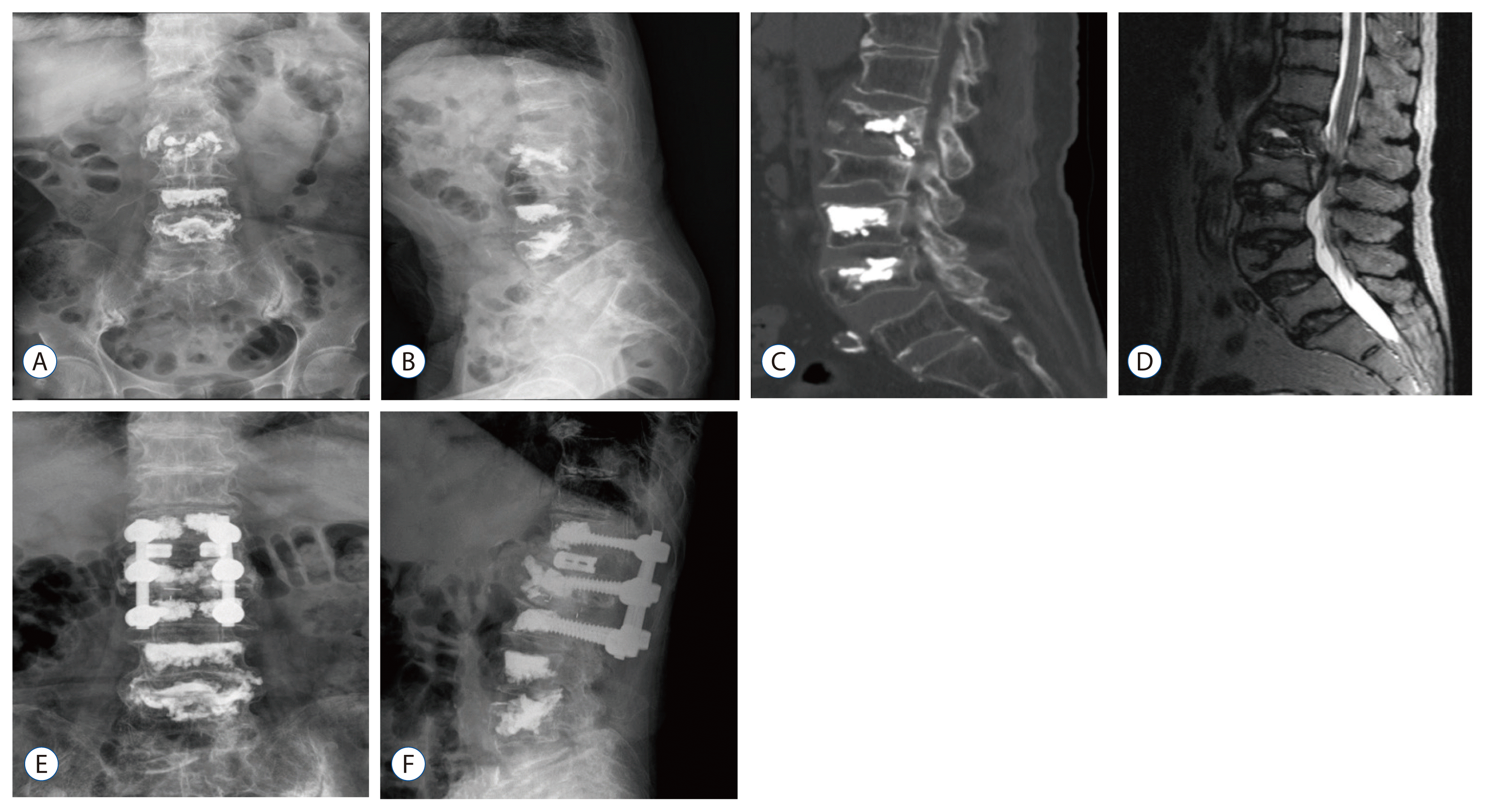
Three patients had progressive kyphosis due to cement dislodgement; two of them had intravertebral cleft (IVC) signs and anterior cortical bone destruction in their pre-VP or pre-KP lateral radiographs. Three patients showed late neurological deterioration after a period of pain relief. Neurological impairment developed insidiously (mean revision time, 3.7 months); the causes of late neurological deterioration were cement leakage or displacement of a fracture fragment from the cemented vertebrae. These patients underwent decompression and posterior screw fixation with an interbody fusion. Removal of the fragmented or dislodged cement was possible through the posterior approach (Figs. 1 and 2).
 | Fig. 1Imaging studies of an 81-year-old woman who underwent percutaneous vertebroplasty at the L1level. A and B: Simple radiographs taken 5 months after percutaneous vertebroplasty show a trabecular pattern of cement distribution at the L1 level. C and D: Computed tomography and T2-weighted magnetic resonance images reveal displacement of bone fragments and severe stenosis at the T12–L1 and L1–L2 levels. E and F: Postoperative simple radiographs show an interbody fusion with posterior instrumentation at the T12–L2 levels. |
 | Fig. 2Imaging studies of a 69-year-old woman who underwent percutaneous vertebroplasty at the L2 and L3 levels at a local clinic. A and B: Simple radiographs taken 2 months after percutaneous vertebroplasty reveal a solid pattern of cement distribution and cement leakage in the left L2 foramen. C: Computed tomography image shows a more prominent cement leakage. D and E: Postoperative simple radiographs show a complete removal of leaked cement with posterior instrumentation inserted in a superior direction. |
Eight patients underwent posterior screw fixation and interbody fusion for spondylolisthesis or foraminal stenosis at the level of the previously augmented vertebrae. They all underwent percutaneous screw fixation instead of an open transpedicular screw fixation (Fig. 3).
 | Fig. 3Imaging studies of an 82-year-old woman who underwent percutaneous vertebroplasty for an L5 osteoporotic compression fracture. A and B: Simple radiographs show a good filling of the bone cement with a solid pattern at the L5 body and spondylolisthesis at the L4–L5 levels. C: Sagittal T2-weighted magnetic resonance image reveals severe stenosis at the L4–L5 levels. D and E: Postoperative simple radiographs show an interbody fusion with cages and percutaneous instrumentation. |
Clinical outcome
At the final follow-up after surgery, 12 patients (86%) showed excellent or good outcomes according to the modified MacNab’s criteria. Prior to surgery, the mean pain VAS score was 7.9; this decreased to 3.3 at the final follow-up. The improvement was statistically significant (p<0.05). No major complications were observed in any study participant. During the follow-up period, two patients sustained fractures after minor injuries. VP or KP with bone cement augmentation was performed to treat these fractures.
Go to : 
DISCUSSION
Osteoporotic compression fractures are a common and major cause of morbidity in the elderly. VP and KP with polymethylmethacrylate for osteoporotic compression fractures have gained widespread popularity owing to their rapid effectiveness and simplicity.
In spite of their advantages with good clinical outcomes and simplicity, they also have complications. Excluding acute complications, such as extravasation of cements, pulmonary embolism through the venous drainage system, and hematoma, late clinical complications, such as infection, delayed adjacent segment fractures, and cement displacement following VP or KP may occur8,9). Moreover, spine surgeons sometimes encounter situations requiring surgical treatments of augmented vertebrae for degenerative spinal diseases, such as spondylolisthesis or foraminal stenosis. In such cases, application of rigid posterior instrumentation and decompression is usually required to stabilize the spine. However, whether screws can penetrate abnormally consolidated cement-augmented vertebrae is yet unclear.
It is well known that a larger volume of bone cement may increase the risk of both cement leakage and adjacent fractures because of overstiffening of the segment and less deformation of the vertebral body1). Oka et al.7) also reported that a solid pattern of cement distribution was related to the presence of an IVC and a higher incidence in new compression fractures and is a poor prognostic factor.
In our consecutive series, two patients with IVC showed cement dislodgement and progressive collapse of the augmented vertebral body. They showed a solid pattern of cement distribution with IVC after the initial VP or KP. It is believed that less interdigitation of cements and progressive bone resorption can be the causes of this delayed complication2). Posterior instrumentation using short screws for cement blockage and posterolateral bone fusion without anterior reconstruction were performed in these patients.
A possible delayed complication related to VP or KP is cement leakage into surrounding tissues during the procedure, potentially causing radiculopathy or myelopathy. Moreover, progressive collapse of an augmented vertebral body and displacement of bone fragments can aggravate neurological complications without a history of trivial injury.
In our study, three patients with cement leakage or fragmentation of the bone developed delayed neurologic deficits after a period of symptom relief following augmentation. Posterior decompression of neural elements via removal of the leaked cements or bone fragments with posterior instrumentation and interbody fusion was performed. In one patient who showed a solid pattern of cement distribution following VP with a lack of interspersion along the trabecular spaces, it was more difficult to insert screws through an ideal trajectory. Although the procedure was successful, the bone cement served as an obstacle to the screw insertion, and the screws were inserted in a more superior direction than what was considered ideal.
Posterior screw fixation was performed percutaneously in eight patients for degenerative spinal diseases, such as spondylolisthesis or foraminal stenosis without difficulties. In the present study, we also performed posterior screw fixation in the previously augmented vertebrae with bone cement in revision surgeries for delayed complications following VP or KP and for degenerative spinal diseases. Regardless of whether the screw insertion technique was percutaneous or open, a trabecular cement pattern made it possible to insert screws without difficulties. Although it was difficult to insert screws in an ideal trajectory when the cement distribution showed a solid pattern, the results of our study showed that posterior screw fixation is not a contraindication and can safely be used even in elderly patients.
However, despite our excellent outcomes, this study is intrinsically limited owing to its retrospective review of a heterogeneous patient group. Moreover, since we did not know the exact amount of the injected cement volume, we could not analyze the correlation between the injected cement volume and surgical outcome. Thus, case-controlled studies on delayed complications of bone cement augmentation procedures with long-term follow-ups are crucial.
Go to : 


 Citation
 Print
Print


 XML Download