Journal List > J Korean Neurosurg Soc > v.59(1) > 1161803

Lee: Chronic Subdural Hematoma in the Aged, Trauma or Degeneration?

Abstract

Chronic subdural hematomas (CSHs) are generally regarded to be a traumatic lesion. It was regarded as a stroke in 17th century, an inflammatory disease in 19th century. From 20th century, it became a traumatic lesion. CSH frequently occur after a trauma, however, it cannot occur when there is no enough subdural space even after a severe head injury. CSH may occur without trauma, when there is sufficient subdural space. The author tried to investigate trends in the causation of CSH. By a review of literature, the author suggested a different view on the causation of CSH. CSH usually originated from either a subdural hygroma or an acute subdural hematoma. Development of CSH starts from the separation of the dural border cell (DBC) layer, which induces proliferation of DBCs with production of neomembrane. Capillaries will follow along the neomembrane. Hemorrhage would occur into the subdural fluid either by tearing of bridge veins or repeated microhemorrhage from the neomembrane. That is the mechanism of hematoma enlargement. Trauma or bleeding tendency may precipitate development of CSH, however, it cannot lead CSH, if there is no sufficient subdural space. The key determinant for development of CSH is a sufficient subdural space, in other words, brain atrophy. The most common and universal cause of brain atrophy is the aging. Modifying Virchow's description, CSH is sometimes traumatic, but most often caused by degeneration of the brain. Now, it is reasonable that degeneration of brain might play pivotal role in development of CSH in the aged persons.

INTRODUCTION

Chronic subdural hematomas (CSHs) are generally regarded to be consequences of head trauma1451). The most commonly accepted pathophysiological explanation of CSH is that mild head trauma leads to tearing of bridging veins with subsequent bleeding, thus creating the hematoma7). However, a history of trauma is absent in about 30–50% of the cases, especially after the age of 65 years1121952). Besides trauma, there were numerous risk factors such as alcohol abuse, seizures, cerebrospinal fluid shunts, and coagulopathies including therapeutic anticoagulant1451).
In 17th century, CSH was a stroke. It became an inflammation in 19th century and finally a traumatic lesion in 20th century. Trauma is the most commonly alluded as a cause, however, it cannot occur when there is no enough subdural space even after a severe head injury. If there is sufficient subdural space, CSH may occur without trauma such as intracranial hypotension. Causation of CSH is an important issue in the accident insurance. The author tried to investigate trends in the causality of CSH.

MATERIALS AND METHODS

A computerized search of the National Library of Medicine (PubMed) database of the up-to-date literature (published from 2010 to 2014) was undertaken. The medical subject headings "chronic subdural hematoma" combined with "causality" yielded 197 citations (excluded non-English language and non-human citations). The abstracts of relevant citations were reviewed and 41 articles were selected. The selected papers have been categorized into one of five categories by the causalities proposed by the authors (Table 1). By a review of literature, the author suggested a different view on the causation of CSH.

RESULTS

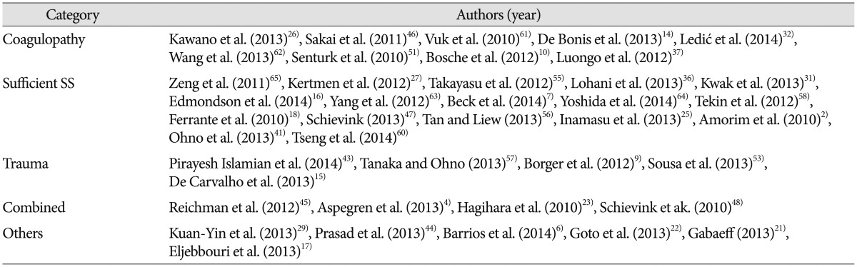
As the cause of CSH, sufficient subdural space was alluded in 19 papers, coagulopathy in 13 papers, and trauma in 8 papers, including the combined categories. Sufficient subdural space included arachnoid cyst, cerebrospinal fluid leak, lumbar drainage, spinal anesthesia, intracranial hypotension, hydrocephalus, excerebral atrophy, and the old age. Coagulopathy included factor X deficiency, factor XIII deficiency, von Willebrand's disease, afibrinogenemia, anticoagulant therapy, and end stage renal disease. Trauma included head-banging, dancing, and any tears in the arachnoid membrane following contusion of the brain or slight bleeding from a bridging vein. Combined category included trauma with coagulopathy or sufficient subdural space with coagulopathy. Others included dural metastasis, lymphoma, infection, antiviral therapy, and birth related complications.

DISCUSSION

Sufficient subdural space was the most commonly alluded as the cause of CSH. The number of papers did not have a value by itself, since papers did not have equal frequency or importance. However, it may represent relative importance or apportionment of certain factors. Recent papers may not represent the whole aspects of the causation. They may have a tendency to publish atypical cases. However, we can explore this curious lesion using the more updated knowledge.
Traditional model of causation, so-called the classic epidemiologic triangle of host, agent, and environment was not suitable for CSH. To evaluate relative importance in multiple causation, web of causation model with directed acyclic graph (Fig. 1) is more suitable. Development of CSH starts from the separation of the dural border cell (DBC) layer, which can occur either by trauma or low intracranial pressure. Severe trauma cannot produce a CSH even in a patient with coagulopathy, when the subdural space is not enough. Trauma cannot be the causality of CSH in this situation. Furthermore, CSH may occur without trauma. Causation implies that there is a true mechanism that leads from exposure to disease. Trauma or bleeding tendency is frequently associated with CSH and they may precipitate development of CSH, however, it cannot lead CSH, if there is no prerequisite. The key determinant for development of CSH is a sufficient subdural space, in other words, cerebral atrophy. The most common and universal cause of cerebral atrophy is the aging. If we classify a disease according to the key determinant, cerebral atrophy is responsible for the CSH instead of trauma.
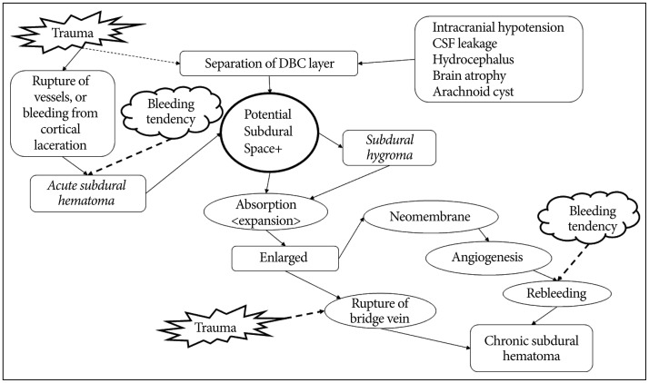
Trauma was so frequently observed that CSH might be a traumatic lesion. The majority of CSHs are suspected by an undiagnosed trivial head injury. This assumption is hard to explain why CSH is rare in the young with severe head injury, while the majority of CSH occur in the old even without trauma33). In patients without trauma history, we presumed that they forgot the trauma, since the CSH is common in the aged and alcoholics. However, there are numerous cases of CSHs without trauma3781011152428303940424748495962), which made trauma as an associated factor instead of the cause.
Bleeding tendency clearly has a significant association with an increased risk of CSH14). Because of the continuing rise of life expectancy and the increasing use of anticoagulants, the incidence of CSH is expected to rise45142035). Anticoagulant and antiplatelet therapy became an important risk factor for development of CSH. However, bleeding tendency cannot lead CSH by itself.
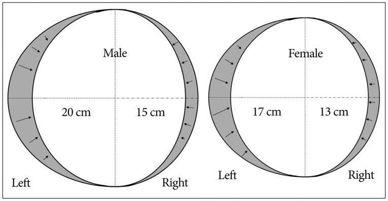
Although about two-thirds (57–78.7%) of CSHs were in persons older than 65 years553), CSH does not occur in all aged person. Since aging and cerebral atrophy are universal, atrophy alone is not enough to explain the causality of CSH. The author suspect the cranial size and morphology as an important predisposing factor. Cerebral atrophy produce negative pressure within the cranium. This negative pressure is constant on each side of the cranium, according to Boyle's law, which the pressure of gas tried to hold at a constant temperature change by inverse with the volume of the gas. As the degree of atrophy is constant, the decreased length of brain diameter on the large hemisphere is longer than on the small hemisphere (Fig. 2). So, the distance from the skull to the cerebral cortex in the shrunken hemisphere is longer on the side of large hemisphere. The bridging vein is more stretched on the large hemisphere, so it may be tore by a trivial injury. If the distance exceeds the length of the arachnoid trabeculae, separation of the DBC layer will occur at the large hemisphere, which will create so-called the subdural space. For example, overall 10% reduction result 2 cm in the left, 1.5 cm in the right in the male cranium. While in the female cranium, 9% reduction (the degree of atrophy is less severe than male) of shorter diameter result about 1.5 cm in the left side. This anatomical difference of the cranium influences the left and male prevalence of CSH. Actual separation of the DBC layer will depends on the force pulling the arachnoid trabeculae, which is the sum of the cranial morphology and the degree of cerebral atrophy.

CONCLUSION

The origin of a CSH is multiple. It can be developed spontaneously, or changed from a subdural hygroma or an acute subdural hematoma. A more important prerequisite is sufficient potential subdural space that is aging, degeneration of the brain. CSH was a stroke in 17th century13), an inflammation in 19th century, and it became a traumatic lesion in 20th century38). Virchow recognized that hematoma durae matris sometimes was traumatic, but he believed that this lesion was most often caused by chronic inflammation (pachymeningitis chronica hemorrhagica) of the dura1354). Modifying Virchow's description, CSH is sometimes traumatic, but most often caused by degeneration of the brain. Now, it would be reasonable that in the aged persons, degeneration of brain might play the most important role in development of CSH.

References

1. Adhiyaman V, Asghar M, Ganeshram KN, Bhowmick BK. Chronic subdural haematoma in the elderly. Postgrad Med J. 2002; 78:71–75. PMID: 11807186.
crossref
2. Amorim JA, Remígio DS, Damázio Filho O, de Barros MA, Carvalho VN, Valença MM. Intracranial subdural hematoma post-spinal anesthesia : report of two cases and review of 33 cases in the literature. Rev Bras Anestesiol. 2010; 60:620–629. 344–349. PMID: 21146058.
crossref
3. Arpino L, Gravina M, Basile D, Franco A. Spontaneous chronic subdural hematoma in a young adult. J Neurosurg Sci. 2009; 53:55–57. PMID: 19546844.
4. Aspegren OP, Åstrand R, Lundgren MI, Romner B. Anticoagulation therapy a risk factor for the development of chronic subdural hematoma. Clin Neurol Neurosurg. 2013; 115:981–984. PMID: 23128014.
crossref
5. Balser D, Farooq S, Mehmood T, Reyes M, Samadani U. Actual and projected incidence rates for chronic subdural hematomas in United States Veterans Administration and civilian populations. J Neurosurg. 2015; 20:1–7.
crossref
6. Barrios L, Clément R, Visseaux G, Bord E, Le Gall F, Rodat O. A case of atypical chronic subdural hematoma : a spontaneous rupture of dural lymphoma nodule. J Forensic Leg Med. 2014; 22:145–147. PMID: 24485439.
crossref
7. Beck J, Gralla J, Fung C, Ulrich CT, Schucht P, Fichtner J, et al. Spinal cerebrospinal fluid leak as the cause of chronic subdural hematomas in nongeriatric patients. J Neurosurg. 2014; 121:1380–1387. PMID: 25036203.
crossref
8. Berhouma M, Houissa S, Jemel H, Khaldi M. Spontaneous chronic subdural hematoma of the posterior fossa. J Neuroradiol. 2007; 34:213–215. PMID: 17572494.
9. Borger V, Vatter H, Oszvald Á, Marquardt G, Seifert V, Güresir E. Chronic subdural haematoma in elderly patients : a retrospective analysis of 322 patients between the ages of 65-94 years. Acta Neurochir (Wien). 2012; 154:1549–1554. PMID: 22772400.
crossref
10. Bosche B, Molcanyi M, Noll T, Kochanek M, Kraus B, Rieger B, et al. Occurrence and recurrence of spontaneous chronic subdural haematoma is associated with a factor XIII deficiency. Clin Neurol Neurosurg. 2013; 115:13–18. PMID: 22541133.
crossref
11. Bosma JJ, Miles JB, Shaw MD. Spontaneous chronic and subacute subdural haematoma in young adults. Acta Neurochir (Wien). 2000; 142:1307–1310. PMID: 11201648.
crossref
12. Cheng SY, Chang CK, Chen SJ, Lin JF, Tsai CC. Chronic subdural hematoma in elderly taiwan patients : a retrospective analysis of 342 surgical cases. Int J Geront. 2014; 8:37–41.
crossref
13. D'Errico AP, German WJ. Chronic Subdural Hematoma. Yale J Biol Med. 1930; 3:11–20. PMID: 21433469.
14. De Bonis P, Trevisi G, de Waure C, Sferrazza A, Volpe M, Pompucci A, et al. Antiplatelet/anticoagulant agents and chronic subdural hematoma in the elderly. PLoS One. 2013; 8:e68732. PMID: 23874740.
crossref
15. De Carvalho D, Almenawer S, Lozej M, Noble H, Murty NK. Spontaneous chronic subdural hematoma in a 22-year-old healthy woman. World Neurosurg. 2013; 80:654.e9–654.e11. PMID: 23182734.
crossref
16. Edmondson L, Upshaw JE, Tuuri RE. A 14-year-old male with a 10-week history of headaches. Pediatr Ann. 2014; 43:220222–223. PMID: 24972416.
crossref
17. Eljebbouri B, Okacha N, Boucetta M. Chronic subdural hematoma infected by Campylobacter fetus. Surg Infect (Larchmt). 2013; 14:563–564. PMID: 23930907.
18. Ferrante E, Arpino I, Citterio A, Wetzl R, Savino A. Epidural blood patch in Trendelenburg position pre-medicated with acetazolamide to treat spontaneous intracranial hypotension. Eur J Neurol. 2010; 17:715–719. PMID: 20050898.
crossref
19. Fogelholm R, Heiskanen O, Waltimo O. Chronic subdural hematoma in adults. Influence of patient's age on symptoms, signs, and thickness of hematoma. J Neurosurg. 1975; 42:43–46. PMID: 1167376.
20. Forster MT, Mathé AK, Senft C, Scharrer I, Seifert V, Gerlach R. The influence of preoperative anticoagulation on outcome and quality of life after surgical treatment of chronic subdural hematoma. J Clin Neurosci. 2010; 17:975–979. PMID: 20580997.
crossref
21. Gabaeff SC. Investigating the possibility and probability of perinatal subdural hematoma progressing to chronic subdural hematoma, with and without complications, in neonates, and its potential relationship to the misdiagnosis of abusive head trauma. Leg Med (Tokyo). 2013; 15:177–192. PMID: 23434514.
crossref
22. Goto T, Ohshima S, Miura K, Shibuya T, Sato W, Dohmen T, et al. A chronic subdural hematoma in a patient receiving combination therapy with pegylated interferon alfa-2b and ribavirin for chronic hepatitis C. Intern Med. 2013; 52:2057–2060. PMID: 24042512.
crossref
23. Hagihara N, Abe T, Kojima K, Watanabe M, Tabuchi K. Coexistence of cranial and spinal subdural hematomas : case report. Neurol Med Chir (Tokyo). 2010; 50:333–335. PMID: 20448430.
crossref
24. Hong JC, Kim MS, Chang CH, Kim SH. Arachnoid cyst with spontaneous intracystic hemorrhage and chronic subdural hematoma. J Korean Neurosurg Soc. 2008; 43:54–56. PMID: 19096549.
crossref
25. Inamasu J, Watabe T, Ganaha T, Yamada Y, Nakae S, Ohmi T, et al. Clinical characteristics and risk factors of chronic subdural haematoma associated with clipping of unruptured cerebral aneurysms. J Clin Neurosci. 2013; 20:1095–1098. PMID: 23669172.
crossref
26. Kawano H, Yamamoto D, Uchihashi Y, Wakahashi K, Kawano Y, Sada A, et al. Severe inhibitor-negative acquired factor XIII/13 deficiency with aggressive subdural haemorrhage. Blood Coagul Fibrinolysis. 2013; 24:638–641. PMID: 23511901.
crossref
27. Kertmen H, Gürer B, Yilmaz ER, Sekerci Z. Chronic subdural hematoma associated with an arachnoid cyst in a juvenile taekwondo athlete : a case report and review of the literature. Pediatr Neurosurg. 2012; 48:55–58. PMID: 22832284.
crossref
28. Kim BW, Jung YJ, Kim MS, Choi BY. Chronic subdural hematoma after spontaneous intracranial hypotension : a case treated with epidural blood patch on c1-2. J Korean Neurosurg Soc. 2011; 50:274–276. PMID: 22102965.
crossref
29. Kuan-Yin T, Dueng-Yuan H, Hsin-I M. Subdural hematoma associated with skull and dural metastasis of gastric carcinoma : a case report. Turk Neurosurg. 2013; 23:796–799. PMID: 24310465.
30. Kuramae T, Inamasu J, Nakagawa Y, Nakatsukasa M. Spontaneous intracranial hypotension presenting without orthostatic headache complicated by acute subdural hematoma after drainage for chronic subdural hematoma--case report. Neurol Med Chir (Tokyo). 2011; 51:518–521. PMID: 21785248.
crossref
31. Kwak YS, Hwang SK, Park SH, Park JY. Chronic subdural hematoma associated with the middle fossa arachnoid cyst : pathogenesis and review of its management. Childs Nerv Syst. 2013; 29:77–82. PMID: 22914923.
crossref
32. Ledić D, Girotto D, Pal S, Kolbah B. Risk factors for subdural bleeding in elderly population. Coll Antropol. 2014; 38:1195–1198. PMID: 25842756.
33. Lee KS, Bae WK, Doh JW, Bae HG, Yun IG. Origin of chronic subdural haematoma and relation to traumatic subdural lesions. Brain Inj. 1998; 12:901–910. PMID: 9839025.
34. Lee KS, Bae WK, Park YT, Yun IG. The pathogenesis and fate of traumatic subdural hygroma. Br J Neurosurg. 1994; 8:551–558. PMID: 7857535.
crossref
35. Lindvall P, Koskinen LO. Anticoagulants and antiplatelet agents and the risk of development and recurrence of chronic subdural haematomas. J Clin Neurosci. 2009; 16:1287–1290. PMID: 19564115.
crossref
36. Lohani S, Robertson RL, Proctor MR. Ruptured temporal lobe arachnoid cyst presenting with severe back pain. J Neurosurg Pediatr. 2013; 12:281–283. PMID: 23829378.
crossref
37. Luongo M, Pizzuti M, Godano U. Bilateral chronic subdural non-traumatic hematoma associated with von Willebrand's type I disease : a case report. Acta Neurochir (Wien). 2012; 154:1087–1088. PMID: 22392015.
crossref
38. Markwalder TM. Chronic subdural hematomas : a review. J Neurosurg. 1981; 54:637–645. PMID: 7014792.
39. Missori P, Domenicucci M, Sassun TE, Tarantino R, Peschillo S. Alterations in the intracranial venous sinuses in spontaneous nontraumatic chronic subdural hematomas. J Clin Neurosci. 2013; 20:389–393. PMID: 23219821.
crossref
40. Mori K, Maeda M. Surgical treatment of chronic subdural hematoma in 500 consecutive cases : clinical characteristics, surgical outcome, complications, and recurrence rate. Neurol Med Chir (Tokyo). 2001; 41:371–381. PMID: 11561347.
crossref
41. Ohno T, Iihara K, Takahashi JC, Nakajima N, Satow T, Hishikawa T, et al. Incidence and risk factors of chronic subdural hematoma after aneurysmal clipping. World Neurosurg. 2013; 80:534–537. PMID: 23072878.
crossref
42. Park HR, Lee KS, Shim JJ, Yoon SM, Bae HG, Doh JW. Multiple densities of the chronic subdural hematoma in CT scans. J Korean Neurosurg Soc. 2013; 54:38–41. PMID: 24044079.
crossref
43. Pirayesh Islamian A, Polemikos M, Krauss JK. Chronic subdural haematoma secondary to headbanging. Lancet. 2014; 384:102. PMID: 24998813.
crossref
44. Prasad BC, Chandra VV, Varaprasad G. Dural metastases in chronic myeloid leukemia presenting as subdural hematoma. Turk Neurosurg. 2012; 22:777–778. PMID: 23208914.
crossref
45. Reichman J, Singer S, Navi B, Reiner A, Panageas K, Gutin PH, et al. Subdural hematoma in patients with cancer. Neurosurgery. 2012; 71:74–79. PMID: 22705720.
crossref
46. Sakai N, Akamine S, Tokuyama T, Sugiyama K, Kanayama N, Namba H. Chronic subdural hematoma in a patient with congenital afibrinogenemia successfully treated with fibrinogen replacement. Neurol Med Chir (Tokyo). 2011; 51:780–783. PMID: 22123482.
crossref
47. Schievink WI. Stroke and death due to spontaneous intracranial hypotension. Neurocrit Care. 2013; 18:248–251. PMID: 23196352.
crossref
48. Schievink WI, Maya MM, Moser FG, Tourje J. Spectrum of subdural fluid collections in spontaneous intracranial hypotension. J Neurosurg. 2005; 103:608–613. PMID: 16266041.
crossref
49. Schievink WI, Maya MM, Pikul BK, Louy C. Spontaneous spinal cerebrospinal fluid leaks as the cause of subdural hematomas in elderly patients on anticoagulation. J Neurosurg. 2010; 112:295–299. PMID: 19199465.
crossref
50. Seçkin H, Kazanci A, Yigitkanli K, Simsek S, Kars HZ. Chronic subdural hematoma in patients with idiopathic thrombocytopenic purpura: a case report and review of the literature. Surg Neurol. 2006; 66:411–414. discussion 414PMID: 17015125.
crossref
51. Senturk S, Guzel E, Bayrak AH, Bukte Y, Guzel A. Factor X deficiency presenting with bilateral chronic subdural hematoma. Pediatr Neurosurg. 2010; 46:54–57. PMID: 20516741.
crossref
52. Sim YW, Min KS, Lee MS, Kim YG, Kim DH. Recent changes in risk factors of chronic subdural hematoma. J Korean Neurosurg Soc. 2012; 52:234–239. PMID: 23115667.
crossref
53. Sousa EB, Brandão LF, Tavares CB, Borges IB, Neto NG, Kessler IM. Epidemiological characteristics of 778 patients who underwent surgical drainage of chronic subdural hematomas in Brasília, Brazil. BMC Surg. 2013; 13:5. PMID: 23452673.
crossref
54. Taarnhoj P. Chronic subdural hematoma; historical review and analysis of 60 cases. Cleve Clin Q. 1955; 22:150–156. PMID: 13261320.
crossref
55. Takayasu T, Harada K, Nishimura S, Onda J, Nishi T, Takagaki H. Chronic subdural hematoma associated with arachnoid cyst. Two case histories with pathological observations. Neurol Med Chir (Tokyo). 2012; 52:113–117. PMID: 22362297.
56. Tan VE, Liew D. A case of chronic subdural hematoma following lumbar drainage for the management of iatrogenic cerebrospinal fluid rhinorrhea : pitfalls and lessons. Ear Nose Throat J. 2013; 92:513–515. PMID: 24170465.
crossref
57. Tanaka Y, Ohno K. Chronic subdural hematoma - an up-to-date concept. J Med Dent Sci. 2013; 60:55–61. PMID: 23918031.
58. Tekin T, Colak A, Kutlay M, Demircan MN. Chronic subdural hematoma after endoscopic third ventriculostomy : a case report and literature review. Turk Neurosurg. 2012; 22:119–122. PMID: 22274985.
59. Thirumal Y, Alugolu R. Spontaneous chronic subdural hematoma following Plasmodium vivax malaria : a rare association. J Vector Borne Dis. 2014; 51:73–74. PMID: 24717209.
60. Tseng JH, Tseng MY, Liu AJ, Lin WH, Hu HY, Hsiao SH. Risk factors for chronic subdural hematoma after a minor head injury in the elderly : a population-based study. Biomed Res Int. 2014; 2014:218646. PMID: 25295251.
61. Vuk A, Stancić V, Rincić G, Ledinsky M, Grbac L, Stancić N. Nontraumatic bilateral subdural hematoma caused by antiaggregation therapy : case report and review of the literature. Acta Clin Croat. 2010; 49:163–168. PMID: 21086734.
62. Wang HS, Kim SW, Kim SH. Spontaneous chronic subdural hematoma in an adolescent girl. J Korean Neurosurg Soc. 2013; 53:201–203. PMID: 23634275.
crossref
63. Yang AI, Balser DS, Mikheev A, Offen S, Huang JH, Babb J, et al. Cerebral atrophy is associated with development of chronic subdural haematoma. Brain Inj. 2012; 26:1731–1736. PMID: 22759238.
crossref
64. Yoshida H, Takai K, Taniguchi M. Leakage detection on CT myelography for targeted epidural blood patch in spontaneous cerebrospinal fluid leaks : calcified or ossified spinal lesions ventral to the thecal sac. J Neurosurg Spine. 2014; 21:432–441. PMID: 24949904.
crossref
65. Zeng T, Shi SS, Lin YF. Chronic subdural hematoma associated with sylvian arachnoid cyst in juvenile athletes : report of two cases and literature review. Chin J Traumatol. 2011; 14:174–177. PMID: 21635806.
Fig. 1

Schematic representation of the web of causation model for causation of chronic subdural hematoma. Development of CSH starts from the separation of the dural border cell (DBC) layer. Trauma or bleeding tendency may precipitate hematoma formation, however, it cannot produce chronic subdural hematoma, when there is no potential subdural space. The real lines mean direct causal relationships, while the dotted lines represent precipitations. Adopted from Lee KS et al.34), with permission from Br J Neurosurg.

jkns-59-1-g001
Fig. 2

Schematic explanation for the difference in the incidence of chronic subdural hematoma. Anatomical difference of the cranium may influence the left and male prevalence of chronic subdural hematoma. The length and degree of atrophy in this figure are not actual data.

jkns-59-1-g002
Table 1

Causality of chronic subdural hematomas in selected papers (2010–2014)

jkns-59-1-i001

SS : subdural space

TOOLS
Similar articles