Journal List > J Korean Neurosurg Soc > v.58(1) > 1161705

Rajesh, Xiaojie, and Xiangying: The Effect of Early Intervention and Rehabilitation in the Expression of Aquaporin-4; and Ultrastructure Changes on Rat's Offspring's Damaged Brain Caused by Intrauterine Infection

Abstract

Objective

To study the effect of early intervention and rehabilitation in the expression of aquaporin-4 and ultrastructure changes on cerebral palsy pups model induced by intrauterine infection.

Methods

20 pregnant Wistar rats were consecutively injected with lipopolysaccharide intraperitoneally. 60 Pups born from lipopolysaccharide group were randomly divided into intervention group (n=30) and non-intervention group (n=30); intervention group further divided into early intervention and rehabilitation group (n=10), acupuncture group (n=10) and consolidate group (n=10). Another 5 pregnant rats were injected with normal saline intraperitoneally; 30 pups born from the normal saline group were taken as control group. The intervention group received early intervention, rehabilitation and acupuncture treatment. The motor functions of all pups were assessed via suspension test and modified BBB locomotor score. Aquaporin-4 expression in brain tissue was studied through immunohistochemical and western-blot analysis. Ultrastructure changes in damaged brain and control group were studied electron-microscopically.

Results

The scores of suspension test and modified BBB locomotor test were significantly higher in the control group than the intervention and non intervention group (p<0.01); higher in the intervention group than the non-intervention group (p<0.01). The expression of Aquaporin-4 was lower in intervention and non intervention group than in the control group (p<0.01); also lower in non-intervention group than the intervention group (p<0.01). Marked changes were observed in ultrastructure of cortex and hippocampus CAI in brain damaged group.

Conclusion

Early intervention and rehabilitation training can improve the motor function in offspring with brain injury and reduce the expression of aquaporin-4 in damaged brain.

INTRODUCTION

Cerebral palsy (CP) describes a group of disorders that affect the development of movement and posture, causing activity limitation, and are attributed to non progressive disturbances that occurred in the developing fetal or infant brain. The motor disorders of CP are often accompanied by disturbances of sensation, cognition, communication, perception, and/or behavior and/or by a seizure disorder4). CP results from asphyxia during delivery as well as intrauterine infection7). The disease is also called periventricular leukomalacia (PVL), hypoxia-ischemia encephalopathy (HIE), white matter injury/damage (WMI/WMD). CP in serious cases exhibits neurobehavioral symptoms. Although asphyxia during delivery is considered an important etiological factor in many cases with PVL, the etiology might be multi-factorial. Infections and inflammation, coagulopathy and genetic background alone or in combination seem to be important15).The rate of CP is about 2 per 1000 in children born at term or near term and about 6 per 100 in children born at less than 32 weeks6). The limitations in activity have led to need of individual rehabilitation throughout life. Impaired control and coordination of voluntary muscles is accompanied by mental retardation or learning disabilities in 50 to 75% of children and by speech disorders (25%), auditory impairments (25%), seizure disorders (25-35%), or abnormalities of vision (40-50%)20). Numerous studies have confirmed that early intervention and rehabilitation through a series of mechanisms can promote neurobehavioral changes in brain injured rats.
The aquaporins (AQPs) are a family of membrane-bound water channel proteins1). Aquaporin-4 (AQP4), the predominant aquaporin in the central nervous system, enabling the influx and efflux of water along hydrostatic and osmotic gradients221). AQP4 has been clearly identified in the brain, and is well known to participate mainly in brain edema22). AQP4 is primarily expressed at the border between brain parenchyma and major fluid compartments, including astrocytes foot processes, glia limitans, as well as ependymal cells and subependymal astrocytes19). This distribution suggests that AQP4 control water fluxes into and out of the brain parenchyma.
Here, we studied the effect of early intervention and rehabilitation in the expression of AQPs and ultrastructure changes on rat's offspring's damaged brain caused by intrauterine infection.

MATERIALS AND METHODS

Study design

Animal experimental study.

Study time and setting

The present experimental study was conducted at Central animal Experimental Laboratory from June 2013 to December 2013.

Animals

A total of 25 female Wister rats weighing 220-250 gm and 10 male Wistar rats weighing 250-300 gm of specific pathogen free grade were provided by the animal experimental center of Dalian medical University, Dalian, China. The study was approved by the animal research ethic committee of Jiamusi University. All experimental procedures were performed in accordance with the guidance and suggestions for care and use of laboratory animals, published by the Ministry of Science and Technology of People's Republic of China. Rats were maintained in well temperature controlled room (21±2℃) on 12 hours light/dark cycle with rodent standard chow and water available ad libitum. Rats were acclimatized to their environment for 1 week before any experimental procedure. All behavioral experiments were performed during the light phase of cycle, i.e., between 8 : 00 am to 3 : 00 pm.

Experimental model preparation and design

Female and male rats were kept in a cage in the ratio of 2 : 1. Vaginal smear was taken and checked for any presence of sperm in the early morning at 8 o'clock. If sperm was detected in the smear then it was considered as 0 day of pregnancy. Initially rats were divided into two groups;
• Lipopolysaccharide (LPS) group (n=20) : LPS (420 µg/kg/day) was injected intraperitoneally in the pregnant rats on the 17th and 18th gestational day.
• Normal saline (NS) group (n=5) : In this group, normal saline (420 µg/kg/day) was injected instead of LPS.
After the delivery of pups, placenta from rats of LPS group was taken for hematoxylin-eosin (HE) staining to confirm its intrauterine infection. 60 pups from LPS group were randomly selected and further divided into two groups : intervention group (n=30) and non intervention group (n=30). Intervention group was again further divided into : early intervention and rehabilitation group (EI&R) (n=10), acupuncture group (n=10) and consolidate group (n=10). 30 pups from NS group rats were taken as control group.
Early intervention begins from the first day after birth in intervention group, acupuncture started from 8th day, while rehabilitation training was given after 2 weeks. Non-intervention group and the control group were kept conventionally.

Early intervention and rehabilitation

Early interventions were started through different intensities and frequencies of sound and light stimulation, soft brush moved uniformly straight on the body, without too much force to prevent pain; once a day, each 20 minutes, continued for 2 weeks. Acupuncture were started from 8th days of age and continued till the age of 28th days. Needles were inserted on acupoint (Bailao, Feng Chi, Qi haishu) every day for 10 minutes. Rehabilitation training started on 15th days with enriched environments stimuli and rehabilitation training; once a day for 30 minutes each. Enriched environments included rotary table, ladder, pipelines, swing, ramp and different colors and sizes of ball games, environments were changed twice a week till the age of 4 weeks. Rehabilitation training included training on balance beam, turn and tumble and rod training. No any intervention was given to pups from non-intervention and control groups.

Motor function analysis

Motor function was assessed by using suspension test17) and modified BBB locomotor function testing5) on 14th days and 28th days old pups from each group. Assessment was done by an observer in a double blind mode.

Histopathology and Immunohistochemical examination

At 28th post-natal day, pups were euthanized by injecting lethal dose of 10% choral hydrate (600 mg/kg) intraperitoneally. Cardiac perfusion was done first with normal saline to wash out blood from blood vasculature, and then 4% paraformaldehyde was perfused intracardiac to fix the tissue. Brain was decapitated and fixed in 4% paraformaldehyde solution at 4℃ for 24 hours. After this, brain was taken out and dissected sagittally into two equal halves, dehydrated in a graded ethanol, and embedded in paraffin. Later, cut 2 mm thick slice of optic chiasm with slicing machine. Free floating sections were mounted on poly-l-amino coated slides, deparaffinized and rehydrated in microwave oven at 85℃ for 20 minutes, cleaned with xylene I, II at 37℃, 100% ethanol I, II for 15 minutes respectively and endogenous peroxidase was blocked with 3% H2O2 and dehydrated with descending 95%, 90%, 80%, and 70% ethanol for 10 minutes respectively. Slides were washed 2 times with distill water for 5 minutes each, 0.01 M citrate buffer (PH 6.0) was added, heated at 'medium high' for 5 minutes and 'medium low' for 15 minutes in microwave oven, allowed to cool at room temperature for an hour and washed 3 times with 0.1 M PBS for 5 minutes respectively, 5% BSA blocking solution was added and incubated for 20 minutes, anti-rabbit AQP4 (1 : 200; Beijing Biosynthesis Biotechnology Co., Ltd., Beijing, China) was added as a primary antibody and stored overnight at 4℃ refrigerator. Washed 3 times with 0.1 M PBS for 5 minutes respectively, secondary antibody (anti-rabbit IgG) was added and incubated at 37℃ for 20 minutes, washed 3 times with 0.1 M PBS for 5 minutes respectively, SABC was added and incubated at 37℃ for 30 minutes, Washed 4 times with 0.1 M PBS for 5 times respectively. Diaminobenzidine (DAB) dye was added until brown colour reaction was observed, then slides were washed 3 times with distill water, stained with hematoxylin for few seconds and again washed 3 times with distil water and dehydrated with ascending concentration (70%, 80%, 90%, 95%, and 100%) of ethanol for 10 minutes respectively. Slides were cleaned with xylene for 10 minutes and cover slipped with an aqueous mounting medium for microscopic study.

Western blot analysis

Pups were euthanized with lethal dose of 10% chloral hydrate and brain was immediately removed out and stored in -80℃ liquid nitrogen chamber. Brain tissue samples were homogenized in ice-cold mammalian tissue extraction reagent (Beijing Leagene Biotechnology Co., Ltd., Beijing, China). Proteins were extracted from brain tissues, and protein concentrations were determined by BCA assay (Beijing Leagene Biotechnology Co., Ltd., Beijing, China). Equal amounts of proteins (50 µg) were loaded on a 12% polyacrylamide gel for electrophoresis, and electro-transferred onto nitrocellulose membrane. Blots were blocked with 5% non-fat milk, and incubated with primary antibodies anti-rabbit AQP4 (1 : 200; Beijing Biosynthesis Biotechnology Co., Ltd., Beijing, China) and β-actin (1 : 200; Beijing Leagene Biotechnology Co., Ltd., Beijing, China) at 4℃ overnight. After washing, the membranes were incubated with secondary antibodies for 1 h. Proteins were detected by ECL chemiluminescence system (Beijing Leagene Biotechnology Co., Ltd., Beijing, China). Densitometry analysis of bands was determined by Image J software (V1.40).

Ultra structure analysis

Pups from brain damaged group and control group were decapitated; brain was dissected out and immediately kept on ice. Overlying tissues on brain surface was stripped out and 0.01 M PBS was used to washout blood from surface of brain. Brain cortex and hippocampus was isolated and frontal cortex and hippocampal CA1 was sliced with dimension of 1×1×1 mm, then tissue specimen was immersed in 2.5% glutaraldehyde at 4℃. After washing specimen with 0.01 M PBS three times for 10 minutes each, 1% Osmium tetroxide was used for 90 minutes to fix it and again washed with PBS for three times 10 minutes each. Specimen was dehydrated in ascending concentration of ethanol then immersed in 100% acetone twice 10 minutes each. It was again immersed in solution containing actone and embedding agent at the ratio 1 : 1 for 60 minutes and at ratio 1 : 2 for 3 hours then left soaked in pure embedding medium overnight. Tissue sample was placed in polymerization embedding plate at 6℃ for 48 hours. Embedded tissue sample was cut in ultrathin slices and double stained with saturated uranyl acetate and lead citrate. Tissue specimen was observed via conventional electron microscope at 46000×magnification. Neuronal morphology, thickness and active length of post-synaptic density zone were studied. Five microscopic fields in specimen sample were observed.

Statistical analysis

Experimental data were expressed as mean±standard deviation. SPSS17.0 (SPSS Inc., Chicago, IL, USA) statistical software was used to calculate the result of experiment. Multiple groups were compared by using one-way ANOVA (group were compared using t-test, when heterogeneity of variance between groups were compared by using t-test), comparison with p<0.05 was considered statistically significant, p<0.01 was considered statistically highly significant.

RESULTS

After injection of lipopolysaccharide in pregnant rats, their feeding behaviour and activity were markedly reduced as compared to that of NS group. The mental state of pregnant rats from LPS group was intensely malaise. Three pregnant rats from LPS group died, two ahead of delivery and the remaining 15 had smooth delivery; a total of 68 full term offsprings were delivered and only 8 were stillbirth. LPS pups showed bruising body color, low body weight, feeding less, depressed and reduced activities. On the other hand, 30 full term pups, delivered from NS group pregnant rats showed no abnormalities with pink body color and normal weight.
The microscopic observation of placenta from LPS group showed large number of neutrophils, vascular congestion and edema, signs of apparent inflammation; whereas no any such abnormality was seen in NS group.

Histopathological examination

The HE examination of brain tissue from LPS group newborn pups showed significant inflammatory changes, such as loose white matter structure, disordered turbulence, glial cell aggregation, nerve cell karyopyknosis, periventricular capillary dilatation, congestion or bleeding, intraventricular hemorrhage. Whereas NS group newborn pups had orderly arranged white matter, fine cells, normal morphology, complete structure, normal nuclear membrane and nucleolus, no significance pathological changes.

Motor function analysis

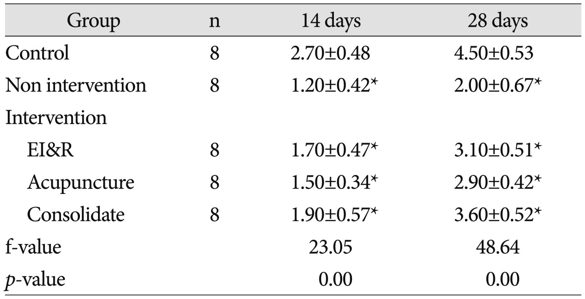
The suspension test score of 14 days old pups of EI&R, acupuncture, consolidate and non treatment groups when compared with control group, the activity of EI&R, acupuncture, consolidate and non treatment groups were significantly decreased, poor balance and coordination, high muscle tone, low strength, unresponsive, short suspension time, the test scores significantly lower (p<0.01); test score of EI&R, acupuncture and consolidate groups were significantly higher than non treatment group (p<0.01), further the test score of consolidate group were significantly higher than EI&R and acupuncture groups (p<0.01). The suspension test score of 28 days old offspring of EI&R, acupuncture, consolidate and non treatment groups were significantly lower than control group (p<0.01); test score of EI&R, acupuncture and consolidate groups were significantly higher than non treatment group (p<0.01), further the test score of consolidate group were significantly higher than EI&R and acupuncture groups (p<0.01) (Table 1).
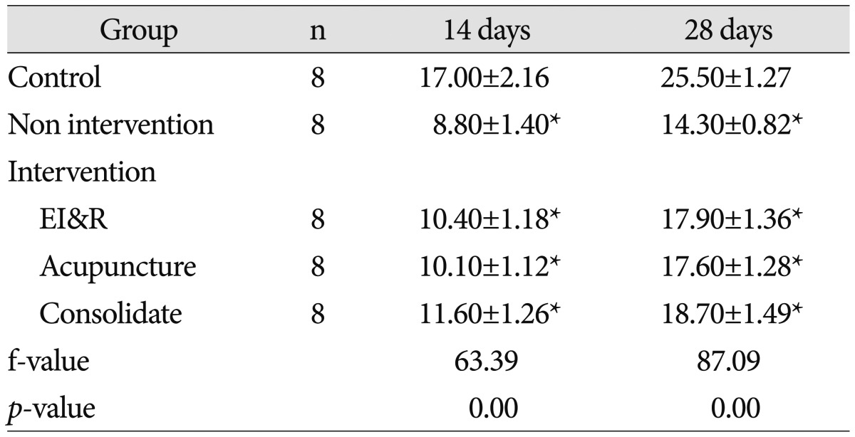
Modified BBB locomotor function test score of 14 days-old pups of control group when compared with EI&R, acupuncture, consolidate and non treatment groups, the BBB test scores of EI&R, acupuncture, consolidate and non treatment groups were lower than control group, the difference was statistically significant (p<0.01); and the BBB test score of EI&R, acupuncture and consolidate groups were significantly higher than the non treatment group, the difference was statistically significant (p<0.01). While, 28 days-old pups of EI&R acupuncture, consolidate and non treatment groups when compared with control group, EI&R, acupuncture, consolidate and non treatment groups test score were still lower than the control group, the difference was statistically significant (p<0.01); and the BBB test score of EI&R, acupuncture and consolidate groups were significantly higher than the non treatment group, the difference was statistically significant (p<0.01) (Table 2).

Immunuhistochemical analysis

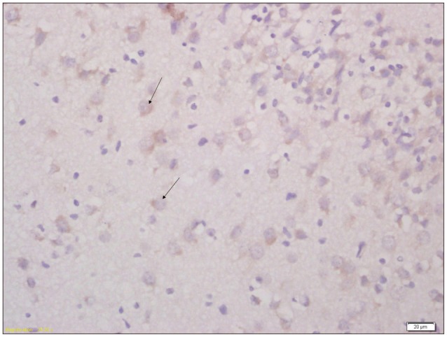
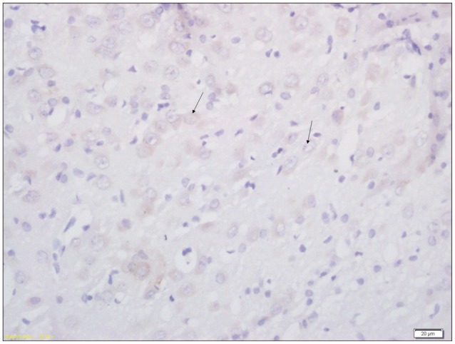
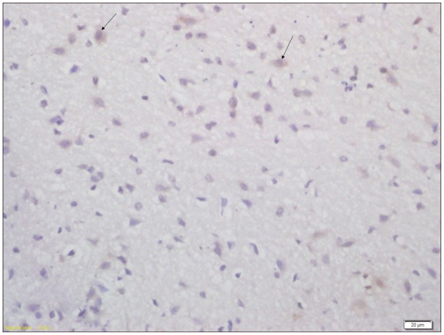
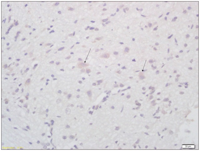
AQP-4 was expressed in the brain tissue of pups from each group; the positive expression was mainly seen in the subependymal, pia mater and perivascular astrocytes. Expression was higher in control group than intervention and non intervention group; the result was statically significant (p<0.01). In non intervention group expression was lower than intervention group (p<0.01), while expression was significantly higher in consolidate group than both EI&R & acupuncture groups (p<0.01) (Table 3, Fig. 1, 2, 3, 4, 5).

Western blot analysis

The results showed that the brain AQP4 protein expression was significantly lower in intervention and non intervention group as compared with the control group (p<0.05), but expression was higher in intervention group than non intervention group (p<0.05); expression of AQP4 protein was significantly higher in consolidate group than both EI&R & acupuncture groups (p<0.05) (Table 4, Fig. 6).

Ultrastructure analysis

In control group pups, the shape of hippocampal and cortical neurons were clear, abundant mitochondria in the cytoplasm, smooth and clear membrane; large and round nucleus, prominent nucleoli projections intertwined to form nerve grains, the large number of synapses nerve grain area, clear and complete synaptic structure; presynaptic swelling seen in many synaptic vesicles, density uniformly distributed within the presynaptic swollen, denser postsynaptic membrane were seen. While as in brain damaged group pups the hippocampal and cortical neurons were seriously damaged, neuronal glial cells were visible and larger capillary gap, lower electron density floc, vacuoles and mitochondrial swelling within the cytoplasm of some neurons; reduced synaptic nerve grain area, presynaptic swelling with indistinct outline, uneven membrane thickness; decreased synaptic vesicles and dissolved vacuolization were seen. The thickness and activity length of postsynaptic density of hippocampus CAI and cortex of brain damaged group were thinner and shorter than that of control group, and difference was statically significant (p<0.001) (Table 5, Fig. 7).

DISCUSSION

Early intervention and rehabilitation training can promote recovery after brain damage by the proliferation of brain cells and regeneration of myelin sheath. Brain's compensatory ability plays a vital role in maximum extension of plasticity9). A number of studies have proved that early enriched environments stimulation in pups after birth can increase dendritic branching in motor cortex and lengthening in visual cortex and can improve blood circulation in the brain tissue that brings structural changes as well as enhance metabolism in the brain23). They also bring improvement in various abilities in brain damaged pups; such as irritability, learning ability, ability to balance and coordination as well as memory and motor function12). In this study it has been observed that the motor function of pups from control group was significantly better than the brain damaged pups. After early intervention, acupuncture and rehabilitation training there was significant improvement in motor function in brain damaged pups, while in non treated pups there was no improvement. Motor function test and suspension test scores were significantly higher in consolidate group (combined treatment with early intervention and rehabilitation and acupuncture) than individual early intervention & rehabilitation and acupuncture group. But at subsequent days as pups grew day by day, motor function was improved. This describes that brain damage in pups caused by intrauterine infection was non-progressive type. After early intervention, acupuncture and rehabilitation training, the activity features of the pups in intervention group were improved significantly than the pups in non-intervention group, which explains that early intervention and rehabilitation training program, as well as combined treatment with early intervention & rehabilitation and acupuncture is superior than individual treatment after this type of central nervous system injury, which plays a significant promoting effect on brain plasticity.
AQP4 is a selective water permeable integral membrane protein. It is distributed in a polarized form in different regions of the brain. They are particularly concentrated in the astrocytic end feet opposed to capillaries and the pia mater 216). Investigations of AQP4-deficient (AQP4-/-) mice have revealed that AQP4 promotes cytotoxic edema but reduces vasogenic edema after brain injury. In addition, AQP4 is a regulator of normal astrocyte growth as well as astrocyte migration and glial scar formation after injury318). In AQP4-/- mice, acute brain injury after focal cerebral ischemia is attenuated as a result of reduced cytotoxic edema14). So AQP4 plays an important role in development of cerebral edema after brain injury. But the regulating mechanism of AQP4 expression in the brain is still not very clear. In this study it has been revealed that the expression of AQP4 in control group was high but it was significantly decreased in non treatment (brain damaged) group, while the expression of AQP4 was significantly increased in the brain damaged pups after treatment. The expression was even higher in the group with the combined treatment than individual treatment, which explains that combined treatment leads to higher expression of AQP4 than in individual treatment and superiority of combined treatment. This suggested that the decrease in the expression of AQP4 leads to increase in brain water content and indicate that AQP4 is involved in the developmental process of brain damaged after cerebral edema. So regulation of AQP4 expressions may provide a new treatment to attenuate cerebral edema.
Brain damage is a diffuse injury; lesions occur mainly in the hippocampus, cortex, striatum, basal ganglia, and thalamus. It is due to the combined effects of multiple mechanisms of biochemical chain reaction.The hippocampus and cortex regions of the brain are important and closely associated with learning, memory and sensory-motor functions. The damage to hippocampus and cerebral cortex result in impairment of learning, memory and sensori-motor functions. The results of this study shows that brain damage can lead to long-term impairment of learning, memory and sensorimotor function. Pathologically hippocampal CA1 area and cortical neuron damage promptly, and neuronal cell quantity decreased significantly, which consisted on Kakizawa et al.10) and Kumral et al.11) studies. Theoretically, reduction in the number of neurons causes the corresponding function disorders. In addition to the number of neurons, synapses also play an important role in the expression of nerve function. Synapse transfers information between neuron and effector cell. Under certain conditions, synaptic function can be adjusted by changing its shape and number. This kind of ability is called synaptic plasticity. Synaptic plasticity mainly includes restoration of function and morphological remodeling-mainly formation of new synapse and changes in synaptic shape, area and thickness of synaptic density and active area length, etc.8). PSD zone was seen under electron microscope and postsynaptic membrane inside cytoplasmic surface and a layer of dense homogeneous high electron density material was observed. It contained neurotransmitter receptors, cell skeleton and a variety of signal molecules. It is most sensitive structure reacting to any brain assault and related to the rate of information transfer and integration. It is one of the important morphological indicators that reflect the synaptic plasticity13). Changes in the thickness and length of the active region of postsynaptic density areas are the most critical synaptic functional changes on morphological basis. So the analysis of synaptic morphology and tracking the activity of PSD thickness and length measurement after brain damage, explain functional remodeling mechanism of brain after its damage, and provide the basis for long-term prognosis and verification of the curative effect. In this study we revealed that hippocampus and cortex were seriously damaged, neuronal glial cells were visible and larger capillary gap, lower electron density floc, vacuoles and mitochondria swelling occur within the cytoplasm of some neurons; reduced synaptic nerve grain area, presynaptic swelling with indistinct outline, uneven membrane thickness, decreased synaptic vesicles and dissolved vacuole were seen in damaged brain of pups.

CONCLUSION

The experimental results show that early intervention and rehabilitation in brain injured pups can significantly improve motor function, enhance the expression of AQP4 suggesting that AQP4 may be important in brain damage recovery processes.

References

1. Agre P, King LS, Yasui M, Guggino WB, Ottersen OP, Fujiyoshi Y, et al. Aquaporin water channels--from atomic structure to clinical medicine. J Physiol. 2002; 542(Pt 1):3–16. PMID: 12096044.
2. Amiry-Moghaddam M, Ottersen OP. The molecular basis of water transport in the brain. Nat Rev Neurosci. 2003; 4:991–1001. PMID: 14682361.
crossref
3. Auguste KI, Jin S, Uchida K, Yan D, Manley GT, Papadopoulos MC, et al. Greatly impaired migration of implanted aquaporin-4-deficient astroglial cells in mouse brain toward a site of injury. FASEB J. 2007; 21:108–116. PMID: 17135365.
crossref
4. Bax M, Goldstein M, Rosenbaum P, Leviton A, Paneth N, Dan B, et al. Proposed definition and classification of cerebral palsy, April 2005. Dev Med Child Neurol. 2005; 47:571–576. PMID: 16108461.
crossref
5. Guo Z, Li X, Pang W. Comparison of two kinds of evaluation for motor function of cerebral palsy neonatal rats caused by intrauterine infection. Chin J Rehabil Theory Pract. 2010; 3:224–227.
6. Hagberg B, Hagberg G, Beckung E, Uvebrant P. Changing panorama of cerebral palsy in Sweden. VIII. Prevalence and origin in the birth year period 1991-94. Acta Paediatr. 2001; 90:271–277. PMID: 11332166.
crossref
7. Hagberg H, Peebles D, Mallard C. Models of white matter injury : comparison of infectious, hypoxic-ischemic, and excitotoxic insults. Ment Retard Dev Disabil Res Rev. 2002; 8:30–38. PMID: 11921384.
crossref
8. Jaworski J, Kapitein LC, Gouveia SM, Dortland BR, Wulf PS, Grigoriev I, et al. Dynamic microtubules regulate dendritic spine morphology and synaptic plasticity. Neuron. 2009; 61:85–100. PMID: 19146815.
crossref
9. Jiulai T. Conductive Education in latest research and trends in cerebral palsy rehabilitation management system. Chin Child Rehabil. 2009; 6:8–9.
10. Kakizawa H, Matsui F, Tokita Y, Hirano K, Ida M, Nakanishi K, et al. Neuroprotective effect of nipradilol, an NO donor, on hypoxic-ischemic brain injury of neonatal rats. Early Hum Dev. 2007; 83:535–540. PMID: 17157452.
crossref
11. Kumral A, Uysal N, Tugyan K, Sonmez A, Yilmaz O, Gokmen N, et al. Erythropoietin improves long-term spatial memory deficits and brain injury following neonatal hypoxia-ischemia in rats. Behav Brain Res. 2004; 153:77–86. PMID: 15219709.
crossref
12. Li X, Lv Z, Sun Z, Gao J. Effects of enriched environmental stimulation on brain development in cerebral palsy rats. Chin J Rehabil Med. 2006; 12:1061–1064.
13. Liu Chuanjun GY, Li Yalu. Early intervention on hypoxic ischemic brain damage and effects of synaptic plasticity on cerebral cortex in newborn rat. Chin Matern Child Health. 2011; 26:1702–1705.
14. Manley GT, Fujimura M, Ma T, Noshita N, Filiz F, Bollen AW, et al. Aquaporin-4 deletion in mice reduces brain edema after acute water intoxication and ischemic stroke. Nat Med. 2000; 6:159–163. PMID: 10655103.
crossref
15. Nelson KB, Willoughby RE. Infection, inflammation and the risk of cerebral palsy. Curr Opin Neurol. 2000; 13:133–139. PMID: 10987569.
crossref
16. Nielsen S, Nagelhus EA, Amiry-Moghaddam M, Bourque C, Agre P, Ottersen OP. Specialized membrane domains for water transport in glial cells : high-resolution immunogold cytochemistry of aquaporin-4 in rat brain. J Neurosci. 1997; 17:171–180. PMID: 8987746.
crossref
17. Pang W, Li XJ, Zhang SL, Guo LM. Effects of early intervention on expression of S-100 protein and neurobehavioral change in offspring rats with brain injury induced by intrauterine infection. J Appl Clin Pediatr. 102; 2:96–98.
18. Saadoun S, Papadopoulos MC, Watanabe H, Yan D, Manley GT, Verkman AS. Involvement of aquaporin-4 in astroglial cell migration and glial scar formation. J Cell Sci. 2005; 118(Pt 24):5691–5698. PMID: 16303850.
crossref
19. Senger D, Cairncross JG, Forsyth PA. Long-term survivors of glioblastoma : statistical aberration or important unrecognized molecular subtype? Cancer J. 2003; 9:214–221. PMID: 12952306.
20. Shepherd R. Cerebral palsy. Physiotherapy in paediatric. Oxford: Butterworth-Heinnemann;1995. p. 110–144.
21. Tang Y, Wu P, Su J, Xiang J, Cai D, Dong Q. Effects of Aquaporin-4 on edema formation following intracerebral hemorrhage. Exp Neurol. 2010; 223:485–495. PMID: 20132816.
crossref
22. Venero JL, Vizuete ML, Machado A, Cano J. Aquaporins in the central nervous system. Prog Neurobiol. 2001; 63:321–336. PMID: 11115728.
crossref
23. Chen Y, Chen Y. Effect of exercise on mouse cerebellar cortex and age-related changes in the spinal cord and the synaptophysin. J Anat. 1994; 25:263–267.
Fig. 1

Immunohistochemical analysis of expression of AQP4 in brain tissue of pups from control group (×400). Arrow indicates the AQP4 positive cell. AQP4 : aquaporin-4.
jkns-58-14-g001
Fig. 2

Immunohistochemical analysis of expression of AQP4 in brain tissue of pups from consolidate group (×400). Arrow indicates the AQP4 positive cell. AQP4 : aquaporin-4.
jkns-58-14-g002
Fig. 3

Immunohistochemical analysis of expression of AQP4 in brain tissue of pups from EI&R group (×400). Arrow indicates the AQP4 positive cell. AQP4 : aquaporin-4.
jkns-58-14-g003
Fig. 4

Immunohistochemical analysis of expression of AQP4 in brain tissue of pups from acupuncture group (×400). Arrow indicates the AQP4 positive cell. AQP4 : aquaporin-4.
jkns-58-14-g004
Fig. 5

Immunohistochemical analysis of expression of AQP4 in brain tissue of pups from non treatment group (×400). Arrow indicates the AQP4 positive cell. AQP4 : aquaporin-4.

jkns-58-14-g005
Fig. 6

Western blot analysis of the expression of AQP4 in brain tissue of pups from different groups. Expression of AQP-4 protein is slightly decreased in consolidate group, further decreased in EI&R group and acupuncture group. The expression is markedly decreased in non-intervention group. Lane A : Control group, Lane B : Consolidate group, Lane C : EI&R group, Lane D : Acupuncture group, Lane E : Non-intervention group. AQP4 : aquaporin-4.

jkns-58-14-g006
Fig. 7

A : Ultra structure study of hippocampus CAl of pups from the control group. B : Ultra structure study of cortex of pups from the control group. In A and B the structure of synapses is clear and intact, many synaptic vesicles were seen in the presynaptic membrane, the postsynaptic density was thick (black arrow). C : Ultra structure study of hippocampus CAl of the brain damaged group. D : Ultra structure study of cortex of the brain damaged group. In C and D the structural of synapses is unclear, the neurons and synapses were significantly reduced, and also the thickness and the activity length of postsynaptic density reduced, the synaptic vesicles were dissolved and became vacuolize (black arrow). CAl : cornus ammonis I.

jkns-58-14-g007
Table 1

Suspension test result of different day old pups from different groups

jkns-58-14-i001

All data are expressed as mean±SD. There were significantly higher test score in control group in comparison to intervention and non-intervention group (p<0.01) and higher score in intervention group in comparison to non-intervention group (p<0.01). *p<0.01, compared with each individual group

Table 2

Modified BBB locomotor test score of different age pups from different groups

jkns-58-14-i002

All data are expressed as mean±SD. There were significantly higher test score in control group in comparison to intervention and non-intervention group (p<0.01) and higher score in intervention group in comparison to non-intervention group (p<0.01). *p<0.01, compared with each individual group

Table 3

Immunohistochemical analysis of expression of AQP4 in brain tissue of pups from different groups

jkns-58-14-i003

All data are expressed as mean±SD. The results were analyzed by one way analysis of variance followed by t-test for multiple comparisons. There were significantly higher expression of AQP4 in pups from control group in comparison to intervention and non-intervention group (p<0.01) and higher expression in intervention group in comparison to non-intervention group (p<0.01). *p<0.01, compared with each individual group. AQP4 : aquaporin-4

Table 4

Western blot analysis of level of AQP4 expression in brain tissue of pups from different groups

jkns-58-14-i004

All data are expressed as mean±SD. The results were analyzed by one way analysis of variance followed by t-test for multiple comparisons. There were significantly higher level of AQP4 in control group in comparison to intervention and non-intervention group (p<0.01) and higher level in intervention group in comparison to non-intervention group (p<0.01). *p<0.01, compared with each individual group. AQP4 : aquaporin-4

Table 5

Comparison of the thickness and the activity length of postsynaptic density of hippocampus CAI and cortex between two groups

jkns-58-14-i005

All data are expressed as mean±SD. The results were analyzed by one way analysis of variance followed by t-test for multiple comparisons. *p<0.001, compared with control group. AQP4 : aquaporin-4

TOOLS
Similar articles