Abstract
It is well known that spontaneous thrombosis in giant cerebral aneurysm is common. However, spontaneous obliteration of a non-giant and unruptured cerebral aneurysm has been reported to be rare and its pathogenic mechanism is not clear. We describe a case with rare vascular phenomenon and review the relevant literatures.
It is well known that saccular or fusiform intracranial aneurysms can often have variable degrees of thrombosis, especially in the giant one. And, spontaneous obliteration of ruptured cerebral aneurysms is not rare8). Several factors, such as geometrical configurations, hemodynamics, biological conditions of the blood vessels around aneurysm have been considered to contribute to the delicate balance between thrombogenesis and thrombolysis within the aneurysmal sac1,4). These partially or completely thrombosed aneurysms may continue to grow in size, recanalize, rupture to develop subarachnoid hemorrhage, compress adjacent neurovascular structures or induce embolic events3,4,13). Spontaneous intra-aneurysmal thrombosis seems to occur more frequently in the ruptured or giant intracranial aneurysms. However, these phenomenon in non-giant or unruptured aneurysms has been reported to be rare. So, its mechanism has also been poorly understood1,4,9,12). We describe a case with spontaneous thrombosis of non-giant and unruptured saccular aneurysm located at the middle cerebral artery bifurcation and review the relevant literatures.
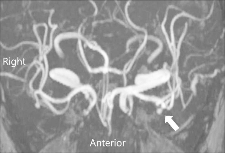
A 69-year-old male patient visited our neurosurgical department to evaluate his chronic headache which has non-throbbing, intermittent, fluctuating characteristics for several days. He has been suffered from a few medical diseases, such as diabetes and hypertension several years ago. He has been stable without any complications. Neurological examination did not show any significant abnormalities. A small saccular unruptured intracranial aneurysm located at the left middle cerebral artery trifurcation was found on the computed tomography angiogram (CTA) incidentally. Aneurysm size, aspect ratio and aneurysm angle were 4.5 mm, 3, and 90 degrees respectively (Fig. 1A). This aneurysm could be classified as small in terms of size. However, it could not be determined whether it is prone to rupture with regard to aspect ratio or aneurysm angle morphometrically. The enhanced axial computed tomography (CT) showed a saccular aneurysm in the left sylvian fissure (Fig. 1B). After 1 year, follow-up magnetic resonance angiogram showed no interval changes in aneurysm size and shape (Fig. 2). After 3 years, an intracranial aneurysm was obliterated with small residual neck remnant without any other vascular changes on CTA (Fig. 3A). Axial CT image showed no high density within the aneurysm sac, suggesting of intra-saccular thrombosis and small neck remnant was seen on the enhanced axial CT image (Fig. 3B).
Complete intra-saccular thrombosis of a ruptured intracranial aneurysm is well known. However, it is uncommon phenomenon with the incidence of 1-2% and this occurrence may rise up to 3% in the cases treated with antifibrinolytic agents7,8). In the ruptured aneurysms, hypotension, severe vasospasm, use of antifibrinolytic agents, giant aneurysms and local injury to the arterial wall have been implicated as the associated factors with spontaneous obliteration of aneurysms8). Calcification within the atherosclerotic wall of aneurysms and loss of elastic lamina, degeneration of the muscular media being typical microscopic features of aneurysms may also induce spontaneous thrombosis in the unruptured one2). In the giant cerebral aneurysms, spontaneous thrombosis of aneurysmal sac is well documented and common event with the incidence of approximately 50%14). However, completeness of the intra-aneurysmal occlusion ranges from 13% to 20% and its incidence diminishes significantly. Of other explanations to induce spontaneous thrombus formation in this condition, blood stagnation, increased viscosity, slow flow, endothelial injury due to turbulent flow and resultant platelet deposition, aggregation are considered to precipitate this phenomenon. Aneurysm size, volume/orifice area of aneurysm sac and height of aneurysm sac/orifice diameter of parent artery may be related with spontaneous thrombosis of unruptured aneurysm2). It has been reported that hemodynamic flow reversal or change of shear stress forces which may induce changes in the morphology of vessel wall could precipitate the spontaneous regression of cerebral aneuryms in the patients with fibromuscular dysplasia9). Unlike giant or ruptured intracranial aneurysm, the spontaneous obliteration of non-giant or unruptured cerebral aneurysms as in our case has been rarely reported and its pathogenic mechanisms are not clear2,4,12). Usually, cerebral aneurysms present with hemorrhagic stroke as subarachnoid hemorrhage. However, the spontaneously thrombosed aneurysm may be asymptomatic or present with ischemic stroke, growing mass, seizure, and recanalization2,6,10,12,13). In our case, any neurological symptoms were not found during the spontaneous obliteration of aneurysmal sac. In a case presenting with the ischemic stroke, parent artery occlusion due to local extension of thrombus, distal embolic occlusion secondary to dislodgement of intra-aneurysmal thrombus or adjacent neurovascular compression due to increased mass effect have been described as possible pathogenic mechanisms. And, this phenomenon has been reported in the thrombosed aneurysm of the supraclinoid internal cerebral artery (ICA), middle cerebral artery, basilar artery, posterior cerebral artery, cavernous ICA, and anterior communicating artery6,10). Medical factors to elevate coagulability or blood viscosity such as dehydration, medical disease have been considered to precipitate the spontaneous thrombosis. It is known that the high incidence of spontaneous thrombosis, especially in giant aneurysms has been related mainly to the ratio between aneurysm volume and aneurysm neck size2). That is, spontaneous thrombosis is prone to occur in aneurysms with narrow neck which result in hemodynamic disturbance in the sac. A saccular aneurysm with long and narrow neck (<4 mm) was observed in our case. It has been reported that rupture risk of aneurysms increases if aneurysm angle is more than 112 degrees5). In addition to several morphological parameters, we thought that aneurysm angle, defined as angle of inclination between aneurysm and its neck plane may also influence the thrombosis although it is not certain that aneurysm angle is associated with thrombosis directly. In our case, rupture risk seemed to be low as the aneurysm angle of about 90 degrees. Other several physiological factors, such as the age of aneurysm, direct distortion of the parent artery by the aneurysmal sac, and the angiographic procedure itself have been also included to explain spontaneous thrombosis1,2,4,9,12,15). Often, it has been recognized that an aneurysm with size ratio or aspect ratio greater than 2 is prone to rupture. However, it may be difficult to explain why cerebral aneurysm of our current case was obliterated spontaneously despite the aspect ratio greater than 3. It seems to be reasonable that other several factors may be related with the balance between thrombosis and thrombolysis. CT or magnetic resonance image may be helpful to document the thrombus in the aneurysm sac and/or real regression of aneurysm size which were not shown by angiography9). We could not identify the high density lesion in the aneurysm sac suggesting thrombus on CT. The thrombosed aneurysmal sac can be asymptomatic, recanalized, continue to grow its size, provoke seizures, or present with ischemic stroke4,11). Therefore, management of thrombosed aneurysms may be debatable, especially in the partially thrombosed aneurysms. If the thrombosed aneurysm presents with mass effect owing to continuous growth, it may be clipped for decompression because blood flow remains in the aneurysm sac. If the thrombosed aneurysm serves as a potential source of emboli leading to cerebral infarction or transient ischemic attacks, it may be treated medically with antiplatelet agents2). However, surgical method of thrombosed aneurysm may be the preferred treatment to alleviate the potential inducing ischemic stroke and reduce risk of subarachnoid hemorrhage by rupture since intra-aneurysmal clot may not protect against aneurysm rupture. In conclusion, close follow-up with radiological images should be required because even completely thrombosed aneurysm may recanalize, continue to grow, or rupture someday.
References
1. Batjer HH, Purdy PD. Enlarging thrombosed aneurysm of the distal basilar artery. Neurosurgery. 1990; 26:695–699. discussion 699-700. PMID: 2330095.

2. Brownlee RD, Tranmer BI, Sevick RJ, Karmy G, Curry BJ. Spontaneous thrombosis of an unruptured anterior communicating artery aneurysm. An unusual cause of ischemic stroke. Stroke. 1995; 26:1945–1949. PMID: 7570753.

3. Cohen JE, Itshayek E, Gomori JM, Grigoriadis S, Raphaeli G, Spektor S, et al. Spontaneous thrombosis of cerebral aneurysms presenting with ischemic stroke. J Neurol Sci. 2007; 254:95–98. PMID: 17258773.

4. Cohen JE, Rajz G, Umansky F, Spektor S. Thrombosis and recanalization of symptomatic nongiant saccular aneurysm. Neurol Res. 2003; 25:857–859. PMID: 14669530.

5. Dhar S, Tremmel M, Mocco J, Kim M, Yamamoto J, Siddiqui AH, et al. Morphology parameters for intracranial aneurysm rupture risk assessment. Neurosurgery. 2008; 63:185–196. discussion 196-197. PMID: 18797347.

6. Fisher M, Davidson RI, Marcus EM. Transient focal cerebral ischemia as a presenting manifestation of unruptured cerebral aneurysms. Ann Neurol. 1980; 8:367–372. PMID: 7436381.

7. Fodstad H, Liliequist B. Spontaneous thrombosis of ruptured intracranial aneurysms during treatment with tranexamic acid (AMCA). Report of three cases. Acta Neurochir (Wien). 1979; 49:129–144. PMID: 517175.

8. Hamilton MG, Dold ON. Spontaneous disappearance of an intracranial aneurysm after subarachnoid hemorrhage. Can J Neurol Sci. 1992; 19:389–391. PMID: 1393850.

9. Hans FJ, Krings T, Reinges MH, Mull M. Spontaneous regression of two supraophthalmic internal cerebral artery aneurysms following flow pattern alteration. Neuroradiology. 2004; 46:469–473. PMID: 15150678.

10. Hoffman WF, Wilson CB, Townsend JJ. Recurrent transient ischemic attacks secondary to an embolizing saccular middle cerebral artery aneurysm. Case report. J Neurosurg. 1979; 51:103–106. PMID: 448403.

11. Lee KC, Joo JY, Lee KS, Shin YS. Recanalization of completely thrombosed giant aneurysm : case report. Surg Neurol. 1999; 51:94–98. PMID: 9952130.
12. Ohta H, Sakai N, Nagata I, Sakai H, Shindo A, Kikuchi H. Spontaneous total thrombosis of distal superior cerebellar artery aneurysm. Acta Neurochir (Wien). 2001; 143:837–842. discussion 842-843. PMID: 11678406.

13. Schaller B, Lyrer P. Focal neurological deficits following spontaneous thrombosis of unruptured giant aneurysms. Eur Neurol. 2002; 47:175–182. PMID: 11914557.

14. Whittle IR, Dorsch NW, Besser M. Spontaneous thrombosis in giant intracranial aneurysms. J Neurol Neurosurg Psychiatry. 1982; 45:1040–1047. PMID: 7175528.

15. Yeh H, Tomsick TA. Obliteration of a giant carotid aneurysm after extracranial-to-intracranial bypass surgery : case report. Surg Neurol. 1997; 48:473–476. PMID: 9352811.

Fig. 1
A : Computed tomography angiogram shows an unruptured, small, saccular aneurysm (white arrow) which is located at the left middle cerebral artery trifurcation. Aneurysm has a relatively long and narrow neck. Aneurysm size, aspect ratio, and aneurysm angle are 4.5 mm, 3, and 90 degrees respectively. B : Small saccular aneurysm (white arrow) in the left sylvian fissure is seen on enhanced axial CT image.

Fig. 2
magnetic resonance angiogram performed at 1 year follow-up shows no interval changes of aneurysm size and shape.

Fig. 3
A : Computed tomography angiogram performed at 3-year follow-up shows a near total obliteration of cerebral aneurysm with small residual neck remnant (white arrow). B : There is no high density lesion suggesting of intra-aneurysmal thrombus and small residual neck remnant (white arrow) are seen on enhanced axial CT image.


 PDF
PDF ePub
ePub Citation
 Print
Print


 XML Download