INTRODUCTION
MATERIALS AND METHODS
Inclusion criteria
RESULTS
Patient’s characteristics
Table 1.
Table 2.
PCA : posterior cerebral artery, MMA : middle meningeal artery, IMA : internal maxillary artery, PA : periauricular artery, OA : occipital artery, B&C : artery of Bernasconi and Cassenari, TS : transverse sinus, SS : sigmoid sinus, IJV : internal jugular vein, MHT : meningiohypophyseal trunk, OPA : ophthalmic artery, SOV : superior ophthalmic vein, IOV : inferior ophthalmic vein, SSS : superior sagittal sinus, SPS : superior petrosal sinus, CS : cavernous sinus, SPiS : sphenoparietal sinus
Technical aspects of TAE
Table 3.
| Case No. | Procedure of TAE | Arterial inflow control using | Microcatheter | Embolization material* |
|---|---|---|---|---|
| 1 | One session of onyx embolization | |||
| 2 | Two session (first double microcatheter interlocking coil embolization & second onyx embolization after 2 weeks) | |||
| 3 | One session of glue embolization | None | Flow guided Sonic | NBCA glue (mixed with tantalum) |
| 4 | One session of glue embolization | None | Flow guided magic | NBCA Glue (mixed with tantalum) |
| 5 | One session of glue embolization | None | Flow guided magic | NBCA Glue (mixed with tantalum) |
| 6 | One session of glue embolization | None | Flow guided magic | NBCA glue (mixed with tantalum) |
| 7 | One session of onyx embolization | Scepter C balloon catheter and pressure cooker technique† | Scepter C balloon catheter | Onyx |
| 8 | One session of glue embolization | None | Flow guided magic | NBCA glue (mixed with tantalum) |
| 9 | One session of glue embolization | None | Flow guided magic | NBCA glue (mixed with tantalum) |
| 10 | One session of onyx embolization | None | Detachable tip Apollo 3 cm | Onyx |
| 11 | One session of onyx embolization | Scepter C balloon catheter | Scepter C balloon catheter | Onyx |
| 12 | One session of onyx embolization | Scepter C balloon catheter | Scepter C balloon catheter | Onyx |
| 13 | One session of onyx embolization | None | Flow guided magic | Onyx |
| 14 | Two session (first squid & second onyx embolization after 1 year) | None | Flow guided sonic | Squid |
| None | Flow guided magic | NBCA glue (mixed with tantalum | ||
| 15 | One session of onyx embolization | None | Detachable tip Apollo 3 cm | Onyx |
Outcomes
Table 4.
Case illustration
Case 1
 | Fig. 1.Case 1. A : Left vertebral artery angiogram (anteroposterior view) demonstrating Cognard type IV DAVF, supplied by pachymeningeal of posterior cerebral artery with early-dilated venous pouch (Transverse sinus). B : Left external carotid angiogram (lateral view) demonstrating Cognard type IV DAVF, supplied by meningeal branches of middle meningeal, posterior auricular and occipital arteries with early-dilated venous pouch (Transverse sinus). C : Magnetic resonance venography (showing hypertrophied dural venous sinuses and thrombosed left sigmoid sinus. DAVF : dural arteriovenous fistula. |
 | Fig. 2.Case 1. A : Final angiogram showing complete occlusion of the DAVF with onyx (white arrow). B : Fluoroscopic unsubtracted image showing the Onyx cast at the fistula point. C : Road map image showing placement of Scepter C balloon catheter in the middle meningeal artery (white arrow). D : Three-dimensional reconstructed angiographic done after 1 year, showing residual filling of DAVF. DAVF : dural arteriovenous fistula. |
Case 2
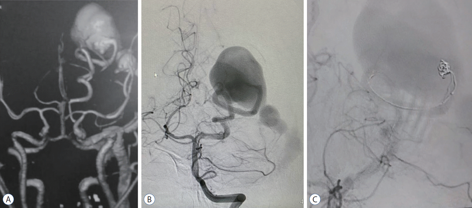
 | Fig. 3.Case 2. A : Computed tomography angiography showing PAVF, supplied by hypertrophied posterior cerebral artery and aneurysmal dilatation of occipital vein with ectatic dural sinuses. B : Left vertebral artery angiogram (anteroposterior view) demonstrating PAVF, supplied by hypertrophied posterior cerebral artery and aneurysmal dilatation of occipital vein with ectatic dural sinuses. C : Left vertebral artery control angiogram (anteroposterior view) demonstrating attempted coil mass placement in the fistulous connection. PAVF : pial arterioveous fistula. |
 | Fig. 4.Case 2. A : Left vertebral artery control angiogram (anteroposterior view) demonstrating onyx embolized arterial feeder and fistulous connection, and deflated HyperGlide balloon proximally placed (white arrow). B : Final angiogram showing complete occlusion of the PAVF with onyx (white arrow). C : Follow up CAT scan of the brain showing onyx and reduced size venous aneurysm with partial thrombosis. D : Three-dimensional reconstructed angiographic done after 6 months, reveal no more filling of the PAVF. PAVF : pial arterioveous fistula. |
Case 7
 | Fig. 5.Case 7. A : Left internal carotid angiogram (anteroposterior view) demonstrating PAVF, supplied by hypertrophied middle cerebral artery and aneurysmal dilatation of cortical draining vein in to superior sagittal sinus. B : Magnetic resonance imaging showing left high parietal PAVF and venous aneurysm. C : Selective angiogram with microcatheter, prior to embolization with n-butyl cyanoacrylate glue mix. D : Final angiogram showing the glue cast (white arrow). PAVF : pial arterioveous fistula. |
Case 11
 | Fig. 6.Case 11. A : CAT scan of the brain showing intracranial hemorrhage secondary to DAVF. B : Left external carotid angiogram (lateral view) demonstrating Cognard type IV DAVF, supplied by meningeal branches of middle meningeal, posterior auricular and occipital arteries draining into transverse sinus. C : Roadmap image showing onyx injection from the microcatheter (white arrow). D : Fluoroscopic unsubtracted roadmap image showing the onyx injection in to the DAVF. DAVF : dural arteriovenous fistula. |


 PDF
PDF Citation
 Print
Print


 XML Download