Abstract
Objective
Retrograde suction decompression (RSD) is an adjuvant technique used for the microsurgical treatment of large and giant internal carotid artery (ICA) aneurysms. In this study, we analyzed the efficacy and safety of the RSD technique for the treatment of large and giant ICA aneurysms relative to other conventional microsurgical techniques.
Methods
The aneurysms were classified into two groups depending on whether the RSD method was used (21 in the RSD group vs. 43 in the non-RSD group). Baseline characteristics, details of the surgical procedure, angiographic outcomes, clinical outcomes, and procedure-related complications of each group were reviewed retrospectively.
Results
There was no significant difference in the rates of complete neck-clipping between the RSD (57.1%) and non-RSD (67.4%) groups. Similarly, there was no difference in the rates of good clinical outcomes (modified Rankin Scale score, 0–2) between the RSD (85.7%) and non-RSD (81.4%) groups. Considering the initial functional status, 19 of 21 (90.5%) patients in the RSD group and 35 of 43 (81.4%) patients in the non-RSD group showed an improvement or no change in functional status, which did not reach statistical significance.
Conclusion
In this study, the microsurgical treatment of large and giant intracranial ICA aneurysms using the RSD technique obtained competitive angiographic and clinical outcomes without increasing the risk of procedure-related complications. The RSD technique might be a useful technical option for the microsurgical treatment of large and giant intracranial ICA aneurysms.
Retrograde suction decompression (RSD) is an adjuvant technique used for the microsurgical treatment of large and giant internal carotid artery (ICA) aneurysms. RSD promotes an adequate degree of relaxation of the aneurysmal dome, enabling the surgeon to fully dissect the ICA with branches and the aneurysm itself. After Flamm [9] published the first technical note on direct aneurysm puncture for suction decompression in 1981, this technique, termed RSD, was modified by Batjer and Samson [3]. Since the introduction of the RSD technique, more than 500 aneurysms have been treated using this approach, and morbidity and mortality rates of 19.9% and 1.3%, respectively, have been achieved [10]. Furthermore, the RSD technique enables the adequate relaxation of the aneurysm dome by applying temporary clipping on the extracranial and intracranial carotid arteries and aspirating blood by direct ICA puncture, which may raise concerns about procedure-related complications such as thromboembolic ischemia, ICA dissection or occlusion, and anemia. Therefore, it is critical to monitor the impact of using RSD techniques on clinical outcomes. Although a number of studies have reported the feasibility of the RSD technique, there has been no objective analysis to compare the efficacy and safety of RSD with other microsurgical techniques. In this study, we analyzed the efficacy and safety of the RSD technique for the treatment of large and giant ICA aneurysms relative to other conventional microsurgical techniques.
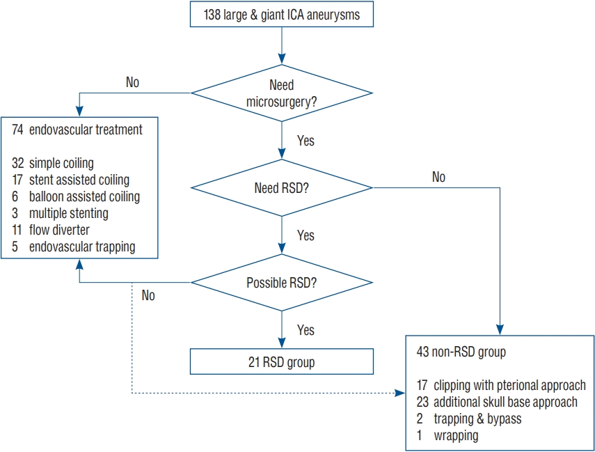
This retrospective study was approved by the Institutional Review Board of Severance Hospital at the Yonsei University College of Medicine and the requirement for informed consent was waived (subject number : 4-2016-0111). From January 2005 to December 2016, 2945 intracerebral aneurysms were treated at our institution. Among them, 259 (10.0%) were large (10–24 mm) and 35 were giant (≥25 mm) intracerebral aneurysms, and 138 were in the ICA. Seventy-four aneurysms were treated by the endovascular method, and 64 ICA aneurysms were treated by the microsurgical method, which were then divided into two groups according to whether the RSD method was used (21 in the RSD group vs. 43 in the non-RSD group).
The treatment strategy for large and giant ICA aneurysms in our institution is summarized in Fig. 1. Cases of aneurysms requiring microsurgical treatment were considered first, and for other cases, endovascular treatment was preferred. When considering microsurgical treatment, we checked whether RSD was necessary, and if microsurgical treatment was possible without RSD, microsurgical treatment was performed without RSD. If RSD was needed, we checked whether RSD was possible. If the microsurgical treatment could not be performed because RSD was not possible despite the need for microsurgical treatment, endovascular treatment was performed, or non-RSD treatment methods, such as trapping and bypass, were considered in selective cases. Through this decisionmaking process, treatment methods for each aneurysm were determined, and patients were assigned to the RSD group or non-RSD group accordingly.
Microsurgical treatment was considered preferentially for the following conditions : 1) lesion that requires immediate decompression due to an accompanying mass effect such as cranial nerve palsy or hydrocephalus; 2) ruptured aneurysm accompanied by a large intraparenchymal hematoma or which increases intracranial pressure, requiring simultaneous clot evacuation and decompression; 3) lesion with a wide neck or high dome-to-neck ratio which may lead to a high chance of recurrence with endovascular treatment; 4) lesion that is expected to be difficult for endovascular treatment due to difficult parent arterial geometry; and 5) multiple intracerebral aneurysms identified on the ipsilateral side that need to be treated simultaneously.
For the microsurgical treatment of large and giant ICA aneurysms, cases where the RSD technique was needed were as follows : 1) if the intracranial proximal control of the aneurysm is difficult because of the juxta-dural ring location of the aneurysmal neck; 2) if large and giant ICA aneurysms occupy a large volume in a narrow surgical field, so atraumatic manipulation of the adjacent neurovascular structure is expected to be difficult without debulking of the aneurysmal sac; 3) if clipping is expected to be difficult due to high intra-aneurysmal pressure and deflation of the aneurysm is necessary; 4) if direct temporary clipping is not possible due to calcification or atherosclerosis of the parent artery; and 5) if intraoperative angiography is required to confirm patency of arteries branched from the ophthalmic and communicating segments of ICA and to confirm complete ligation of the aneurysmal neck after clipping.
The following cases were considered unsuitable for the RSD technique : 1) if the aneurysm was located in the infraophthalmic region so the proximal aneurysmal neck could not be exposed, even with RSD; 2) calcified or thrombosed aneurysms that could not be debulked by RSD; 3) despite using the RSD technique, clipping would be expected to be hindered due to the positional relationship between the anatomic structures around the aneurysm, such as the optic nerve, and the aneurysmal neck; 4) if aspiration of the blood was expected to be difficult due to extensive stenosis of cervical ICA; 5) temporary clipping for RSD was expected to cause ischemic complications in the ipsilateral hemisphere due to the lack of crosscollaterals from the anterior communicating artery (AcomA) and posterior communicating artery (PcomA); and 6) temporary clipping for RSD was expected to cause ischemic complications in the contralateral hemisphere due to stenosis or occlusion of a large artery in the contralateral hemisphere.
Because of the characteristics of large and giant ICA aneurysms, including a wide portion of the distal ICA that causes deformation of normal anatomical structures, it was not appropriate to classify large and giant ICA aneurysms using the conventional classification system, which categorizes ICA aneurysms in relation to anatomical structures such as the anterior clinoid process (ACP). Therefore, the location of the large and giant ICA aneurysms of patients enrolled in this study were classified into three groups according to their positional relationship with the ophthalmic artery as follows : infraophthalmic, paraophthalmic, and supraophthalmic. Furthermore, in terms of the direction of the aneurysm, the location was divided into four groups in relation to the cross-section of the ICA : lateral, medial, ventral, and dorsal [15].
Three-dimensional computed tomography (CT) angiography was performed to assess the geographical relationship between lesions and the surrounding anatomical structures and the characteristics of aneurysms such as calcification or thrombosis. Bone window-setting CT was performed to assess the pneumatization of ACP to avoid cerebrospinal fluid leakage and postoperative infection. Digital subtraction angiography (DSA) was routinely conducted to check the cross-collaterals from the AcomA and PcomA. The height of the bifurcation of the common carotid artery (CCA) and the degree of stenosis in the cervical ICA was also checked by DSA. Balloon test occlusion was performed in selective cases of unruptured aneurysm to determine whether bypass surgery was necessary.
Cerebrospinal fluid drainage such as spinal drainage or intraoperative extraventricular drainage through Paine’s point was performed to facilitate adequate brain relaxation during the operation if necessary. Intraoperative monitoring of motor-evoked potentials was prepared to watch for ischemic events during surgery, while a compressed spectral array was adopted to check for burst suppression on electroencephalograms during temporary occlusion. Indocyanine green videoangiography and/or intraoperative DSA was prepared to confirm complete clipping and preservation of blood flow in the parent artery. Microvascular Doppler ultrasonography was used regularly to assess the blood flow of the parent and branch vessels.
The patient was positioned supine with the head rotated about 30° to 45° to the contralateral side. To facilitate exposure of the cervical carotid artery, the head was slightly elevated prior to fixation and the neck was slightly extended. A standard pterional craniotomy was performed in most cases and the orbitopterional or the orbitozygomatic approach was considered if wider exposure was needed. Interfacial dissection was completed for extensive soft tissue dissection, sparing the frontotemporal branch of the facial nerve. After dural opening, a microsurgical dissection of the Sylvian fissure and arachnoid cisterns was accomplished for distal control of the aneurysmal segment. A site for distal temporary clipping was prepared proximal to the PcomA. Anterior clinoidectomy was performed if necessary, to expose the proximal aneurysmal neck, either intradurally or extradually depending on the case, although most of the anterior clinoidectomy procedures performed in this case series were intradural at the clinician’s discretion.
Simultaneously, the CCA, cervical ICA, and external carotid artery (ECA) were exposed routinely for RSD and intraoperative angiography. A vascular occlusive clamp was applied to the CCA and ECA but not the ICA so as to avoid direct intimal injury of the ICA. Then, the ICA was punctured with an 18-gauge angiocatheter. Following application of a temporary clamp to the CCA and ECA, an extension line was connected to the angiocatheter. A three-way stopcock was added to the extension set and RSD was performed using a 20-mL syringe after a gentle test aspiration of blood to avoid intimal dissection.
The aneurysm was trapped temporarily by placing a temporal clip on the intracranial ICA distal to the aneurysm neck and proximal to PcomA. If the lesion was visualized to originate from PcomA, a temporal clip was applied to the PcomA proper and proximal to the anterior choroidal artery. After clamping of the CCA and ECA, blood was aspirated through the catheter introduced into the cervical ICA, resulting in the collapse of the aneurysm, enabling the surgeon to complete dissection and neck-clipping. Temporary occlusion for RSD was initiated after confirming burst suppression on electroencephalogram by compressed spectral array monitoring, which was accomplished using pentothal sodium. The occlusion time was limited to within 5 minutes, even though there were no obvious changes in motor-evoked potentials. RSD was repeated after declamping the arteries for a period that was longer than each occlusion time. These procedures were repeated until complete clipping was confirmed.
Intraoperative DSA through the catheter placed in the cervical ICA was used to confirm complete clipping without stenosis of the parent artery. Complete hemostasis at the puncture site was achieved by suturing.
Details of the surgical procedure, angiographic outcomes, clinical outcomes, and procedure-related complications were reviewed retrospectively. The details of surgery in each group such as operation time, total blood loss during surgery, and temporary occlusion time were compared to review whether performing the RSD technique led to a significant difference in any aspect of the surgical procedure. Postoperative angiographic results were categorized as complete occlusion, partial neck remnant, and incomplete occlusion based on the results of postoperative angiography. Clinical outcomes were assessed using the modified Rankin Scale (mRS), which was evaluated 1 month after the surgical procedure. Procedure-related complications were defined as any symptomatic or asymptomatic events after the surgical procedure. RSD-related complications were defined as any complications that were directly attributed to the RSD technique itself such as cervical ICA injury, ischemic complication related with temporary clipping, and anemia caused by aspirating too much blood for RSD. All data and outcomes of patients with complications were summarized.
All statistical analyses were performed using SPSS Statistics version 22.0 (IBM Corp., Armonk, NY, USA). An independent-sample t-test and Mann-Whitney U-test were used for numeric variables, and the chi-squared test or Fisher’s exact test were used for nominal variables. A p-value less than 0.05 for a 95% confidence interval was considered statistically significant.
A comparison of the RSD and non-RSD groups indicated no significant differences in baseline characteristics of the patients or aneurysms including the ratio of ruptured aneurysms, and the size, location, and direction of aneurysms. The RSD group included four men and 17 women with a mean age of 54.4±10.1 years (range, 37–67). Five (23.8%) of the 21 patients in the RSD group had ruptured aneurysms and presented with subarachnoid hemorrhage. Among 16 patients with unruptured aneurysms, seven cases (33.3%) developed visual symptoms, one (4.8%) developed ptosis due to oculomotor nerve palsy, six (28.5%) developed headache, one (4.8%) developed hearing symptoms, and one (4.8%) was an incidental finding.
The mean aneurysm size in the RSD group was 18.0±7.2 mm (range, 10.0–34.6) and there were 16 (76.2%) large (10–24 mm) and five (23.8%) giant (≥25 mm) aneurysms. The mean aneurysm size in the non-RSD group was 16.7±7.8 mm (range, 10.0–50.0) and there were 39 (90.7%) large and four (9.3%) giant aneurysms. Although there was no statistical significance in aneurysm frequency between groups, giant aneurysms tended to be more frequent in the RSD group whereas most non-RSD groups had large aneurysms. According to aneurysm location, the RSD group consisted of four (19.0%) paraophthalmic and 17 (81.0%) supraophthalmic segment aneurysms and there were no infraophthalmic aneurysms in the RSD group. In the non-RSD group, there were three (7.0%) infraophthalmic, one (2.3%) paraophthalmic, and 39 (90.7%) supraophthalmic segment aneurysms. Paraophthalmic aneurysms tended to be more frequent in the RSD group and most non-RSD groups were composed of supraophthalmic aneurysms even though there was no statistical significance between groups. The baseline characteristics of the included patients and aneurysms are summarized in Table 1.
The operation time and temporary occlusion time were both longer in the RSD group compared with the non-RSD group, but the total amount of blood loss during surgery was not significantly different between the two groups even though the aspirated blood volume when using the RSD technique was included in the total blood loss of the RSD group. The mean operation time and mean temporary occlusion time for the RSD group were 616.0±70.9 minutes (range, 480–720) and 2113.3±1366.7 seconds (range, 50–3523), respectively. Total blood loss during the operation including aspirated blood in the RSD group was 1229.0±1009.6 mL (range, 310–4700), while the mean volume of aspirated blood was 368.1±209.5 mL (range, 90–750). Operative details are summarized in Table 2.
The outcomes and complications in the two groups are compared and summarized in Table 3. The mean follow-up times of the RSD group and the non-RSD group were 54.9±39.7 months (range, 7–127) and 59.4±52.1 months (range, 7–156), respectively. There was no significant difference in the rates of complete neck-clipping between the RSD (57.1%) and non-RSD (67.4%) groups. Similarly, there was no difference in the rates of good clinical outcomes (mRS score, 0–2) between the RSD (85.7%) and non-RSD (81.4%) groups. Considering initial functional status, 19 of 21 (90.5%) patients in the RSD group and 35 of 43 (81.4%) patients in the non-RSD group showed an improvement or no change in functional status and there was no statistically significant difference.
Two of the 21 patients in the RSD group showed decreased function compared with their initial status. One patient with a left supra-ophthalmic segment ICA aneurysm whose neck incorporated the anterior choroidal artery suffered postoperative right hemiparesis because of deliberate sacrifice of the anterior choroidal artery for complete neck-clipping (postoperative mRS score, 3). Another patient with a right para-ophthalmic segment ICA aneurysm suffered rebleeding 1 month after clipping and became comatose (postoperative mRS score, 6). An additional six patients in the RSD group had complications. Three were asymptomatic (two venous infarction and one subdural hematoma) and the other three had symptomatic but transient complications (one oculomotor nerve palsy, one right hemiparesis, and one partial seizure). There was no complication that was attributable to the RSD technique itself. The overall morbidity and mortality rates of the RSD group were 33.3% and 4.8%, respectively. Details of postoperative complications observed in the RSD group are summarized in Table 4.
The results of this study showed that there was no statistical difference in angiographic outcomes and clinical outcomes between the RSD and non-RSD groups. Furthermore, 21 patients were treated using the RSD technique and most of these patients (19/21; 90.5%) showed good clinical outcomes, suggesting RSD might be an effective and safe adjuvant technique for the microsurgical treatment of large and giant intracranial ICA aneurysms without increasing the risk of procedure-related complications.
Large and giant intracranial aneurysms, which constitute 3.0% to 13.5% of all intracranial aneurysms [7,28], require special attention because they show several different features compared with usual small aneurysms. First, large and giant intracranial aneurysms impart a much higher risk of rupture as the size increases [32,34]. Also, in addition to subarachnoid hemorrhage due to rupture of the aneurysm, patients experience characteristic clinical manifestations such as pain or cranial nerve palsy due to the mass effect [18]. Large and giant aneurysms often present with thrombus formation into the sac, causing thromboembolic symptoms and hindering the complete obliteration of the aneurysm by microsurgical clipping as well as endovascular treatment, causing poor clinical outcomes [5,12]. Finally, relative to small aneurysms, the rate of complete occlusion is low and the risks of recanalization, morbidity, and mortality after recanalization and rebleeding are high as the size of the aneurysm increases [6,20]. For these reasons, a different approach for the treatment of large and giant intracranial aneurysms is needed.
The treatment of large and giant ICA aneurysms has remained challenging because of the complex proximity of surrounding anatomical structures such as the optic nerve, proximal and distal dural rings, and bony structures including the ACP. The outcomes of direct surgery for large and giant ICA aneurysm were not favorable until the 1990s [2,14]. With the progression of technology, endovascular treatment has become another treatment option. However, even though endovascular treatment is less invasive and achieves lower morbidity and mortality rates when compared with microsurgical treatment, large and giant aneurysms treated by the endovascular method still experience a high rate of recanalization [13,26]. Flow diverters have gained increasing attention as good alternatives to microsurgery and conventional endovascular treatments but these have disadvantages in cases of large and giant aneurysms with low rates of complete occlusion and nonsignificantly high rates of complications [17,21]. Moreover, the use of a flow diverter for the treatment of a ruptured aneurysm can cause considerable problems due to the increased risk of hemorrhagic and thromboembolic complications inherent when using antiplatelet medications [31]. Furthermore, the potential risk of delayed rupture remains until the complete obliteration of the aneurysm [33]. In addition to their clinical disadvantages, other disadvantages of flow diverters in terms of financial aspects include the high cost of the device and restrictions on the use of flow diverters due to medical insurance issues in some countries [27]. In this regard, there is no absolute correct method for the treatment of large and giant intracranial ICA aneurysms and, therefore, microsurgical treatment might still be useful as a means to complement the shortcomings of endovascular treatments.
Two important aspects of aneurysm surgery are proximal control of the parent artery and visualization of the aneurysmal neck. However, large and giant ICA aneurysms tend to occupy a large volume in a narrow space, and the proximal portion of intracranial ICA aneurysms is surrounded by complex anatomical structures, so sufficient arachnoid dissection for proximal control and neck exposure is not possible in all cases. In addition, even if the aneurysm neck dissection is sufficient, the intra-aneurysmal pressure of the large and giant aneurysm is high, so applying a clip is usually difficult unless the aneurysm is decompressed sufficiently. To solve these problems, several special strategies are required such as adenosine-induced transient asystole or RSD.
Adenosine-induced transient asystole is the method for decompression of the aneurysm by inducing blood flow arrest due to temporary cardiac arrest by injecting adenosine intravenously [16,25]. The major benefit of this technique is that the aneurysm can be decompressed without additional surgical procedure. Moreover, this temporary flow arrest techinque can be used for controlling intraoperative bleeding. However, adenosine injection can cause some medical complication such as transient atrial fibrillation, hypotension and bronchospasm. Another disadvantage of adenosine injection is that the effect on a single injection is relatively short, about 30 seconds.
RSD is another useful adjuvant technique for the treatment of large and giant ICA aneurysms. RSD was designed for the adequate relaxation of the aneurysm dome, enabling the surgeon to fully dissect between the ICA and its branches, allowing adequate visualization of the aneurysm neck. In cases of large and giant ICA aneurysms, the neck of the aneurysm is often located in the juxtadural portion of the distal dural ring. Therefore, intracranial proximal control is often impossible without direct cavernous sinus exploration, which has a significant risk for complications [10]. For this reason, temporary clipping for proximal control is usually performed on cervical ICA. However, due to the retrograde flow through the ophthalmic artery and cavernous ICA branches, pressure reduction of the aneurysm is often inadequate with temporary clipping of the cervical and communicating segment of the ICA alone [23]. Repeated attempts to place clips in such a situation can lead to clip slippage or thromboembolic complications. Therefore, if such a problem is expected, RSD should be considered as the first option. Sufficient relaxation of the aneurysmal sac and better visualization of the aneurysmal neck through immediate retrograde aspiration of ICA flow using RSD enables the successful clipping of large and giant ICA aneurysms. Additionally, exposed cervical ICA for RSD provides a route for intraoperative angiography to confirm complete occlusion of the aneurysm during surgery and for high-flow bypass in selected cases. In cases of large and giant ICA aneurysm surgery, clip reconstruction is often required with many clips, which hinders confirmation of complete aneurysmal neck ligation or patency of the parent artery and branching artery. In particular, the use of fenestration clips due to the direction of the aneurysm can increase the risk of flow compromise or ICA or incomplete clipping [24]. Therefore, intraoperative angiography can improve the success rate of surgery, which is another advantage of using RSD.
Some researchers think RSD may cause anemia and morbidity of the patient by causing the aspiration of too much blood. However, in this study, RSD did not lead to the aspiration of a significant volume of blood, and even when the expected amount of blood was aspirated in conjunction with RSD, no patient suffered any postoperative complication correlated with hypovolemia. In addition, when there is concern about complications due to large blood loss during RSD, blood loss can be minimized by intraoperative autologous blood transfusion using a blood salvage system or blood-returning circuit [4,19]. Other concerns when performing RSD include the increased length of operation time and temporary occlusion time. Although the operation time and temporary occlusion time were longer in the RSD group, the rate of procedure-related complications was not different between groups. Furthermore, burst suppression using pentothal sodium for temporary clipping can be deployed safely to reduce the risk of ischemic complications. Another concern is that RSD causes complications linked to the exposure of and access to the ICA such as ICA dissection or occlusion, thromboembolic complication, and soft-tissue hematoma. However, no complications related to ICA injury were noted in this study and the procedure is rarely clinically problematic. Moreover, several alternative RSD techniques that avoid direct ICA puncture by puncturing ECA or CCA have been introduced [8,22,29,30] and might be applied to reduce the risk of ICA injury. As endovascular techniques continue to improve, recent studies reported the use of endovascular RSD using a double-lumen balloon catheter, which obviates the need for cervical dissection and reduces the risk of ICA injury [1,11]. Overall, eight complications were reported in this study, but six of eight were asymptomatic or transient, and only two cases (9.5%) were fatal complications (Table 4). Moreover, there were no complications attributable to the RSD technique itself. Considering the relative high morbidity and mortality of large and giant ICA aneurysms, this rate of complications might be acceptable when deciding whether to perform the RSD technique.
This study had some limitations. First, it was designed retrospectively and included a small number of subjects from a single center. However, considering the low prevalence of large and giant ICA aneurysms, recruiting sufficient numbers of RSD cases for a prospective study might take considerable time. Moreover, because the number of RSD cases reported worldwide since the introduction of RSD is only 540 to date, the collection of 21 RSD cases from a single institution is by no means a small number. Therefore, the results of this study should be of considerable value for studies of the RSD technique. Second, due to the nature of this study conducted in a single institution, it is difficult to conclude that all the treatment strategies used in this study and the outcomes derived from the treatment were the best. However, decisions on treatment and specific plans for each case in this study were always determined by multidisciplinary discussions between neurosurgeons, neurointerventionists, and radiologists. Thus, the result of this study and indications for microsurgery of large and giant ICA aneurysms and for use of the RSD technique presented in this study will be useful as a reference in the decision making when selecting a treatment modality.
In this study, microsurgical treatment of large and giant intracranial ICA aneurysms using the RSD technique showed competitive angiographic and clinical outcomes relative to those obtained using other conventional microsurgical techniques. Moreover, using the RSD technique did not lead to a higher complication rate. The RSD technique might be a useful technical option for the microsurgical treatment of large and giant intracranial ICA aneurysms.
References
1. Arnautović KI, Al-Mefty O, Angtuaco E. A combined microsurgical skull-base and endovascular approach to giant and large paraclinoid aneurysms. Surg Neurol. 50:504–518. discussion 518-520. 1998.
2. Batjer HH, Kopitnik TA, Giller CA, Samson DS. Surgery for paraclinoidal carotid artery aneurysms. J Neurosurg. 80:650–658. 1994.
3. Batjer HH, Samson DS. Retrograde suction decompression of giant paraclinoidal aneurysms. Technical note. J Neurosurg. 73:305–306. 1990.
4. Cataldi S, Bruder N, Dufour H, Lefevre P, Grisoli F, François G. Intraoperative autologous blood transfusion in intracranial surgery. Neurosurgery. 40:765–771. discussion 771-772. 1997.
5. Cho YD, Park JC, Kwon BJ, Han MH. Endovascular treatment of largely thrombosed saccular aneurysms: follow-up results in ten patients. Neuroradiology. 52:751–758. 2010.
6. Cognard C, Weill A, Castaings L, Rey A, Moret J. Intracranial berry aneurysms: angiographic and clinical results after endovascular treatment. Radiology. 206:499–510. 1998.
7. Drake CG. Giant intracranial aneurysms: experience with surgical treatment in 174 patients. Clin Neurosurg. 26:12–95. 1979.
8. Fan YW, Chan KH, Lui WM, Hung KN. Retrograde suction decompression of paraclinoid aneurysm--a revised technique. Surg Neurol. 51:129–131. 1999.
9. Flamm ES. Suction decompression of aneurysms. Technical note. J Neurosurg. 54:275–276. 1981.
10. Flores BC, White JA, Batjer HH, Samson DS. The 25th anniversary of the retrograde suction decompression technique (Dallas technique) for the surgical management of paraclinoid aneurysms: historical background, systematic review, and pooled analysis of the literature. J Neurosurg. 130:902–916. 2018.
11. Fulkerson DH, Horner TG, Payner TD, Leipzig TJ, Scott JA, Denardo AJ, et al. Endovascular retrograde suction decompression as an adjunct to surgical treatment of ophthalmic aneurysms: analysis of risks and clinical outcomes. Neurosurgery. 64; (3 Suppl):ons107–ons111. discussion ons111-ons112. 2009.
12. Güresir E, Wispel C, Borger V, Hadjiathanasiou A, Vatter H, Schuss P. Treatment of partially thrombosed intracranial aneurysms: single-center series and systematic review. World Neurosurg. 118:e834–e841. 2018.
13. Jo KI, Yang NR, Jeon P, Kim KH, Hong SC, Kim JS. Treatment outcomes with selective coil embolization for large or giant aneurysms : prognostic implications of incomplete occlusion. J Korean Neurosurg Soc. 61:19–27. 2018.
14. Kattner KA, Bailes J, Fukushima T. Direct surgical management of large bulbous and giant aneurysms involving the paraclinoid segment of the internal carotid artery: report of 29 cases. Surg Neurol. 49:471–480. 1998.
15. Kyoshima K, Kobayashi S, Nitta J, Osawa M, Shigeta H, Nakagawa F. Clinical analysis of internal carotid artery aneurysms with reference to classification and clipping techniques. Acta Neurochir (Wien). 140:933–942. 1998.
16. Lee SH, Kwun BD, Kim JU, Choi JH, Ahn JS, Park W, et al. Adenosineinduced transient asystole during intracranial aneurysm surgery: indications, dosing, efficacy, and risks. Acta Neurochir (Wien). 157:1879–1886. discussion 1886. 2015.
17. Liu JM, Zhou Y, Li Y, Li T, Leng B, Zhang P, et al. Parent artery reconstruction for large or giant cerebral aneurysms using the tubridge flow diverter: a multicenter, randomized, controlled clinical trial (PARAT). AJNR Am J Neuroradiol. 39:807–816. 2018.
18. Luzzi S, Gallieni M, Del Maestro M, Trovarelli D, Ricci A, Galzio R. Giant and very large intracranial aneurysms: surgical strategies and special issues. Acta Neurochir Suppl. 129:25–31. 2018.
19. Matano F, Mizunari T, Kominami S, Suzuki M, Fujiki Y, Kubota A, et al. Retrograde suction decompression of a large internal carotid aneurysm using a balloon guide catheter combined with a blood-returning circuit and STA-MCA bypass: a technical note. Neurosurg Rev. 40:351–355. 2017.
20. Ogilvy CS, Carter BS. Stratification of outcome for surgically treated unruptured intracranial aneurysms. Neurosurgery. 52:82–87. discussion 87-88. 2003.
21. Oishi H, Teranishi K, Yatomi K, Fujii T, Yamamoto M, Arai H. Flow diverter therapy using a pipeline embolization device for 100 unruptured large and giant internal carotid artery aneurysms in a single center in a Japanese population. Neurol Med Chir (Tokyo). 58:461–467. 2018.
22. Otani N, Wada K, Toyooka T, Fujii K, Ueno H, Tomura S, et al. Retrograde suction decompression through direct puncture of the common carotid artery for paraclinoid aneurysm. Acta Neurochir. Suppl 123:51–56. 2016.
23. Parkinson RJ, Bendok BR, Getch CC, Yashar P, Shaibani A, Ankenbrandt W, et al. Retrograde suction decompression of giant paraclinoid aneurysms using a No. 7 French balloon-containing guide catheter. Technical note. J Neurosurg. 105:479–481. 2006.
24. Payner TD, Horner TG, Leipzig TJ, Scott JA, Gilmor RL, DeNardo AJ. Role of intraoperative angiography in the surgical treatment of cerebral aneurysms. J Neurosurg. 88:441–448. 1998.
25. Sheen JJ, Park W, Kwun BD, Park JC, Ahn JS. Microsurgical treatment strategy for large and giant aneurysms of the internal carotid artery. Clin Neurol Neurosurg. 177:54–62. 2019.
26. Shimizu K, Imamura H, Mineharu Y, Adachi H, Sakai C, Sakai N. Endovascular treatment of unruptured paraclinoid aneurysms: single-center experience with 400 cases and literature review. AJNR Am J Neuroradiol. 37:679–685. 2016.
27. Shin DS, Carroll CP, Elghareeb M, Hoh BL, Kim BT. The evolution of flow-diverting stents for cerebral aneurysms; historical review, modern application, complications, and future direction. J Korean Neurosurg Soc. 63:137–152. 2020.
29. Takeuchi S, Tanikawa R, Goehre F, Hernesniemi J, Tsuboi T, Noda K, et al. Retrograde suction decompression for clip occlusion of internal carotid artery communicating segment aneurysms. World Neurosurg. 89:19–25. 2016.
30. Tamaki N, Kim S, Ehara K, Asada M, Fujita K, Taomoto K, et al. Giant carotid-ophthalmic artery aneurysms: direct clipping utilizing the “trapping-evacuation” technique. J Neurosurg. 74:567–572. 1991.
31. Ten Brinck MFM, Jäger M, de Vries J, Grotenhuis JA, Aquarius R, Mørkve SH, et al. Flow diversion treatment for acutely ruptured aneurysms. J Neurointerv Surg. 12:283–288. 2020.
32. UCAS Japan Investigators, Morita A, Kirino T, Hashi K, Aoki N, Fukuhara S, et al. The natural course of unruptured cerebral aneurysms in a Japanese cohort. N Engl J Med. 366:2474–2482. 2012.
Table 1.
Variable | RSD group (n=21) | Non-RSD group (n=43) | p-value |
---|---|---|---|
Age (years) | 54.4±10.1 | 56.5±11.2 | 0.480 |
Female | 17 (81.0) | 39 (90.1) | 0.422* |
Hypertension | 8 (38.1) | 21 (48.8) | 0.418 |
Diabetes | 1 (4.8) | 2 (4.7) | 0.999* |
Smoking | 1 (4.8) | 0 (0.0) | 0.328* |
Initial presentation | 0.597* | ||
Hemorrhage | 5 (23.8) | 13 (30.3) | |
Visual symptom | 7 (33.3) | 11 (25.6) | |
Ptosis | 1 (4.8) | 5 (11.6) | |
Headache | 6 (28.5) | 9 (20.9) | |
Hearing symptom | 1 (4.8) | 0 (0.0) | |
Incidental | 1 (4.8) | 5 (11.6) | |
Aneurysm size (mm) | 18.0±7.2 | 16.7±7.8 | 0.300† |
Large (10–24 mm) | 16 (76.2) | 39 (90.7) | 0.275* |
Giant (≥25 mm) | 5 (23.8) | 4 (9.3) | |
Aneurysm direction, right : left | 7 : 14 | 19 : 24 | 0.407 |
Aneurysm location | 0.052* | ||
Infraophthalmic | 0 (0.0) | 3 (7.0) | |
Paraophthalmic | 4 (19.0) | 1 (2.3) | |
Supraophthalmic | 17 (81.0) | 39 (90.7) | |
Aneurysm direction | 0.566* | ||
Dorsal | 6 (28.6) | 11 (25.6) | |
Ventral | 8 (38.1) | 21 (48.8) | |
Medial | 5 (23.8) | 4 (9.3) | |
Lateral | 2 (9.5) | 6 (14.0) | |
Fusiform | 0 (0.0) | 1 (2.3) |
Table 2.
Variable | RSD group (n=21) | Non-RSD group (n=43) | p-value† |
---|---|---|---|
Operation time (minutes) | 616.0±70.9 | 529.3±215.1 | 0.024 |
Temporary occlusion time (seconds) | 2113.3±1366.7 | 1978.5±2077.2 | 0.027 |
Total blood loss (mL)* | 1229.0±1009.6 | 985.8±1164.7 | 0.091 |
Aspirated blood volume for RSD (mL) | 368.1±209.5 | - | - |
Table 3.
Variable | RSD group (n=21) | Non-RSD group (n=43) | p-value |
---|---|---|---|
Follow-up time (months) | 54.9±39.7 | 59.4±52.1 | 0.723* |
Angiographic outcomes (%) | 0.076† | ||
Complete | 12 (57.1) | 29 (67.4) | |
Neck remnant | 8 (38.1) | 6 (14.0) | |
Incomplete | 1 (4.8) | 8 (18.6) | |
Clinical outcomes (%) | 0.999† | ||
mRS 0–2 | 18 (85.7) | 35 (81.4) | |
mRS 3–6 | 3 (14.3) | 8 (18.6) | |
mRS change (%) | 0.938† | ||
Improved mRS | 6 (28.6) | 13 (30.2) | |
No change in mRS | 13 (61.9) | 22 (51.2) | |
Worsened mRS | 2 (9.5) | 8 (18.6) | |
Procedure-related complications (%) | 0.176† | ||
Asymptomatic | 2 (9.5) | 0 (0.0) | |
Symptomatic | 6 (28.6) | 13 (30.2) |