Journal List > J Korean Neurosurg Soc > v.64(6) > 1160927

Kim, Hyun, and Kim: Systematic Review of Reciprocal Changes after Spinal Reconstruction Surgery : Do Not Miss the Forest for the Trees

Abstract

The purpose of this review was to synthesize the research on global spinal alignment and reciprocal changes following cervical or thoracolumbar reconstruction surgery. We carried out a search of PubMed, EMBASE, and Cochrane Library for studies through May 2020, and ultimately included 11 articles. The optimal goal of a truly balanced spine is to maintain the head over the femoral heads. When spinal imbalance occurs, the human body reacts through various compensatory mechanisms to maintain the head over the pelvis and to retain a horizontal gaze. Historically, deformity correction has focused on correcting scoliosis and preventing scoliotic curve progression. Following substantial correction of a spinal deformity, reciprocal changes take place in the flexible segments proximal and distal to the area of correction. Restoration of lumbar lordosis following surgery to correct a thoracolumbar deformity induces reciprocal changes in T1 slope, cervical lordosis, pelvic shift, and lower extremity parameters. Patients with cervical kyphosis exhibit different patterns of reciprocal changes depending on whether they have head-balanced or trunk-balanced kyphosis. These reciprocal changes should be considered to in order to prevent secondary spine disorders. We emphasize the importance of evaluating the global spinal alignment to assess postoperative changes.

INTRODUCTION

Sagittal spinal alignment has been extensively investigated using radiographs, computed tomography, and magnetic resonance imaging, and several important findings have been reported. In recent years, many surgeons have focused on alignment of the whole skeleton. The optimal goal of a well-balanced spine is to maintain the head over the femoral heads [30]. When spinal imbalance occurs, the human body reacts through various compensatory mechanisms to maintain an erect posture, with the head over the pelvis, and to retain a horizontal gaze [4,12]. These compensations occur not only through mobile spine segments, but also pelvic tilt (PT) and the lower extremities [9]. In primary thoracolumbar deformities, the loss of lumbar lordosis (LL) is accompanied by an increased PT, cervical hyperlordosis, knee flexion, and ankle dorsiflexion. To evaluate and treat these aspects of global alignment, a whole skeletal image is needed, not a whole spinal image.
Surgical correction of spinopelvic malalignment can result in compensatory changes in spinal alignment beyond the fused spinal segment, which are termed reciprocal changes. A single change in one segment induces a change in the reciprocal segment due to the flexibility of the spine [37]. Alteration of cervical or thoracolumbar lordosis is associated with pain and accelerated disc degeneration [32]. However, some surgeons overlook global spinal alignment (GSA) changes after cervical or thoracolumbar reconstruction surgery. In reconstruction surgery for conditions such as cervical spine deformity (CSD) or degenerative thoracolumbar spine, there has been a tendency to focus on the regional curve at the operative level using intraoperative positioning, cages, rods, and osteotomy procedures [36]. Some studies have investigated reciprocal changes in cervical and lower extremity alignment after thoracolumbar surgery or in thoracolumbar alignment after cervical surgery [7,16,26,33]. However, no review article on this topic has yet been published. This study was designed to provide a comprehensive overview of reciprocal changes and to emphasize the importance of evaluating global skeletal changes after surgery using EOS (EOSTM Imaging, Paris, France).

MATERIALS AND METHODS

We carried out a search of PubMed, EMBASE, and Cochrane Library for studies published through May 2020, using “spinal deformity,” “sagittal alignment,” “sagittal balance,” and “change” as search terms. In total, 75 studies were found. Case reports and articles that did not focus on reciprocal changes in GSA after deformity correction were excluded. Eleven articles were ultimately included in this review (Table 1).

Importance of global sagittal alignment in treating spine deformity

Sagittal malalignment causes pain and diminished health-related quality of life (HRQOL) in spinal deformity patients [10,14,21,45]. Historically, deformity correction has focused on correcting scoliosis and preventing scoliotic curve progression. Spinopelvic and lower extremity parameters have been relatively in frequently evaluated when treating spinal deformities. Compensatory mechanisms are recruited not only in the spinal segment, but also in the pelvis and lower extremities to maintain a horizontal gaze with an erect spine [4,25]. To evaluate sagittal plane alignment, regional/global spinal-, spinopelvic alignment, and compensated knee and ankle f lexion should be evaluated [19]. The pelvis is important as an interpolated unit between the spine and the lower extremities. Failure to account for pelvic alignment when treating a spinal deformity increases the risk of spinal misalignment, decompensation, and treatment failure [2]. The reciprocal change of cervical lordosis (CL) following deformity progression in the thoracolumbar spine is significantly correlated with axial pain and accelerated disc degeneration [16]. Therefore, these changes of the mobile cervical spine in patients with a thoracolumbar deformity should be considered in order to prevent secondary cervical spine disorders.
Some realignment goals have been established for deformity correction surgery, including C7 sagittal vertical axis (SVA) (sagittal distance between the C7 plumb line (PL) and the vertical line through the postero-superior corner of the S1 endplate) <50 mm, PT <22°, and pelvic incidence (PI) – LL <±9° [15]. However, since there are no clear goals to be achieved in terms of CL, and it has become a generally accepted rule to correct cervical kyphosis to be as close to neutral as possible. A trend has emerged to define cervical sagittal alignment parameters similar to the accepted C7 SVA, which is traditionally used to measure sagittal alignment of the thoracolumbar spine [39]. T1 slope (TS), thoracic inlet angle, the C2 PL, and chin-brow to vertical angle (CBVA) are also being used with increasing frequency [38,42]. The CBVA is defined as the angle subtended between a line drawn from the patient’s chin to brow and a vertical line. Evaluating the CBVA requires a clinical whole skeletal photograph of the patient standing with hips and knees extended while the neck is in the neutral position [23,24].

EOSTM imaging

EOS is a system that uses a gas particle detector using a multiwire proportional chamber, for which the Nobel Prize was awarded in 1992. The chamber, which generates a secondary flow of electrons, is placed between the X-ray and the distal detector. This in turn stimulates the distal detector that gives rise to the digital image. This secondarily increase in photons explains how a low-dose primary X-ray beam is sufficient to display a high-quality digital radiograph [46]. Conventional radiography causes distortion between the center and the edges of the radiograph, which increases the risk of size measurement error for structures located far from the central region [8]. The major advantage of the EOS system is that it enables single-shot X-ray imaging of the spine, pelvis, and lower extremities under weight-bearing condition without image distortion. The image distortion and standard error of measurement of the EOS only accounted for 0.5% to 1.5% of the total length measurement. Thus this variability would likely have minimal clinical impact in comparison to 15% in conventional X-ray [40]. This system takes simultaneous anteroposterior and lateral two-dimensional images of the entire body and makes it possible to obtain a three-dimensional reconstruction of the bones with a relatively low dose of radiation (50–80% less than that of conventional X-rays) [31].

Assessment of the whole axial skeleton using EOS imaging

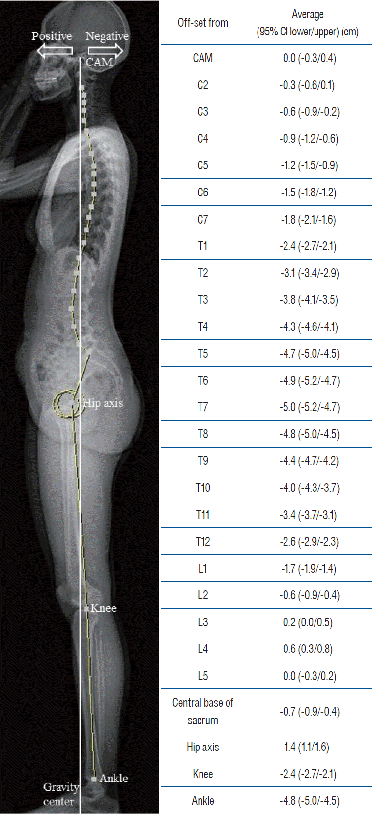
The “cone of economy” concept refer to a stable region in the standing posture that requires a minimum of muscle activity in normal conditions [11]. Hasegawa et al. [17] reported the standing sagittal alignment of whole axial skeletons using EOS in 158 healthy human volunteers (mean age, 39.7 years; range, 20–69 years). They defined the center of the head as the center of acoustic meatus (CAM) (Fig. 1). The gravity line (GL), defined as a line through the center of vertical pressure, was measured by a force plate (ANIMA®, Tokyo, Japan). The mean offset of the CAM was 0. The mean offset of the cervical and thoracic vertebrae was posterior to the GL with the apex of thoracic kyphosis at T7. Thoracic kyphosis changed to LL at the level of L2. The alignment changed to posterior through the apex of the LL at L4, and the base of the sacrum was just posterior to the GL. The hip axis was 1.4 cm anterior to the GL, the knee was 2.4 cm posterior to the GL, and the ankle was 4.8 cm posterior to the GL.
Posterior shifting of the lower lumbar vertebrae and sacrum increases with age, inducing loss of lordosis and an increase in PT. The C7 SVA increases with age. No correlation has been found between CAM–GL and age. Aging induces trunk stooping, but the global alignment compensates for by an increase in CL, PT, PI and knee flexion. This suggests that the horizontal gaze remains constant with age in healthy humans despite trunk stooping by compensatory changes in the cervical region, pelvis, and lower extremities.
Previously, PI has been believed to remain consistent with age. PI reflects the anatomical configuration of the pelvis and is an important parameter of sagittal balance that provides a target for optimal LL in thoracolumbar reconstruction surgery. Since the sacroiliac joint is thought not to move, PI has been considered to stable with age. However, some recent studies found that PI tended to increase with age [17,44]. It is thought that movement of the sacroiliac joint might increase due to osteoarthritic changes, but further study is needed.

Thoracolumbar reconstruction surgery affects GSA

Patients with thoracolumbar malalignment exhibit compensatory changes, including cervical hyperlordosis, pelvic shift, knee flexion, hip extension and pelvic retroversion [29]. Ha et al. [16] studied 49 patients undergoing thoracolumbar spine surgery results in reciprocal changes in GSA. They suggested that cervical alignment is affected by thoracic regional alignment, and that GSA and reciprocal changes are correlated with the magnitude of sagittal deformities. TS is correlated with the CL and the C7 SVA. Changes in the C7 SVA after surgery induce changes in TS, which may serve as a driving force for inducing reciprocal changes in cervical alignment. TS has been demonstrated to be the radiologic parameter most closely correlated with the C7 SVA in other studies [22]. Patients with a low preoperative C7 SVA group (C7 SVA ≤6 cm) tended to have hypolordotic cervical curvatures (low TS and CL) and to have increased CL after surgery, whereas those with a high C7 SVA group (C7 SVA ≥9 cm) had a high TS and CL preoperatively and decreased CL after surgery. CL increases after larger iatrogenic reductions in thoracolumbar spine surgery.
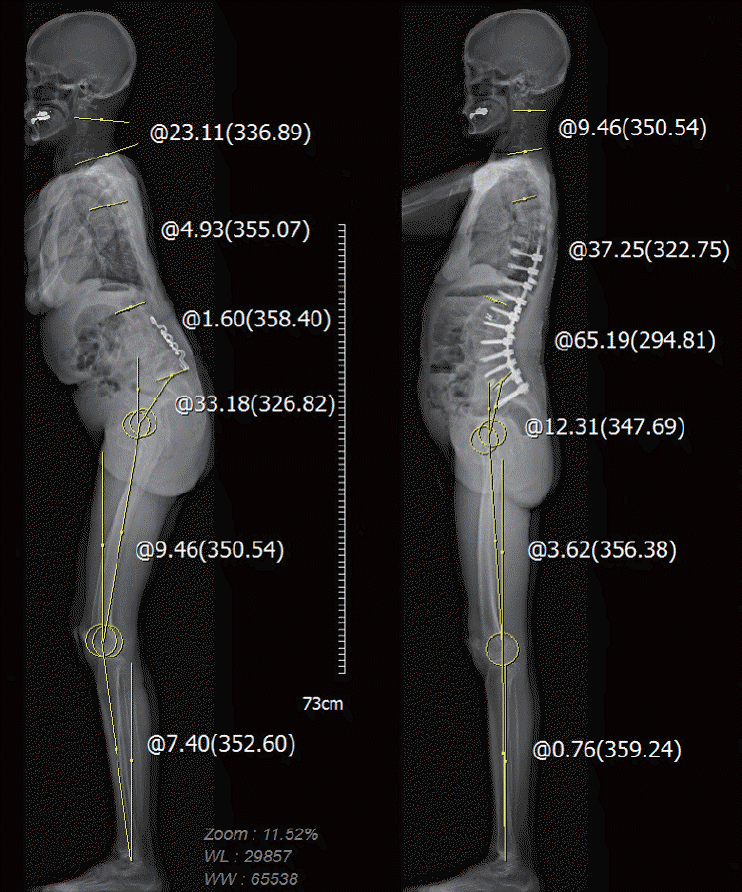
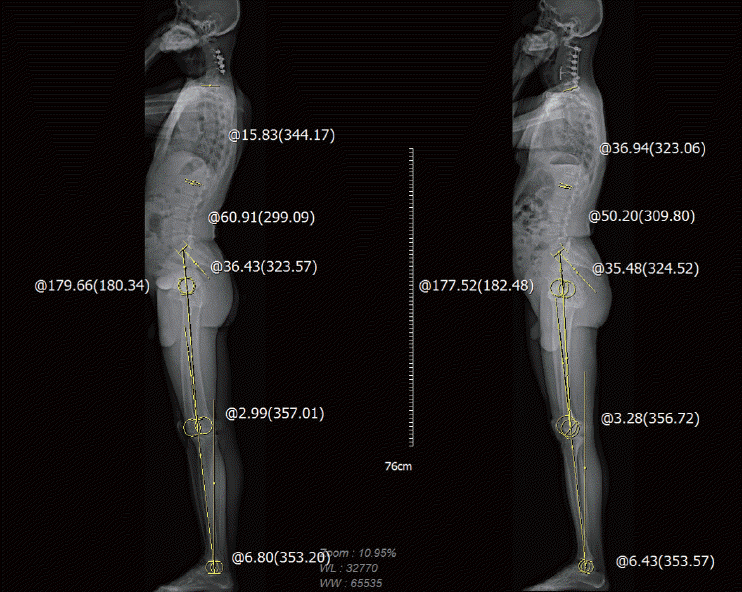
There is a reciprocal relationship between LL and thoracic kyphosis. Thoracic kyphosis and pelvic retroversion can corrected after restoration of LL [5]. Reciprocal reduction occurs in PT and C7 SVA in response to increases in LL. Posterior pelvic shift, knee flexion and hip extension also decrease when sagittal malalignment is corrected. The magnitude of the T1 pelvis angle is strongly correlated with reciprocal changes in the knee angle, hip extension, and posterior pelvic shift (Fig. 2) [7].
Several studies have investigated spontaneous improvements in cervical alignment after lumbar pedicle subtraction osteotomy (PSO) [6,35,36,41]. Lumbar PSO significantly improves LL. LL restoration induces a decreased need for compensation at the thoracic spine and pelvis. After lumbar PSO, thoracic kyphosis and sacral slope increase, and PT, PI–LL, knee flexion, and SVA decrease. In the cervical area, reciprocal changes lead to reductions in CL and TS following surgery. There is a significant decrease in distal CL because there is no more need for compensation within the distal cervical spine to maintain horizontal gaze, whereas there is no significant decrease in proximal CL following surgery.

Assessment of cervical spinal deformity

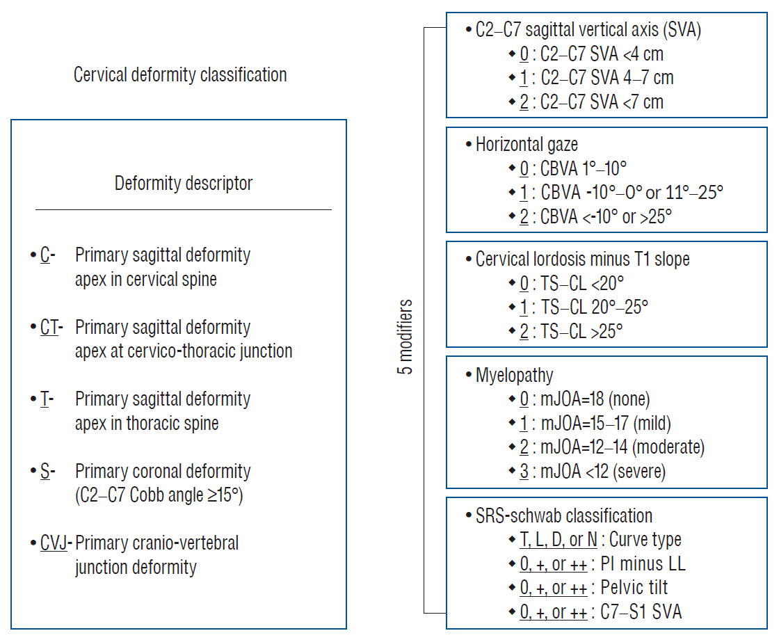
Ames and colleagues [1] made a meaningful attempt to create a comprehensive classification of CSD using a modified version of the Delphi approach. The proposed classification includes a basic deformity descriptor and modifiers based on clinical relevance and impact on patient HRQOL. This classification provides a mechanism to assess CSD with GSA and clinically relevant parameters. The authors defined C2–C7 SVA, horizontal gaze, TS–CL, myelopathy and the Scoliosis Research Society (SRS)-schwab classification as five modifiers. First, regarding C2–C7 SVA, Tang and colleagues [43] reported that C2–C7 SVA was negatively correlated with SF-36 physical component scores and positively correlated with Neck disability index (NDI) scores. Second, for horizontal gaze, significant correlations were found between C2–C7 SVA and C1–C2 lordosis [43]. C1–C2 alignment is the terminal link between the cranium and the cervical spine to regulate the angle of the gaze. A CBVA of 10° has been described as an optimal target [39]. Third, for TS–CL, the relationship between TS and CL is similar to the relationship between PI and LL, in that a greater TS requires a greater CL for harmonious alignment [27]. Hyun et al. [18] reported that TS–CL was strongly correlated with disability. Fourth, myelopathy may occur in patients with CSD due to direct compression resulting from spondylotic changes and the deformity itself. The development of myelopathy has been associated with progressive cervical kyphosis and positive sagittal cervical malalignment (C2–C7 SVA) [39]. Fifth, regarding the SRS-Schwab classification, cervical deformities contribute to thoracolumbar deformities. Thus, it is apparent that surgeons should not only focus on cervical alignment, but also on GSA. Adults with positive sagittal spinopelvic malalignment tend to compensate with abnormally increased CL in an effort to maintain horizontal gaze. The assessment and classification of CSD should not occur in isolation, which is why the alignment of a thoracolumbar spinal deformity was selected as a modifier for the CSD classification. Therefore, a full-length lateral radiograph is needed even when evaluating a cervical deformity.
Recently, Hyun et al. [18] suggested revised modifiers (Fig. 3). The authors investigated the validity of a CSD classification system using long-term follow-up data. They studied 30 consecutive patients with average follow-up period for 7.3 years. They demonstrated that C2–C7 SVA values of 4 cm and 7 cm were correlated with moderate and severe disability based on NDI scores. The authors found that C2–C7 SVA values of 4 cm and 7 cm corresponded to TS–CL values of 20° and 25°, respectively. TS–CL mismatch was significantly associated with disability and cervical malalignment. Therefore, a TS–CL less than 20° might be used for preoperative surgical planning, similar to a PI–LL less than 9°.

Cervical reconstruction surgery affects GSA

Only a few authors have analyzed the effect of GSA changes after cervical reconstruction surgery [26,33]. Cervical sagittal imbalance causes significant impairment due to the inability to gaze horizontally and is associated with poorer HRQOL [20,28].
Mizutani et al. [33] recently reported on the effects of cervical reconstruction surgery on thoracolumbar alignment changes. The C7 PL and GL are usually concordant, and GL is measured as the center of gravity of the head plumb line (COG PL). A distance between COG PL and C7 PL over 30 mm is defined as occiput-trunk (OT)-discordance. Compensation in primary cervical kyphosis occurs via posterior shifting of C7 SVA, a small TS, and large LL. The goal of cervical reconstruction surgery is therefore to achieve OT-concordance and cervical sagittal balance. COG PL and C7 PL move onto the femoral head as needed to achieve OT-concordance [34]. Subsequent thoracolumbar alignment changes occur as needed to harmonize GSA.
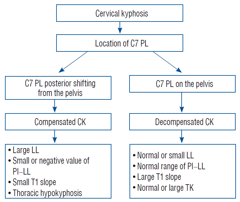
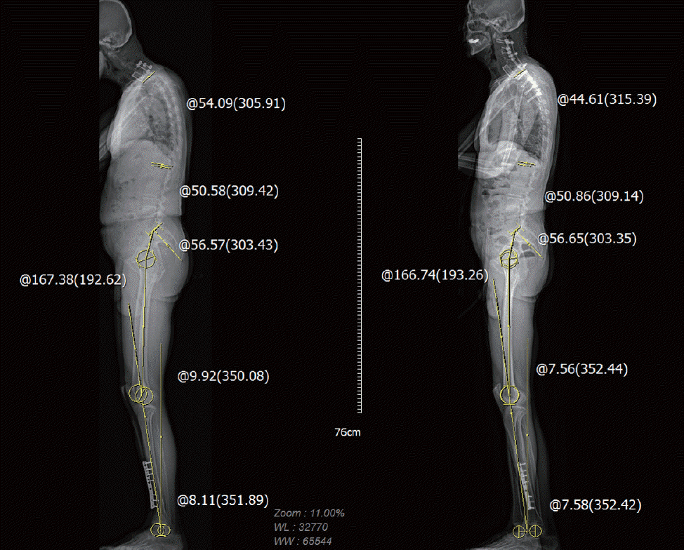
Reciprocal changes after cervical reconstruction surgery depend on whether patients have head-balanced (compensated) or trunk-balanced (decompensated) kyphosis (Fig. 4). Headbalanced kyphosis is characterized by a head position on the femoral head with posterior shifting of the C7 PL, hypokyphotic thoracic curve, straightened thoracolumbar junction, and lumbar hyperlordosis. The preoperative posterior shifted C7 PL moves anteriorly after surgery, and subsequently TS and the thoracic curve increase while LL decreases. However, pelvic and lower extremity parameters do not change after kyphosis correction (Fig. 5). In the trunk-balanced kyphosis, the COG PL is anteriorly shifted, which causes the C7 PL to remain anterior (located on the pelvis) compared to head-balanced kyphosis. These decompensated patients have a large preoperative TS and low LL. The LL is at the upper limit of PI–LL harmonization, which is the result of lumbar extension with maximum effort or lumbar degeneration. There are no significant changes in thoracolumbar alignment in trunk-balanced patients after kyphosis correction (Fig. 6). While TS and thoracic kyphosis decrease, spinopelvic and lower extremity parameters do not change after kyphosis correction. The COG PL moves posteriorly to achieve better GSA after surgery.

Systematic review of previous studies

We reviewed 11 articles to synthesize the available information on GSA and reciprocal changes in healthy humans and patients with deformities. Only recently has there been widespread recognition of the importance of global imaging of the spine in the setting of deformities, and only a few studies have sought to define reciprocal changes in terms of GSA. In some studies, surgical procedures were not uniform and the follow-up was short-term. The CSD classification was derived from a modified Delphi approach and expert opinion. Thus, although initial steps have been taken provide a mechanism to assess CSD within the framework of GSA, further cohort studies are needed to validate the classification.
Sagittal malalignment after surgery is a major source of pain and disability. Positive sagittal malalignment was identified as the radiographic parameter most closely correlated with adverse health status outcomes [15]. There is a significant correlation between C7 SVA and TS, as well as between LL and thoracic kyphosis [5,16]. Furthermore, reciprocal changes are affected by preoperative sagittal alignment. Therefore, when patients undergo corrective surgery for a deformity, it is important to evaluate whole-body images both preoperatively and postoperatively.
Radiographs produced by conventional X-ray, computed tomography and the EOS can provide reliable and accurate Cobb angle measurements for scoliosis assessments. None of the methods systemically underestimates or overestimates the Cobb angle measurement. However, the static global balance of a patient cannot be evaluated properly on a conventional radiograph or computed tomography in which all the elements of the chain of balance (lower limbs, cervical spine and head) are not taken into account [13]. EOS evaluation for the correction of a deformity would make a big difference in the preoperative planning and postoperative evaluation.

CONCLUSION

Following the correction of a deformity, reciprocal changes occur in the flexible segments proximal and distal to the area of correction. These changes occur not only through mobile spine segments, but also PT and the lower extremities in order to maintain a horizontal gaze and the GL [3,41]. Restoration of LL induces reciprocal changes in TS, CL, pelvic shift, and lower extremity parameters following surgery to correct a thoracolumbar deformity. Patients with cervical kyphosis exhibit different reciprocal changes depending on whether they have head-balanced or trunk-balanced kyphosis.
The proverb, “Do not miss the forest for the trees” is helpful when evaluating these reciprocal global alignment changes. It is better to take “whole-body” films rather than “whole-spine” images to understand global alignment and the status of compensation.

Notes

CONFLICTS OF INTEREST

No potential conflict of interest relevant to this article was reported.

INFORMED CONSENT

This type of study does not require informed consent.

AUTHOR CONTRIBUTIONS

Conceptualization : SJH, KJK

Data curation : SJH, CWK

Formal analysis : SJH, KJK

Methodology : SJH, CWK

Project administration : SJH, CWK

Visualization : SJH, CWK

Writing - original draft : CWK

Writing - review & editing : SJH, KJK

References

1. Ames CP, Smith JS, Eastlack R, Blaskiewicz DJ, Shaffrey CI, Schwab F, et al. Reliability assessment of a novel cervical spine deformity classification system. J Neurosurg Spine. 23:673–683. 2015.
crossref
2. Ames CP, Smith JS, Scheer JK, Bess S, Bederman SS, Deviren V, et al. Impact of spinopelvic alignment on decision making in deformity surgery in adults: a review. J Neurosurg Spine. 16:547–564. 2012.
crossref
3. Barrey C, Roussouly P, Le Huec JC, D’Acunzi G, Perrin G. Compensatory mechanisms contributing to keep the sagittal balance of the spine. Eur Spine J 22 Suppl. 6:S834–S841. 2013.
crossref
4. Barrey C, Roussouly P, Perrin G, Le Huec JC. Sagittal balance disorders in severe degenerative spine. Can we identify the compensatory mechanisms? Eur Spine J 20 Suppl. 5:626–633. 2011.
5. Blondel B, Lafage V, Schwab F, Farcy JP, Bollini G, Jouve JL. Reciprocal sagittal alignment changes after posterior fusion in the setting of adolescent idiopathic scoliosis. Eur Spine J. 21:1964–1971. 2012.
crossref
6. Choi HY, Hyun SJ, Kim KJ, Jahng TA, Kim HJ. Radiographic and clinical outcomes following pedicle subtraction osteotomy : minimum 2-year follow-up data. J Korean Neurosurg Soc. 63:99–107. 2020.
crossref
7. Day LM, Ramchandran S, Jalai CM, Diebo BG, Liabaud B, Lafage R, et al. Thoracolumbar realignment surgery results in simultaneous reciprocal changes in lower extremities and cervical spine. Spine (Phila Pa 1976). 42:799–807. 2017.
crossref
8. Deschênes S, Charron G, Beaudoin G, Labelle H, Dubois J, Miron MC, et al. Diagnostic imaging of spinal deformities: reducing patients radiation dose with a new slot-scanning X-ray imager. Spine (Phila Pa 1976). 35:989–994. 2010.
9. Diebo BG, Ferrero E, Lafage R, Challier V, Liabaud B, Liu S, et al. Recruitment of compensatory mechanisms in sagittal spinal malalignment is age and regional deformity dependent: a full-standing axis analysis of key radiographical parameters. Spine (Phila Pa 1976). 40:642–649. 2015.
crossref
10. Dru AB, Lockney DT, Vaziri S, Decker M, Polifka AJ, Fox WC, et al. Cervical spine deformity correction techniques. Neurospine. 16:470–482. 2019.
crossref
11. Dubousset J. Three-dimensional analysis of the scoliotic deformity. In : Weinstein SL, editor. The pediatric spine: principles and practice, ed 1. New York: Raven Press;1994. p. 479–496.
12. Ferrero E, Liabaud B, Challier V, Lafage R, Diebo BG, Vira S, et al. Role of pelvic translation and lower-extremity compensation to maintain gravity line position in spinal deformity. J Neurosurg Spine. 24:436–446. 2016.
crossref
13. Glaser DA, Doan J, Newton PO. Comparison of 3-dimensional spinal reconstruction accuracy: biplanar radiographs with EOS versus computed tomography. Spine (Phila Pa 1976). 37:1391–1397. 2012.
14. Glassman SD, Berven S, Bridwell K, Horton W, Dimar JR. Correlation of radiographic parameters and clinical symptoms in adult scoliosis. Spine (Phila Pa 1976). 30:682–688. 2005.
crossref
15. Glassman SD, Bridwell K, Dimar JR, Horton W, Berven S, Schwab F. The impact of positive sagittal balance in adult spinal deformity. Spine (Phila Pa 1976). 30:2024–2029. 2005.
crossref
16. Ha Y, Schwab F, Lafage V, Mundis G, Shaffrey C, Smith J, et al. Reciprocal changes in cervical spine alignment after corrective thoracolumbar deformity surgery. Eur Spine J. 23:552–559. 2014.
crossref
17. Hasegawa K, Okamoto M, Hatsushikano S, Shimoda H, Ono M, Homma T, et al. Standing sagittal alignment of the whole axial skeleton with reference to the gravity line in humans. J Anat. 230:619–630. 2017.
crossref
18. Hyun SJ, Han S, Kim KJ, Jahng TA, Kim HJ. Assessment of T1 slope minus cervical lordosis and C2-7 sagittal vertical axis criteria of a cervical spine deformity classification system using long-term follow-up data after multilevel posterior cervical fusion surgery. Oper Neurosurg (Hagerstown). 16:20–26. 2019.
crossref
19. Hyun SJ, Jung JM. Spinal deformity surgery : it becomes an essential part of neurosurgery. J Korean Neurosurg Soc. 61:661–668. 2018.
crossref
20. Iyer S, Nemani VM, Nguyen J, Elysee J, Burapachaisri A, Ames CP, et al. Impact of cervical sagittal alignment parameters on neck disability. Spine (Phila Pa 1976). 41:371–377. 2016.
crossref
21. Kang J, Hosogane N, Ames C, Schwab F, Hart R, Burton D, et al. Diversity in surgical decision strategies for adult spine deformity treatment: the effects of neurosurgery or orthopedic training background and surgical experience. Neurospine. 15:353–361. 2018.
crossref
22. Knott PT, Mardjetko SM, Techy F. The use of the T1 sagittal angle in predicting overall sagittal balance of the spine. Spine J. 10:994–998. 2010.
crossref
23. Kuntz C 4th, Levin LS, Ondra SL, Shaffrey CI, Morgan CJ. Neutral upright sagittal spinal alignment from the occiput to the pelvis in asymptomatic adults: a review and resynthesis of the literature. J Neurosurg Spine. 6:104–112. 2007.
crossref
24. Lafage R, Challier V, Liabaud B, Vira S, Ferrero E, Diebo BG, et al. Natural head posture in the setting of sagittal spinal deformity: validation of chin-brow vertical angle, slope of line of sight, and McGregor’s slope with health-related quality of life. Neurosurgery. 79:108–115. 2016.
crossref
25. Lafage V, Schwab F, Skalli W, Hawkinson N, Gagey PM, Ondra S, et al. Standing balance and sagittal plane spinal deformity: analysis of spinopelvic and gravity line parameters. Spine (Phila Pa 1976). 33:1572–1578. 2008.
26. Lee DH, Ha JK, Chung JH, Hwang CJ, Lee CS, Cho JH. A retrospective study to reveal the effect of surgical correction of cervical kyphosis on thoraco-lumbo-pelvic sagittal alignment. Eur Spine J. 25:2286–2293. 2016.
crossref
27. Lee SH, Kim KT, Seo EM, Suk KS, Kwack YH, Son ES. The influence of thoracic inlet alignment on the craniocervical sagittal balance in asymptomatic adults. J Spinal Disord Tech. 25:E41–E47. 2012.
crossref
28. Lee SH, Son DW, Lee JS, Kim DH, Sung SK, Lee SW, et al. Differences in cervical sagittal alignment changes in patients undergoing laminoplasty and anterior cervical discectomy and fusion. Neurospine. 15:91–100. 2018.
crossref
29. Makhni MC, Shillingford JN, Laratta JL, Hyun SJ, Kim YJ. Restoration of sagittal balance in spinal deformity surgery. J Korean Neurosurg Soc. 61:167–179. 2018.
crossref
30. McClendon J Jr, Graham RB, Sugrue PA, Smith TR, Thompson SE, Koski TR. Cranial center of mass compared to C7 plumb line alignment in adult spinal deformity. World Neurosurg. 91:199–204. 2016.
crossref
31. Melhem E, Assi A, El Rachkidi R, Ghanem I. EOS(®) biplanar X-ray imaging: concept, developments, benefits, and limitations. J Child Orthop. 10:1–14. 2016.
crossref
32. Miyazaki M, Hymanson HJ, Morishita Y, He W, Zhang H, Wu G, et al. Kinematic analysis of the relationship between sagittal alignment and disc degeneration in the cervical spine. Spine (Phila Pa 1976). 33:E870–E876. 2008.
crossref
33. Mizutani J, Strom R, Abumi K, Endo K, Ishii K, Yagi M, et al. How cervical reconstruction surgery affects global spinal alignment. Neurosurgery. 84:898–907. 2019.
crossref
34. Mizutani J, Verma K, Endo K, Ishii K, Abumi K, Yagi M, et al. Global spinal alignment in cervical kyphotic deformity: the importance of head position and thoracolumbar alignment in the compensatory mechanism. Neurosurgery. 82:686–694. 2018.
crossref
35. Obeid I, Boniello A, Boissiere L, Bourghli A, Pointillart V, Gille O, et al. Cervical spine alignment following lumbar pedicle subtraction osteotomy for sagittal imbalance. Eur Spine J. 24:1191–1198. 2015.
crossref
36. Park JH, Hyun SJ, Kim KJ, Jahng TA. Comparative study between pedicle subtraction osteotomy (PSO) and closing-opening wedge osteotomy (fish-mouth PSO) for sagittal plane deformity correction. Spine (Phila Pa 1976). 42:E899–E905. 2017.
crossref
37. Roussouly P, Pinheiro-Franco JL. Sagittal parameters of the spine: biomechanical approach. Eur Spine J 20 Suppl. 5:578–585. 2011.
crossref
38. Scheer JK, Ames CP, Deviren V. Assessment and treatment of cervical deformity. Neurosurg Clin N Am. 24:249–274. 2013.
crossref
39. Scheer JK, Tang JA, Smith JS, Acosta FL Jr, Protopsaltis TS, Blondel B, et al. Cervical spine alignment, sagittal deformity, and clinical implications: a review. J Neurosurg Spine. 19:141–159. 2013.
40. Sharp J, Bouton D, Anabell L, Degan T, Sienko S, Welborn MC. Image distortion in biplanar slot scanning: part 2 technology-specific factors. J Pediatr Orthop. 40:587–591. 2020.
crossref
41. Smith JS, Shaffrey CI, Lafage V, Blondel B, Schwab F, Hostin R, et al. Spontaneous improvement of cervical alignment after correction of global sagittal balance following pedicle subtraction osteotomy. J Neurosurg Spine. 17:300–307. 2012.
crossref
42. Suk KS, Kim KT, Lee SH, Kim JM. Significance of chin-brow vertical angle in correction of kyphotic deformity of ankylosing spondylitis patients. Spine (Phila Pa 1976). 28:2001–2005. 2003.
crossref
43. Tang JA, Scheer JK, Smith JS, Deviren V, Bess S, Hart RA, et al. The impact of standing regional cervical sagittal alignment on outcomes in posterior cervical fusion surgery. Neurosurgery. 71:662–669. discussion 669. 2012.
crossref
44. Vrtovec T, Janssen MM, Pernuš F, Castelein RM, Viergever MA. Analysis of pelvic incidence from 3-dimensional images of a normal population. Spine (Phila Pa 1976). 37:E479–E485. 2012.
crossref
45. Wui SH, Hyun SJ, Kang B, Kim KJ, Jahng TA, Kim HJ. Bicortical screw purchase at upper instrumented vertebra (UIV) can cause UIV fracture after adult spinal deformity surgery: a finite element analysis study. Neurospine. 17:377–383. 2020.
crossref
46. Wybier M, Bossard P. Musculoskeletal imaging in progress: the EOS imaging system. Joint Bone Spine. 80:238–243. 2013.
crossref

Fig. 1.
Normative offset distance between bony landmarks and the gravity line [17]. Positive means anterior to the gravity line and negative means posterior to the gravity line. CAM : center of acoustic meatus, CI : confidence interval.
jkns-2020-0234f1.tif
Fig. 2.
Whole body image of thoracolumbar malalignment (left) exhibiting cervical hyperlordosis, posterior pelvic shift and knee flexion. Postoperative whole body image of the same patient (right) exhibiting restoration of lumbar lordosis (from 1.6° to 65.2°) with reciprocal changes in thoracic kyphosis (from -4.9° to 37.2°) cervical lordosis (from 23.1° to 9.5°), pelvic tilt (from 33.1° to 12.3°), knee flexion (from 9.5° to 3.6°) and ankle dorsiflexion (from 7.4° to 0.7°).
jkns-2020-0234f2.tif
Fig. 3.
Revised cervical spinal deformity classification system [18], which consists of a deformity descriptor and five modifiers. T : thoracic major, CBVA : chin-brow to vertical angle, TS : T1 slope, CL : cervical lordosis, mJOA : modified Japanese Orthopaedic Association, SRS : Scoliosis Research Society, L : lumbar major, D : double, N : no scoliosis, LL : lumbar lordosis.
jkns-2020-0234f3.tif
Fig. 4.
Compensation mechanisms in patients with symptomatic primary cervical kyphosis. PL : plumb line, CK : cervical kyphosis, LL : lumbar lordosis, PI : pelvic incidence, TK : thoracic kyphosis.
jkns-2020-0234f4.tif
Fig. 5.
Whole body images of a head-balanced (compensated) patient. While T1 slope, thoracic kyphosis, and lumbar lordosis changed, spinopelvic- and lower extremity parameters did not change following cervical kyphosis correction.
jkns-2020-0234f5.tif
Fig. 6.
Whole body images of a trunk-balanced (decompensated) patient. While T1 slope and thoracic kyphosis decreased, lumbar lordosis, spinopelvic- and lower extremity parameters did not change following cervical kyphosis correction.
jkns-2020-0234f6.tif
Table 1.
Summary of the literatures included in this systemic review
Study Sample population, duration Object Inclusion criteria Conclusion
Ames et al. [2] (2012) Review using modified Delphi approach. Reliability study was conducted using 10 clinical cases by 20 spinal deformity surgeons To develop CSD classification system 10 cervical spine deformity cases, broadly representative of the proposed classification system Proposed classification provides a mechanism to assess CSD within the frame work of global spinopelvic alignment
Hyun et al. [18] (2019) 30; average follow-up period, 7.3 years To revise and validate CSD classification system Patients treated with multilevel (≥3) posterior cervical fusion C2–C7 SVA (4 cm and 7 cm) and TS–CL (20° and 25°) correlated with moderate and severe disability
Ha et al. [16] (2014) 39 To identify changes in cervical parameters following thoracolumbar spine surgery Patients treated with multilevel (≥5) thoracolumbar fusion Reciprocal changes are correlated with sagittal deformity
Mizutani et al. [33] (2019) 78 (head-balanced group, 2; trunk-balanced group, 36) To elucidate effects of cervical reconstruction for GSA Cervical kyphosis patients who were recommended surgery The primary goal of cervical reconstruction surgery is to achieve occiput-trunk concordance
Obeid et al. [35] (2015) 31, 3-month follow-up To investigate cervical alignment after lumbar PSO Sagittal deformity patients operated by lumbar PSO Lumbar PSO improve LL and decrease the need of compensation at the thoracic spine and pelvis
Mizutani et al. [34] (2018) 222 (cervical kyphosis patients, 103; thoracolumbar deformity patients, 119) To elucidate GSA and compensatory mechanisms in primary cervical kyphosis patients Cervical kyphosis, thoracolumbar deformity patients who underwent surgery Compensation in cervical kyphosis is via posterior shifting of SVA C7, small T1 slope and large LL
Smith et al. [41] (2012) 75 To evaluate reciprocal changes within the cervical spine following lumbar PSO Patients treated using lumbar PSO, preoperative C7–S1 plumb line greater than 5 cm Surgical correction of sagittal malalignment results in improvement of cervical hyperlordosis
Hasegawa et al. [17] (2017) 136 To clarify standing sagittal alignment of whole axial skeletons Volunteers with no history of treatment for spinal disease Aging induces trunk stooping, but the GSA is compensated for by an increased CL, PT and knee flexion
Lee et al. [26] (2016) 55 To assess effect of surgical correction of cervical kyphosis on thoracolumbo-pelvic alignment Patients who underwent ≥2 level cervical fusions for cervical radiculopathy or myelopathy Surgical correction of cervical kyphosis affects T1 slope and thoracic kyphosis, but not lumbo-pelvic alignment
Day et al. [7] (2017) 87 To assess cervical spine and lower extremity compensatory changes in thoracolumbar alignment Patients who underwent thoracolumbar fusion without cervical spine fusion Thoracolumbar malalignment patients exhibit compensatory changes in cervical hyperlordosis, pelvic shift, knee flexion, and pelvic retroversion
Ferrero et al. [12] (2016) 336 To analyze the relationship between spinopelvic- and lower extremity parameters Patients with spinal deformity and full-body EOS images Significant correlations exist between lower-limb parameters and pelvic shift, PT and SVA

CSD : cervical spine deformity, SVA : sagittal vertical axis, TS : T1 slope, CL : cervical lordosis, GSA : global spinal alignment, PSO : pedicle subtraction osteotomy, LL : lumbar lordosis, PT : pelvic tilt

TOOLS
Similar articles